. 2017 Jul 25;8(30):48545-48562.
doi: 10.18632/oncotarget.18153.

ZNF131 suppresses centrosome fragmentation in glioblastoma stem-like cells through regulation of HAUS5

Affiliations

ZNF131 suppresses centrosome fragmentation in glioblastoma stem-like cells through regulation of HAUS5

Yu Ding et al. Oncotarget. .

Abstract

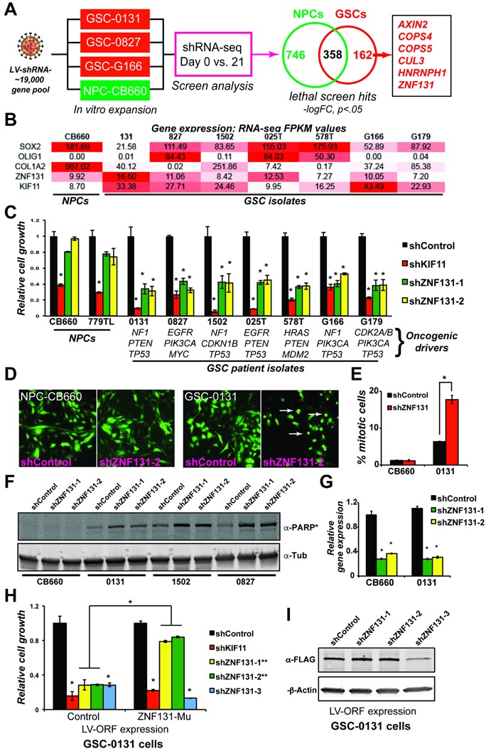
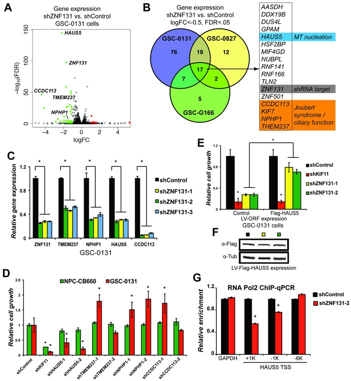
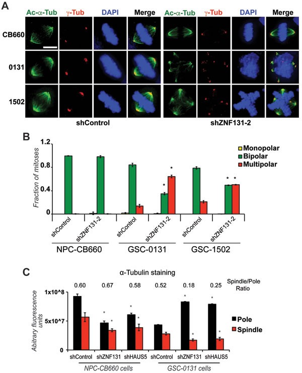
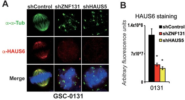
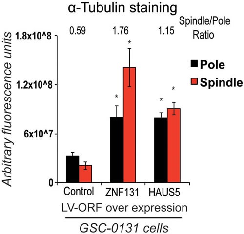
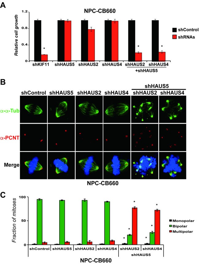
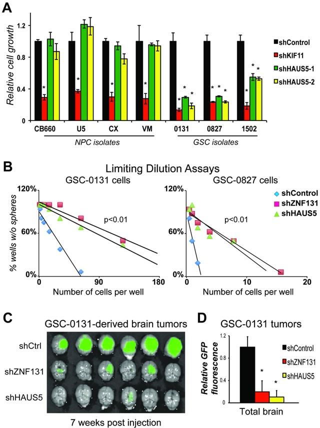
Zinc finger domain genes comprise ~3% of the human genome, yet many of their functions remain unknown. Here we investigated roles for the vertebrate-specific BTB domain zinc finger gene ZNF131 in the context of human brain tumors. We report that ZNF131 is broadly required for Glioblastoma stem-like cell (GSC) viability, but dispensable for neural progenitor cell (NPC) viability. Examination of gene expression changes after ZNF131 knockdown (kd) revealed that ZNF131 activity notably promotes expression of Joubert Syndrome ciliopathy genes, including KIF7, NPHP1, and TMEM237, as well as HAUS5, a component of Augmin/HAUS complex that facilitates microtubule nucleation along the mitotic spindle. Of these genes only kd of HAUS5 displayed GSC-specific viability loss. Critically, HAUS5 ectopic expression was sufficient to suppress viability defects of ZNF131 kd cells. Moreover, ZNF131 and HAUS5 kd phenocopied each other in GSCs, each causing: mitotic arrest, centrosome fragmentation, loss of Augmin/HAUS complex on the mitotic spindle, and loss of GSC self-renewal and tumor formation capacity. In control NPCs, we observed centrosome fragmentation and lethality only when HAUS5 kd was combined with kd of HAUS2 or HAUS4, demonstrating that the complex is essential in NPCs, but that GSCs have heightened requirement. Our results suggest that GSCs differentially rely on ZNF131-dependent expression of HAUS5 as well as the Augmin/HAUS complex activity to maintain the integrity of centrosome function and viability.

Keywords: Augmin/HAUS complex; HAUS5; ZNF131; cancer therapeutics; glioblastoma.


Conflict of interest statement

CONFLICTS OF INTEREST

The authors disclose no potential conflicts of interest.

Figures

Figure 1
Figure 1. ZNF131 is a candidate GSC-lethal gene
A. Overview of GSCs and NPCs shRNA screens that gave rise to ZNF131 as a candidate GSC-lethal gene. B. ZNF131 expression among NPC and GSC isolates. Values are from FPKM normalized RNA-seq data from self-renewing in vitro cultures. C. Short term growth assays showing differential viability requirement for ZNF131 among GSC and NPC isolates. Cell growth assays were performed 7 days after selection for LV-shRNAs in monolayer culture (n ≥ 3). D. Fluorescent micrographs of shRNA transduced cells (GFP+). Arrows show GSC cells displaying mitotic arrest phenotype observed with ZNF131 kd. E. Quantification of mitotic cells from D.. F. Western blot to detect cleavage of PARP protein, an indicator of apoptotic cell death, in NPC-CB660 and three GSC isolates after knockdown of ZNF131. Cells were assayed 48hrs post-selection after LV-shRNA infection (n = 3). G. RT-qPCR analysis of ZNF131 kd in NPCs and GSCs. Cells were assayed 48hrs post-selection after LV-shRNA infection (n = 3). H. Suppression of growth defects caused by shZNF131 using shRNA resistant ZNF131 ORF. Cells were first infected with LV containing control or shRNA resistant ORF followed by LV-shRNA and assayed for cell growth in monolayer culture 7 days post selection (n = 3). Target sites for ZNF131 shRNAs #1 and #2 where both mutated in the ORF construct and thus made resistant, while the site for shRNA #3 was left unchanged. I. Western blots of shRNA resistant ORF expression. *indicates p < .01 student's t-test.
Figure 2
Figure 2. ZNF131 regulates expression of the HAUS5 gene to promote expansion of GSCs
A. A volcano plot of changes in mRNA-sequencing resulting from ZNF131 kd in GSC-0131 cells. B. A Venn diagram summary of overlap in genes whose expression is down regulated after ZNF131 kd in 0131, 0827, and G166 GSC isolates. RNA-seq data is available in Supplementary Table S2. C. RT-qPCR confirmation of RNA-seq results from A. and B. for ZNF131, TMEM237, NPHP1, HAUS5, and CCDC113. D. Retests of a subset of genes from B. in knockdown growth assays in GSC-0131 and NPC-CB660 cells, including for ZNF131, TMEM237, NPHP1, HAUS5, and CCDC113. E. Suppression of ZNF131 kd lethality by stable ectopic expression of the HAUS5 ORF. Cells were first infected with LV containing control or HAUS5 ORF followed by LV-shRNA and assayed for cell growth in monolayer culture 7 days post selection (n = 3). F. Western blot analysis of GSC-0131 cells expressing HAUS5 ORF used for E.. G. Chromatin-immunoprecipitation of RNA polymerase II at the HAUS5 or GAPDH (control) promoter with and without ZNF131 kd. *indicates p < .01 student's t-test.
Figure 3
Figure 3. ZNF131 kd causes centrosome fragmentation, multipolar spindle formation, and loss of MT-nucleation
A. Examination of spindle polarity and centrosome morphology in NPCs and GSCs with and without ZNF131 kd. Cells were stained for Acetylated-α-Tubulin and γ-Tubulin to reveal changes to spindle and centrosome, respectively, and analyzed using deconvolved 3D images from z-stacked sections (0.2μm intervals). Scale bar indicates 5 μm. B. Quantification of spindle polarity in A. (n > 20). C. Quantification of α-tubulin staining at spindle pole and along spindle in NPCs and GSCs with and without ZNF131 and HAUS5 kd (n = 5).
Figure 4
Figure 4. Knockdown of ZNF131 and HAUS5 results in loss of Augmin/HAUS complex along the mitotic spindle
A. HAUS6 staining along the mitotic spindle of GSCs and NPCs with and without ZNF131 and HAUS5 kd. B. Quantification of A. using deconvolved 3D images from z-stacked sections (0.2μm intervals) as detailed in methods details. *indicates p < .01 student's t-test.
Figure 5
Figure 5. Overexpression of either ZNF131 or HAUS5 kd cause increased MT-nucleation along the mitotic spindle and at spindle poles
Quantification of α-tubulin staining at spindle pole and along spindle was performed 48hrs after selection for LV-ZNF131 and LV-HAUS5 in GSC-0131 cells as detailed in methods (n = 30 for each group) (using devolved 3D images aquired by a Deltavision imaging station). *indicates p-value < .001, as compared to control values.
Figure 6
Figure 6. Combinatorial kd of HAUS5+HAUS2 or HAUS5+HAUS4 cause centrosome fragmentation and multipolar spindle formation in NPC-CB660 cells
A. Combination of HAUS5+HAUS2 or HAUS5+HAUS4 kd are synthetic lethal in NSC-CB660 cells, based on growth assays 7 days post selection with LV-shRNAs in monolayer culture. Experiments were performed as detailed in Methods (n = 3) using dual expression shRNA LV vectors. B. Representative images of of NSC-CB660 cells after knockdown of HAUS2, HAUS4, and/or HAUS5. Cells were stained for pericentrin and tubulin 48hrs after selection for LV-shRNAs according to Methods. C. Examination of spindle polarity from B. (n > 20). *indicates p-value < .01.
Figure 7
Figure 7. HAUS5 kd phenocopies ZNF131 kd in GSC growth, self-renewal, and tumorigenesis assays
A. Short term growth assays showing differential viability requirement for HAUS5 among GSC and NPC isolates, performed as in Figure 1 (n = 3). B. Tumorisphere limiting dilution assays with and without ZNF131 and HAUS5 kd in GSC-131 and GSC-0827 isolates. C. GSC-0131 brain tumorigenesis assays in immunocompromised mice with and without ZNF131 and HAUS5 kd in GSC-0131 (n = 6), seven weeks post-injection. D. Quantification of GFP intensity from (n = 6) C.. *indicates p < .01 student's t-test.

Similar articles

Cited by

References

    1. American Cancer Society American Cancer Society: Cancer Facts and Figures. 2010.
    1. Stupp R, Mason WP, van den Bent MJ, Weller M, Fisher B, Taphoorn MJ, Belanger K, Brandes AA, Marosi C, Bogdahn U, Curschmann J, Janzer RC, Ludwin SK, et al. Radiotherapy plus concomitant and adjuvant temozolomide for glioblastoma. N Engl J Med. 2005;352:987–96. doi: 10.1056/NEJMoa043330. - DOI - PubMed
    1. Hemmati HD, Nakano I, Lazareff JA, Masterman-Smith M, Geschwind DH, Bronner-Fraser M, Kornblum HI. Cancerous stem cells can arise from pediatric brain tumors. Proc Natl Acad Sci U S A. 2003;100:15178–83. - PMC - PubMed
    1. Singh SK, Clarke ID, Terasaki M, Bonn VE, Hawkins C, Squire J, Dirks PB. Identification of a cancer stem cell in human brain tumors. Cancer Res. 2003;63:5821–8. - PubMed
    1. Singh SK, Hawkins C, Clarke ID, Squire JA, Bayani J, Hide T, Henkelman RM, Cusimano MD, Dirks PB. Identification of human brain tumour initiating cells. Nature. 2004;432:396–401. - PubMed

MeSH terms