. 2017 Jun 8;7(1):3086.
doi: 10.1038/s41598-017-02771-4.

Leukocyte Bim deficiency does not impact atherogenesis in ldlr -/- mice, despite a pronounced induction of autoimmune inflammation

Affiliations

Leukocyte Bim deficiency does not impact atherogenesis in ldlr -/- mice, despite a pronounced induction of autoimmune inflammation

Lieve Temmerman et al. Sci Rep. .

Abstract

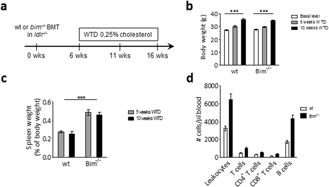
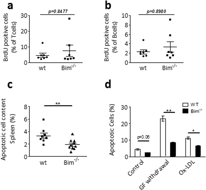
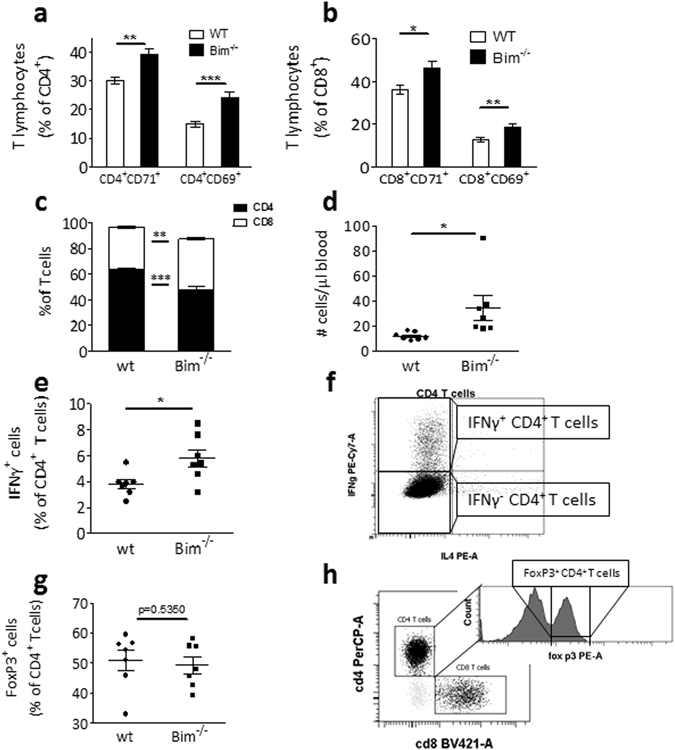
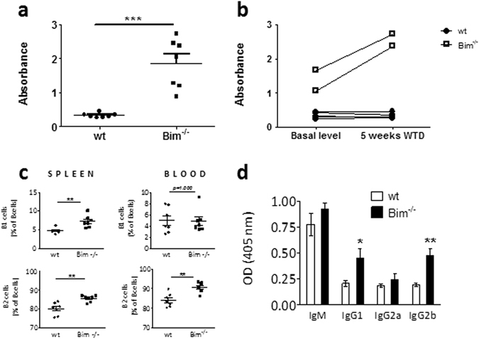
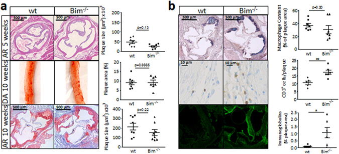
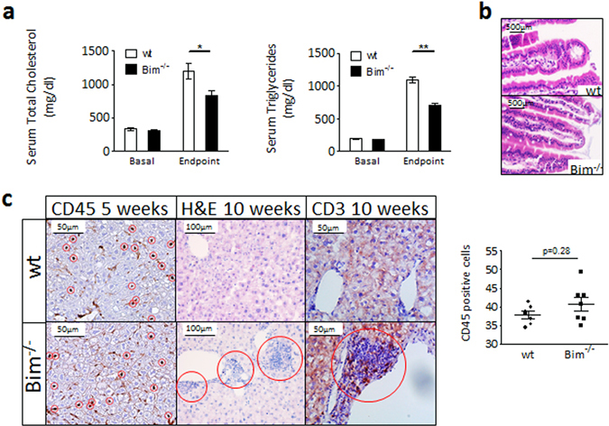
Proapoptotic Bcl-2 family member Bim is particularly relevant for deletion of autoreactive and activated T and B cells, implicating Bim in autoimmunity. As atherosclerosis is a chronic inflammatory process with features of autoimmune disease, we investigated the impact of hematopoietic Bim deficiency on plaque formation and parameters of plaque stability. Bim -/- or wild type bone marrow transplanted ldlr -/- mice were fed a Western type diet (WTD) for 5 or 10 weeks, after which they were immunophenotyped and atherosclerotic lesions were analyzed. Bim -/- transplanted mice displayed splenomegaly and overt lymphocytosis. CD4+ and CD8+ T cells were more activated (increased CD69 and CD71 expression, increased interferon gamma production). B cells were elevated by 147%, with a shift towards the pro-atherogenic IgG-producing B2 cell phenotype, resulting in a doubling of anti-oxLDL IgG1 antibody titers in serum of bim -/- mice. Bim -/- mice displayed massive intraplaque accumulation of Ig complexes and of lesional T cells, although this did not translate in changes in plaque size or stability features (apoptotic cell and macrophage content). The surprising lack in plaque phenotype despite the profound pro-atherogenic immune effects may be attributable to the sharp reduction of serum cholesterol levels in WTD fed bim -/- mice.


Conflict of interest statement

The authors declare that they have no competing interests.

Figures

Figure 1
Figure 1
Bim −/− chimeric mice have splenomegaly and lymphocytosis. (a) Lethally irradiated ldlr −/− mice were reconstituted with wt or bim −/− bone marrow, and after 6 weeks recovery, put on a Western Type Diet containing 0.25% cholesterol for 5 (n = 7) or 10 weeks (n = 12). (b) Body weight of wt and bim −/− chimeric mice. Data is presented as mean ± SEM. ***p < 0.001 in two-way ANOVA. (c) Relative spleen weight of wt and bim −/− chimeric mice at sacrifice. Data is presented as mean ± SEM. ***p < 0.001 in two-way ANOVA. (d) TruCount tubes were used for quantitative analysis of leukocyte subsets in blood of wt and bim −/− chimeric mice after 5 weeks of WTD. Data is presented as mean ± SEM. p < 0.001 between wt and bim −/− groups in two-way ANOVA.
Figure 2
Figure 2
Bim −/− leukocytes are more resistant towards apoptosis. (a,b) After 5 weeks of WTD, wt and bim −/− chimeric mice were injected with BrdU 24 and 12 hrs before sacrifice to quantify proliferating cells. BrdU incorporation in T (a) and B (b) cell subsets was measured by flow cytometry. Mean ± SEM are indicated. (c) Cyrosections of wt and bim −/− spleens after 10 weeks of WTD were analyzed by TUNEL staining to quantify apoptotic cells (n = 8 for wt, n = 10 for bim −/−). Data is presented as mean ± SEM. **p < 0.01 in student’s t Test. (d) wt and bim −/− bone marrow derived macrophages (n = 5) were exposed to different apoptotic stimuli (growth factor withdrawal or oxLDL 40 μg/ml) and percentage of apoptotic cells was quantified by flow cytometry based on AnnexinV and Propidium Iodide signals. Body weight of wt and bim −/− chimeric mice. Data is presented as mean ± SEM. *p < 0.05, **p < 0.01 in Mann-Whitney U Test.
Figure 3
Figure 3
Bim −/− T cells are more activated. (a,b) After 10 weeks of WTD, CD69 and CD71 expression on CD4+ and CD8+ T cells, respectively, were quantified by flow cytometry (n = 12). (c) The CD4+/CD8+ T cell ratio is disturbed in bim −/− chimeric mice. Flow cytometry measurements on blood after 5 weeks of WTD (n = 7). (d) Bim −/− chimeric mice have more Tbet+ CD4+ T cells. TruCount flow cytometry measurements after 5 weeks of WTD (n = 7). (e) Wt and bim −/− splenocytes were harvested after 5 weeks of WTD, stimulated in vitro with PMA and ionomycin and IFNγ production in T cells was quantified using flow cytometry (n = 7). (f) Gatings for the IFNγ positive cells quantified in (e). (g) Regulatory T cell populations are similar between bim −/− and wt chimeric mice. Flow cytometry measurements on bone marrow after 5 weeks of WTD (n = 7). (h) Gatings for the FoxP3+ Treg cells cells quantified in (g). Data is presented as mean ± SEM. *p < 0.05, **p < 0.01, ***p < 0.001 in Mann-Whitney U Tests.
Figure 4
Figure 4
Bim deficiency alters the humoral response in atherosclerotic mice. (a) After 5 weeks of WTD, anti-dsDNA autoantibodies in sera of wt and bim −/− chimeric mice (n = 7) were quantified by ELISA. (b) Paired anti-dsDNA autoantibody determination in sera before start of WTD and after 5 weeks of WTD in 5 wt and 2 bim −/− chimeric mice. 2-way ANOVA test shows highly significant effects of both the bim −/− phenotype as well as the induction of the diet (p < 0.0001). (c) B1 and B2 cells in the blood and spleen of wt or bim −/− chimeric mice after 5 weeks of WTD were defined as CD19+, CD220+, CD11b+ (B1) and CD19+, CD220+, CD5, CD11b (B2) respectively using flow cytometry (n = 7). (d) oxLDL specific antibody titers were measured by ELISA in sera of wt or bim −/− chimeric mice after 10 weeks of WTD (n = 12). Data is presented as mean ± SEM. *p < 0.05, **p < 0.01, ***p < 0.001.
Figure 5
Figure 5
Plaques of bim −/− chimeras are marked by T cell accumulation and large Ig deposits. (a) Representative pictures of aortic root sections after 5 weeks and 10 weeks of WTD (top and bottom panel respectively), and descending aorta after 10 weeks of WTD (middle panel) reveal no differences in plaque size between wt and bim −/− chimeric mice. N = 7 (5 weeks WTD), n = 12 (10 weeks WTD). Data is shown as mean ± SEM. (b) Phenotypic analysis of atherosclerotic plaque composition in wt and bim −/− chimeric mice after 10 weeks of WTD (n = 12). Macrophage content was quantified by MOMA staining (top panel, blue), CD3 staining was used to identify T cells (middle panel, brown), and Ig complexes were visualized using FITC-labeled anti-mouse Ig. Data is presented as mean ± SEM. *p < 0.05, **p < 0.01.
Figure 6
Figure 6
Lower cholesterol levels and liver infiltrates in bim −/− chimeric mice. (a) Cholesterol and Triglyceride levels in serum of wt and bim−/− chimeric mice after before (Basal) and after 10 weeks of WTD (Endpoint). N = 12. Mean + SEM are indicated. *p < 0.05, **p < 0.01 (b) Representative images of H&E stained wt and bim−/− duodena show intacρt villi. (c) Liver sections of wt and bim−/− chimeric mice were analyzed for leukocyte infiltration at 5 weeks WTD (brown CD45 staining on left panels, CD45+ cells encircled in red were quantified as is shown by the graph). After 10 weeks WTD, infiltrates are clearly visible on H&E stainings (middle panels, red circles) and contain high amounts of T cells (brown CD3 staining on right panels).

Similar articles

Cited by

  • Apolipoprotein A1 deficiency increases macrophage apoptosis and necrotic core development in atherosclerotic plaques in a Bim-dependent manner.
    Qian AS, Kluck GEG, Yu P, Gonzalez L, Balint E, Trigatti BL. Qian AS, et al. J Lipid Res. 2025 May;66(5):100782. doi: 10.1016/j.jlr.2025.100782. Epub 2025 Mar 20. J Lipid Res. 2025. PMID: 40120762 Free PMC article.
  • Gene Expression Profiling of Apoptotic Proteins in Circulating Peripheral Blood Mononuclear Cells in Type II Diabetes Mellitus and Modulation by Metformin.
    Al Dubayee M, Alshahrani A, Aljada D, Zahra M, Alotaibi A, Ababtain I, Alnaim M, Alahmari A, Aljarallah A, Elahi MA, Fakhoury HMA. Al Dubayee M, et al. Diabetes Metab Syndr Obes. 2021 Mar 15;14:1129-1139. doi: 10.2147/DMSO.S300048. eCollection 2021. Diabetes Metab Syndr Obes. 2021. PMID: 33758522 Free PMC article.
  • The role of immunoglobins in atherosclerosis development; friends or foe?
    Zhang L, Li P, Li Y, Qu W, Shi Y, Zhang T, Chen Y. Zhang L, et al. Mol Cell Biochem. 2025 May;480(5):2737-2747. doi: 10.1007/s11010-024-05158-y. Epub 2024 Nov 27. Mol Cell Biochem. 2025. PMID: 39592554 Review.
  • Low human and murine Mcl-1 expression leads to a pro-apoptotic plaque phenotype enriched in giant-cells.
    Fontaine MAC, Westra MM, Bot I, Jin H, Franssen AJPM, Bot M, de Jager SCA, Dzhagalov I, He YW, van Vlijmen BJM, Gijbels MJJ, Reutelingsperger CP, van Berkel TJC, Sluimer JC, Temmerman L, Biessen EAL. Fontaine MAC, et al. Sci Rep. 2019 Oct 10;9(1):14547. doi: 10.1038/s41598-019-51020-3. Sci Rep. 2019. PMID: 31601924 Free PMC article.
  • Apoptotic cell death in disease-Current understanding of the NCCD 2023.
    Vitale I, Pietrocola F, Guilbaud E, Aaronson SA, Abrams JM, Adam D, Agostini M, Agostinis P, Alnemri ES, Altucci L, Amelio I, Andrews DW, Aqeilan RI, Arama E, Baehrecke EH, Balachandran S, Bano D, Barlev NA, Bartek J, Bazan NG, Becker C, Bernassola F, Bertrand MJM, Bianchi ME, Blagosklonny MV, Blander JM, Blandino G, Blomgren K, Borner C, Bortner CD, Bove P, Boya P, Brenner C, Broz P, Brunner T, Damgaard RB, Calin GA, Campanella M, Candi E, Carbone M, Carmona-Gutierrez D, Cecconi F, Chan FK, Chen GQ, Chen Q, Chen YH, Cheng EH, Chipuk JE, Cidlowski JA, Ciechanover A, Ciliberto G, Conrad M, Cubillos-Ruiz JR, Czabotar PE, D'Angiolella V, Daugaard M, Dawson TM, Dawson VL, De Maria R, De Strooper B, Debatin KM, Deberardinis RJ, Degterev A, Del Sal G, Deshmukh M, Di Virgilio F, Diederich M, Dixon SJ, Dynlacht BD, El-Deiry WS, Elrod JW, Engeland K, Fimia GM, Galassi C, Ganini C, Garcia-Saez AJ, Garg AD, Garrido C, Gavathiotis E, Gerlic M, Ghosh S, Green DR, Greene LA, Gronemeyer H, Häcker G, Hajnóczky G, Hardwick JM, Haupt Y, He S, Heery DM, Hengartner MO, Hetz C, Hildeman DA, Ichijo H, Inoue S, Jäättelä M, Janic A, Joseph B, Jost PJ, Kanneganti TD, Karin M, Kashkar H, Kaufmann T, Kelly … See abstract for full author list ➔ Vitale I, et al. Cell Death Differ. 2023 May;30(5):1097-1154. doi: 10.1038/s41418-023-01153-w. Epub 2023 Apr 26. Cell Death Differ. 2023. PMID: 37100955 Free PMC article. Review.

References

    1. Ruparelia, N., Chai, J. T., Fisher, E. A. & Choudhury, R. P. Inflammatory processes in cardiovascular disease: a route to targeted therapies. Nat Rev Cardiol, doi:10.1038/nrcardio.2017.33 (2017). - PMC - PubMed
    1. Binder CJ. Naturally occurring IgM antibodies to oxidation-specific epitopes. Adv Exp Med Biol. 2012;750:2–13. doi: 10.1007/978-1-4614-3461-0_1. - DOI - PubMed
    1. Merched AJ, Daret D, Li L, Franzl N, Sauvage-Merched M. Specific autoantigens in experimental autoimmunity-associated atherosclerosis. FASEB J. 2016;30:2123–34. doi: 10.1096/fj.201500131. - DOI - PubMed
    1. Lutgens E, et al. Biphasic pattern of cell turnover characterizes the progression from fatty streaks to ruptured human atherosclerotic plaques. Cardiovasc Res. 1999;41(2):473–479. doi: 10.1016/S0008-6363(98)00311-3. - DOI - PubMed
    1. van Vlijmen BJ, et al. Macrophage p53 deficiency leads to enhanced atherosclerosis in APOE*3-Leiden transgenic mice. Circ Res. 2001;88(8):780–786. doi: 10.1161/hh0801.089261. - DOI - PubMed

MeSH terms