. 2017 Jun 8;7(1):3068.
doi: 10.1038/s41598-017-03036-w.

Infectious pancreatic necrosis virus enters CHSE-214 cells via macropinocytosis

Affiliations

Infectious pancreatic necrosis virus enters CHSE-214 cells via macropinocytosis

Jorge Levican et al. Sci Rep. .

Abstract

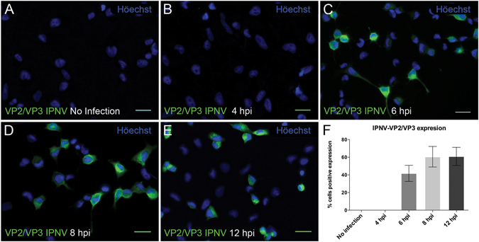
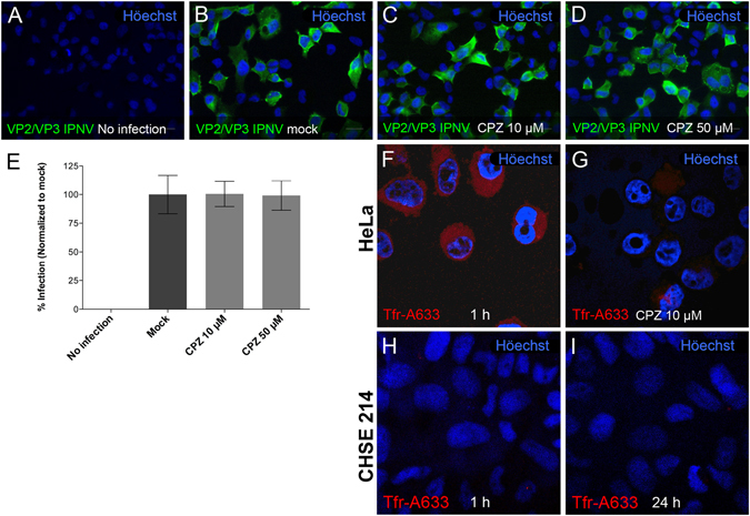
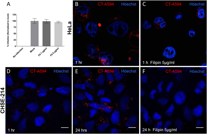
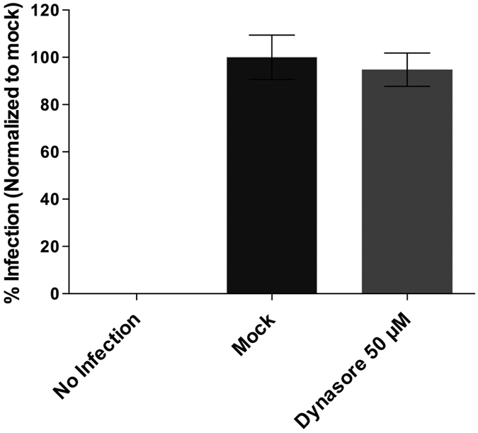
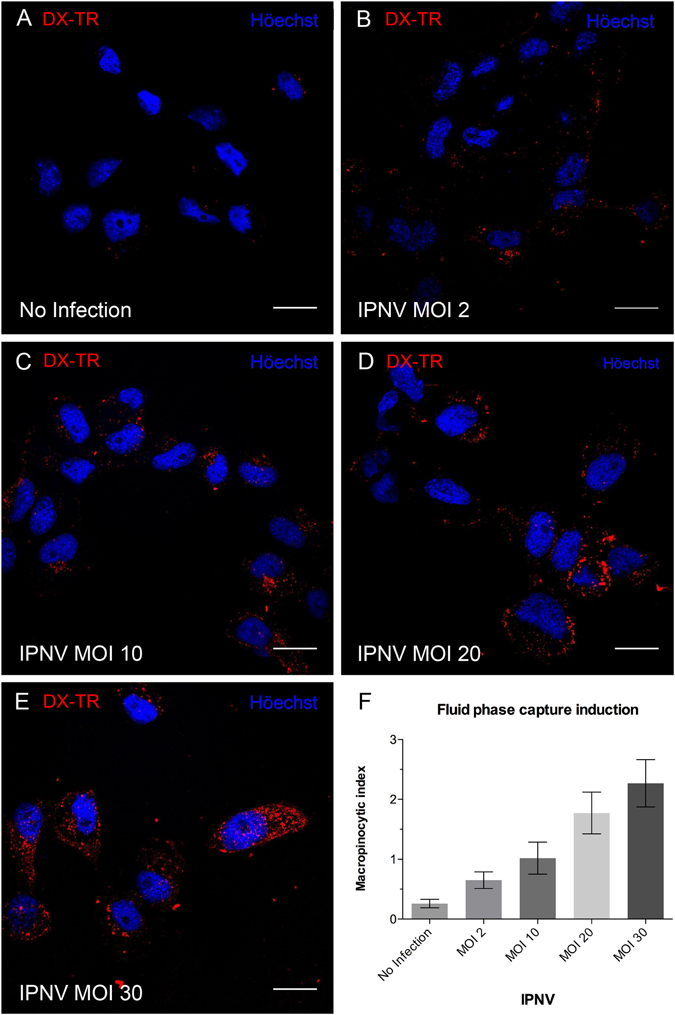
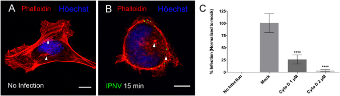
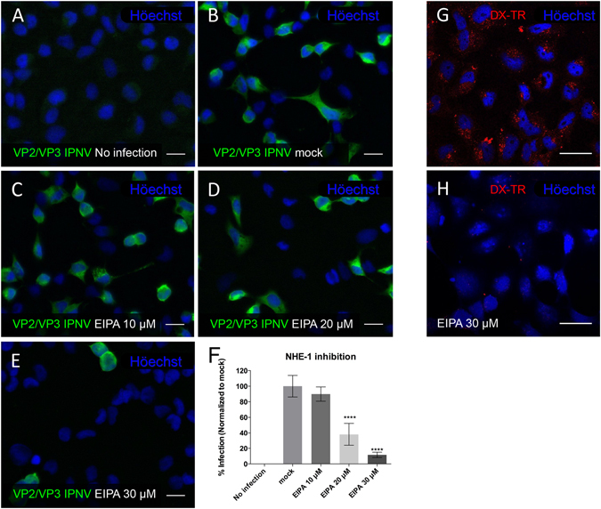
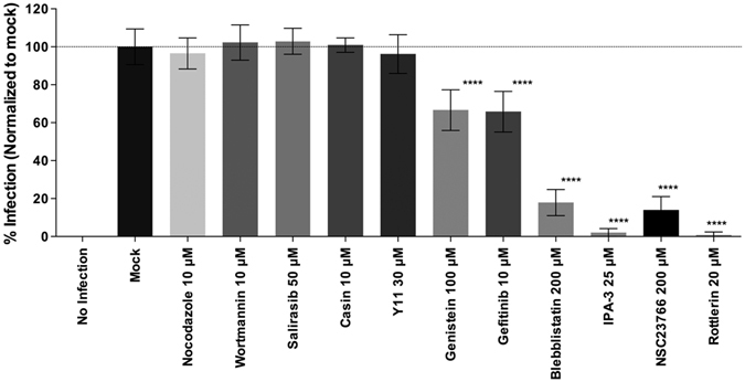
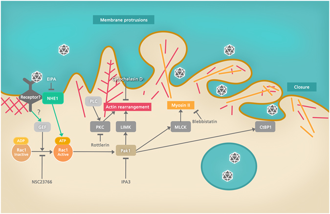
Infectious pancreatic necrosis virus (IPNV) is a non-enveloped virus belonging to the Birnaviridae family. IPNV produces an acute disease in salmon fingerlings, with high mortality rates and persistent infection in survivors. Although there are reports of IPNV binding to various cells, the viral receptor and entry pathways remain unknown. The aim of this study was to determine the endocytic pathway that allows for IPNV entry. We observed that IPNV stimulated fluid uptake and virus particles co-localysed with the uptake marker dextran in intracellular compartments, suggesting a role for macropinocytosis in viral entry. Consistent with this idea, viral infection was significantly reduced when the Na+/H+ exchanger NHE1 was inhibited with 5-(N-Ethyl-N-isopropyl) amiloride (EIPA). Neither chlorpromazine nor filipin complex I affected IPNV infection. To examine the role of macropinocytosis regulators, additional inhibitors were tested. Inhibitors of the EGFR pathway and the effectors Pak1, Rac1 and PKC reduced viral infection. Together, our results indicate that IPNV is mainly internalized into CHSE-214 cells by macropinocytosis.


Conflict of interest statement

The authors declare that they have no competing interests.

Figures

Figure 1
Figure 1
Timecourse of IPNV infection of CHSE-214 cells. VP2/VP3 expression was determined by immunofluorescence after 4 (B), 6 (C), 8 (D) and 12 (E) h post infection (hpi) in CHSE-214 cells. Cells were propagated at 60–70% confluence in 24-well plates with round glass coverslips. IPNV was inoculated at a MOI of 1 for 1 h. After 4, 6, 8 and 12 h of incubation at 20 °C, cells were processed by indirect immunofluorescence (IFI) using mouse oligoclonal anti-VP2/VP3 IPNV antibody and Alexa 488-conjugated donkey anti-mouse antibody. Representative images, recorded with an Olympus Spinning Disk IX81 microscope, are shown (bar scale = 20 μm). At each incubation time, 250 cells were counted. Number of VP2/VP3-expressing cells is shown as a percentage, normalized to mock cells (F).
Figure 2
Figure 2
Chlorpromazine does not affect IPNV infection in CHSE-214 cells. CHSE-214 cells were propagated to 70–80% confluence in 24-well plates with round glass coverslips. IPNV was inoculated for 1 h in the presence of 0 (B), 10 (C) or 50 (D) μM CPZ. After 8 h of incubation at 20 °C, cells were processed by indirect immunofluorescence (IFI) for IPNV VP2/VP3 and images were recorded using a Olympus Spinning Disk IX81 microscope. Representative images are shown (scale bar = 20 µm). For each condition, 250 cells were counted. Number of VP2/VP3-expressing cells is shown as percentage, normalized to mock cells (E). Control HeLa cells were pre-incubated for 1 h with 0 (F) or 10 μM CPZ (G), and 5 μg/ml of Alexa Fluor 633-conjugated transferrin (Trf-A633) was added. After 15 min of incubation cells were processed for confocal fluorescence microscopy. Trf-A633 was added to CHSE-214 and cells were incubated for 1 (H) and 24 h (I). Representative images are shown (scale bar = 10 μm).
Figure 3
Figure 3
Filipin does not affect IPNV infection of CHSE-214 cells. CHSE-214 cells were infected with IPNV at an MOI of 1 for 1 h with 1 or 5 μg/ml of filipin (Fil). After 12 h of incubation at 20°, cells were processed by indirect immunofluorescence (IFI) for IPNV VP2/VP3. Images were recorded with an Olympus Spinning Disk IX81 microscope. For each condition, 250 cells were counted. Number of VP2/VP3-expressing cells is shown as a percentage, normalized to mock cells (A). Control HeLa cells were pre-incubated for 1 h with 0 (B) or 5 μg/ml (C) filipin and 2.5 μg/ml Alexa Fluor 594-conjugated cholera toxin B (CTB-A594) subunit were added. After 30 min, cells were processed for confocal fluorescence microscopy. CHSE-214 cells were preincubated 1 h in the absence (D,E) or with 5 μg/ml filipin (F). CTB-A594 (2.5 μg/ml) was added and cells were incubated for 1 h (D) or 24 h (E and F). Images were recorded with a C2 Plus Eclipse TI Nikon confocal microscope (scale bar = 10 µm). Representative images are shown.
Figure 4
Figure 4
IPNV infection is independent of dynamin. CHSE-214 cells were propagated at 70–80% confluence in 24-well plates with round glass coverslips and pre incubated with 0 or 50 µM dynasore for 1 h at 20 °C. IPNV was inoculated at an MOI of 1 and led the infection proceed for 12 h at 20 °C in absence (mock) or presence of the inhibitor. Cells were processed by indirect immunofluorescence (IFI) for VP2/VP3 IPNV proteins. Imaging of fixed slides was performed with an Olympus Spinning Disk IX81 microscope, and 250 cells were counted at each condition. Number of VP2/VP3-expressing cells is shown as a percentage, normalized to mock cells.
Figure 5
Figure 5
IPNV induces fluid phase capture in CHSE-214 cells. CHSE-214 cells were grown to 70% confluence in 24-well plates with round coverslips in MEM with 2% FBS. Cells were left for 16 h in MEM without FBS. IPNV at MOI of 0, 2, 10, 20 and 30 (AE) was inoculated, then left to adsorb for 1 h at 4 °C. Dextran Texas red (250 µg/ml) was added, and cells were incubated at 20 °C for 45 min. Cells were fixed and stained with Höechst. Images were recorded with a C2 Plus Eclipse TI Nikon confocal microscope. Representative images are shown (scale bar = 10 μm). A graph of the macropinocytic index vs MOI is presented in F. The macropinocytic index was determined as described in Materials and Methods.
Figure 6
Figure 6
IPNV infection is dependent on actin polymerization dynamics. A. CHSE-214 cells were infected with IPNV at a MOI of 10 and adsorbed for 1 h at 4 °C. The supernatant was removed and MEM medium added at 20 °C. Cells were incubated at 20 °C for 15 min, fixed and stained with rhodamine-conjugated phalloidin. Images were recorded with a C2 Plus Nikon spectral confocal microscope, (scale = 10 μm). (A) no infection (B) 15 min post infection (white arrow heads indicate stress fibers in A and disorganized actin in B). (C) CHSE-214 cells were preincubated with 1 or 2 µM cytochalasin D (Cyto D) for 1 h and infected with IPNV at a MOI of 1 in the presence of Cyto D. Cells were incubated at 20 °C for 6 h and processed by indirect immunofluorescence (IFI) for IPNV VP2/VP3. For each condition, 250 cells were counted. The number of VP2/VP3-expressing cells is shown as a percentage, normalized to mock cells (****p < 0.0001).
Figure 7
Figure 7
IPNV infection of CHSE-214 cells is blocked by EIPA. CHSE-214 cells were propagated to 70–80% confluence in 24-well plates with round glass coverslips and infected with IPNV at a MOI of 1 for 1 h in the presence of 0 (B), 10 (C), 20 (D), 30 (E) µM EIPA. No infection (A). After 12 h of incubation at 20 °C, cells were processed by indirect immunofluorescence (IFI). Representative images recorded with an Olympus Spinning Disk IX81 microscope are shown (bar scale = 20 µm). For quantification, 250 cells were counted at each condition and the number of VP2/VP3-expressing cells are shown as a percentage, normalized to mock cells (F) (****p < 0.0001). Control CHSE-214 cells pre-incubated for 1 h in the presence (H) or absence (G) of 30 µM EIPA and 1 mg/ml of Dx-Texas Red 70 kDa (Dx-TR) were added. After 30 min of incubation, cells were washed, fixed with 3.7% PFA-4% sucrose for 30 min and stained with Höechst solution. Images were recorded with a C2 Plus Eclipse TI Nikon confocal microscope. Representative images are shown (bar scale = 20 µm).
Figure 8
Figure 8
IPNV particles co-localize with Dextran-TR. IPNV at a MOI of 10 was adsorbed to serum-starved CHSE-214 cells at 4 °C for 1 h and pulsed with dx-TR for 45 min at 20 °C without (AD) or with EIPA 20 μM (EH). After washing, cells were processed for immunofluorescence using oligoclonal anti VP2/Vp3 IPNV antibodies and Alexa fluor®466 secondary antibody and Höechst solution for nuclear staining. Z-stacks were recorded by confocal microscopy and representative maximum Z projections are shown (scale bar = 5 µm). Analyses of co-localisation were conducted with Image J software and resulting co-localised pixel map are shown for each condition (colocalised pixels are shown as white spots) (D and H).
Figure 9
Figure 9
Cellular requirements for IPNV infection. IPNV infection of CHSE-214 cells was carried out in the presence of inhibitors of structural and functional cell components: nocodazole (10 μM), wortmannin (10 μM), salirasib (50 μM), casin (10 μM), Y11 (30 μM), genistein (100 μM) gefitinib (10 μM), blebbistatin (200 μM), 3-indole propionic acid (IPA-3 25 μM), NSC23766 (200 μM), rottlerin (20 μM), or no infection. CHSE-214 cells were propagated at 70–80% confluence in 24-well plates with round glass coverslips. IPNV was inoculated at an MOI of 1 for 1 h in the presence of the respective inhibitor. After 12 h of incubation at 20 °C, cells were processed by indirect immunofluorescence (IFI). Imaging of fixed slides was performed with an Olympus Spinning Disk IX81 microscope, and 250 cells were counted at each condition. Number of VP2/VP3-expressing cells is shown as a percentage, normalized to mock cells.
Figure 10
Figure 10
Cellular factors possibly involved in IPNV macropinocytosis. The scheme shows a commonly found factors associated to macropinocytosis pathways. Inhibition of the Rho GTPase Rac1, Pak1, PKC, myosin II and NHE1 affected IPNV infection. Inhibition sites of the drugs and efector proteins (MLCK, LIMK, CtBP1) are shown. MLCK: myosin light chain kinase, LIMK: LIM kinase, CtBP1: C-terminal binding protein 1.

Similar articles

Cited by

References

    1. Kibenge FS, Godoy MG, Fast M, Workenhe S, Kibenge MJ. Countermeasures against viral diseases of farmed fish. Antiviral Res. 2012;95:257–281. doi: 10.1016/j.antiviral.2012.06.003. - DOI - PubMed
    1. Smail DA, et al. Infectious pancreatic necrosis virus in Atlantic salmon, Salmo salar L., post-smolts in the Shetland Isles, Scotland: virus identification, histopathology, immunohistochemistry and genetic comparison with Scottish mainland isolates. J Fish Dis. 2006;29:31–41. doi: 10.1111/j.1365-2761.2005.00678.x. - DOI - PubMed
    1. Dobos P. Protein-primed RNA synthesis in vitro by the virion-associated RNA polymerase of infectious pancreatic necrosis virus. Virology. 1995;208:19–25. doi: 10.1006/viro.1995.1125. - DOI - PubMed
    1. Hong JR, Gong HY, Wu JL. IPNV VP5, a novel anti-apoptosis gene of the Bcl-2 family, regulates Mcl-1 and viral protein expression. Virology. 2002;295:217–229. doi: 10.1006/viro.2001.1336. - DOI - PubMed
    1. Somogyi P, Dobos P. Virus-specific RNA synthesis in cells infected by infectious pancreatic necrosis virus. J Virol. 1980;33:129–139. - PMC - PubMed

Publication types