. 2017 Jun 8;7(1):3093.
doi: 10.1038/s41598-017-03234-6.

Transgenic overexpression of GTP cyclohydrolase 1 in cardiomyocytes ameliorates post-infarction cardiac remodeling

Affiliations

Transgenic overexpression of GTP cyclohydrolase 1 in cardiomyocytes ameliorates post-infarction cardiac remodeling

Yanan Liu et al. Sci Rep. .

Abstract

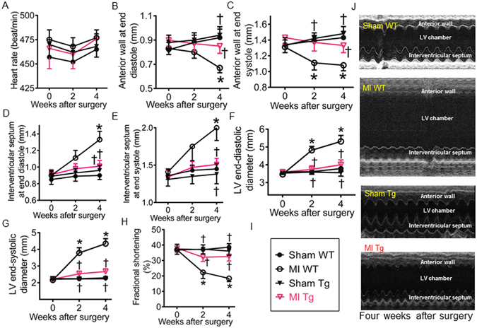
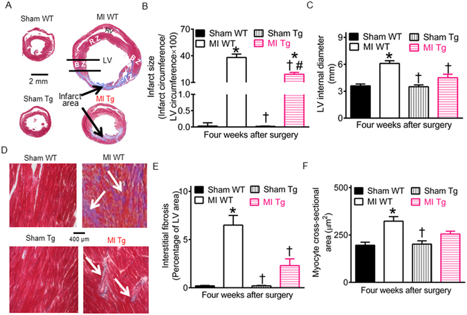
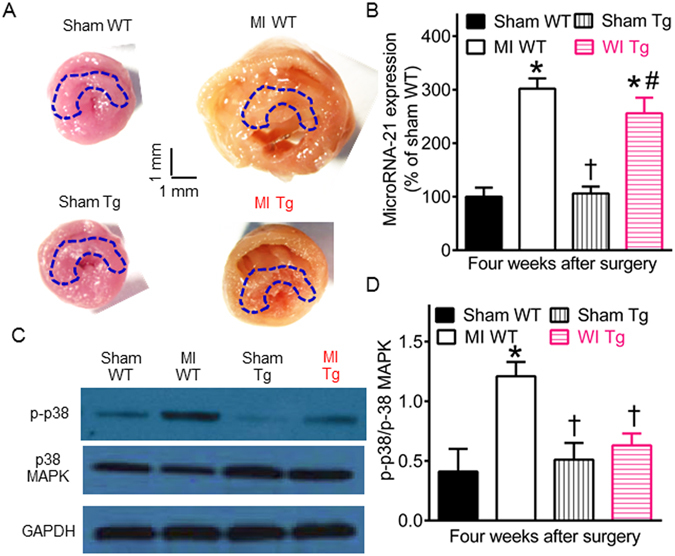
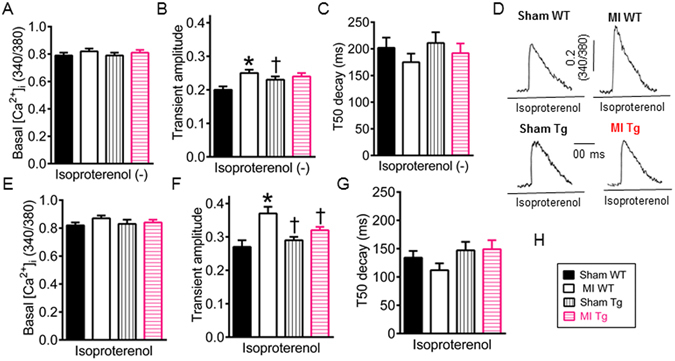
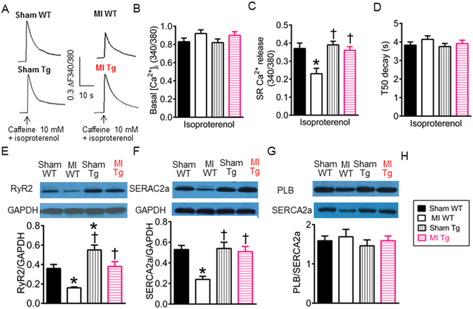
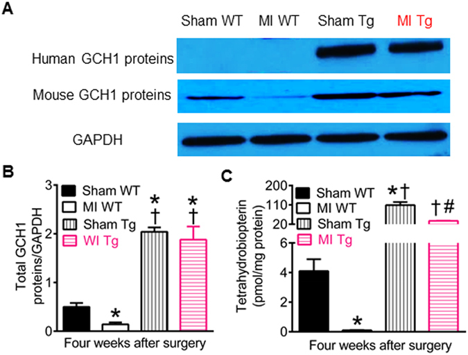
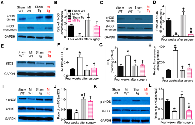
GTP cyclohydrolase 1 (GCH1) and its product tetrahydrobiopterin play crucial roles in cardiovascular health and disease, yet the exact regulation and role of GCH1 in adverse cardiac remodeling after myocardial infarction are still enigmatic. Here we report that cardiac GCH1 is degraded in remodeled hearts after myocardial infarction, concomitant with increases in the thickness of interventricular septum, interstitial fibrosis, and phosphorylated p38 mitogen-activated protein kinase and decreases in left ventricular anterior wall thickness, cardiac contractility, tetrahydrobiopterin, the dimers of nitric oxide synthase, sarcoplasmic reticulum Ca2+ release, and the expression of sarcoplasmic reticulum Ca2+ handling proteins. Intriguingly, transgenic overexpression of GCH1 in cardiomyocytes reduces the thickness of interventricular septum and interstitial fibrosis and increases anterior wall thickness and cardiac contractility after infarction. Moreover, we show that GCH1 overexpression decreases phosphorylated p38 mitogen-activated protein kinase and elevates tetrahydrobiopterin levels, the dimerization and phosphorylation of neuronal nitric oxide synthase, sarcoplasmic reticulum Ca2+ release, and sarcoplasmic reticulum Ca2+ handling proteins in post-infarction remodeled hearts. Our results indicate that the pivotal role of GCH1 overexpression in post-infarction cardiac remodeling is attributable to preservation of neuronal nitric oxide synthase and sarcoplasmic reticulum Ca2+ handling proteins, and identify a new therapeutic target for cardiac remodeling after infarction.


Conflict of interest statement

The authors declare that they have no competing interests.

Figures

Figure 1
Figure 1
Wild-type (WT) mice developed cardiac remodeling and lost GTP cyclohydrolase 1 (GCH1) proteins following myocardial infarction (MI). (A) Time-dependent changes in mouse hearts after MI or sham surgery (n = 6–8 mice/group). Top: representative images of mouse hearts at baseline (0), 1, 2, 4, 8, and 12 weeks after operation. The vertical scale bar indicates 4 mm, and the horizontal scale bar indicates 2 mm. Bottom: the area of the hearts after MI or sham surgery; (B) The anterior wall thickness of the left ventricule after MI or sham surgery (n = 8–10 mice/group); (C) The thickness of interventricular septum following MI or sham surgery (n = 8–10 mice/group); (D) Representative PCR bands showing the expression of GCH1 mRNA and the housekeeping gene, glyceraldehyde 3-phosphate dehydrogenase (GAPDH), at 0, 1, 2, 4, 8, and 12 weeks after MI; (E) GCH1 mRNA normalized to GAPDH (n = 6 mice/group); (F) Representative Western blot bands showing the expression of cardiac mouse GCH1 proteins and GAPDH proteins in WT mice following MI; (G) Expression of mouse GCH1 proteins normalized to GAPDH proteins (n = 6 mice/group). *P < 0.05 versus sham WT groups at corresponding time points (A,B and C) or 0 weeks (G).
Figure 2
Figure 2
Effects of GTP cyclohydrolase 1 (GCH1) overexpression on cardiac remodeling 4 weeks after myocardial infarction (MI). (A) Heart rate; (B) The anterior wall thickness of the left ventricle (LV) at end diastole: (C) The anterior wall thickness of the LV at end systole; (D) The interventricular septum thickness of the LV at end diastole: (E) The interventricular septum thickness of the LV at end systole; (F) LV end-diastolic diameter; (G) LV end-systolic diameter; (H) Fractional shortening; (I) Figure legend: sham WT (wild-type), MI WT, sham Tg (GCH1 transgene), and MI Tg; (J) representative M-mode echocardiograms showing anterior wall, LV chamber, and interventricular septum in the mice 4 weeks after MI. *P < 0.05 versus sham WT groups; P < 0.05 versus MI WT groups (n = 8–12 mice/group).
Figure 3
Figure 3
Effects of GTP cyclohydrolase 1 (GCH1) overexpression on myocardial infarct size and interstitial fibrosis after myocardial infarction (MI). (A) Representative Masson’s trichrome-stained cross section of the heart showing infarct area with fibrosis. The scale bar shows 2 mm, and the arrows indicate infarct area with fibrosis. BZ, border zone; IZ, infarct zone; LV, left ventricle; RV, right ventricle; RZ, remote zone; (B) Infarct size expressed as a percentage of infarct circumference/total circumference of the LV; (C) LV internal diameters at the papillary muscle levels; (D) Masson’s trichrome-stained myocardium showing interstitial fibrosis. The scale bar shows 400 µm, and the arrows indicate fibrosis; (E) Interstitial fibrosis expressed as a percentage of LV area; (F) Myocyte cross-sectional area. Mouse hearts were stained with Masson’s trichrome in wild-type (WT) and transgenic (Tg) mice 4 weeks after MI or sham surgery. *P < 0.05 versus sham WT groups; P < 0.05 versus MI WT groups; #P < 0.05 versus sham Tg groups (n = 8–10 mice/group).
Figure 4
Figure 4
Effects of GTP cyclohydrolase 1 (GCH1) overexpression on microRNA-21 and p38 mitogen-activated protein kinase (MAPK) after myocardial infarction (MI). (A) Representative transverse sections of fresh mouse hearts showing the positon of tissue sampling for measurements of microRNA-21 and p38 MAPK 4 weeks after (MI) or sham surgery. The drawing areas indicate the position of tissue sampling, and the scales indicate 1 mm. (B) The expression of microRNA-21 mRNA; (C) representative Western blot bands showing the expression of phosphorylated p38 (p-p38) MAPK, total p38 MAPK, and GAPDH; (D) The ratio of phosphorylated p38 (p-p38) MAPK/p38 MAPK. *P < 0.05 versus sham WT groups; P < 0.05 versus MI WT groups; #P < 0.05 versus sham Tg groups (n = 8–10 mice/group).
Figure 5
Figure 5
GTP cyclohydrolase 1 (GCH1) overexpression improves cardiac function 4 weeks after myocardial infarction (MI). (A) Left ventricular (LV) end-systolic pressure; (B) The rate of LV pressure rise (+dP/dt); (C) The LV end-diastolic pressure-volume relationship; (D) Heart weight normalized to body weight; (E) LV weight normalized to body weight: (F) Lung weight normalized to body weight. Figure legend: sham WT, wild-type mice receiving sham surgery; MI WT, wild-type mice undergoing myocardial infarction; sham Tg, transgenic GCH1 mice receiving sham surgery; MI Tg, transgenic GCH1 mice undergoing myocardial infarction. *P < 0.05 versus sham WT groups; P < 0.05 versus MI WT groups (n = 8–10 mice/group).
Figure 6
Figure 6
Effects of GTP cyclohydrolase 1 (GCH1) overexpression on intracellular [Ca2+]i after myocardial infarction (MI). (A) Basal Ca2+ concentrations in the absence of isoproterenol; (B) The amplitude of Ca2+ transients elicited by electrical stimulation in the absence of isoproterenol; (C) Time to 50% decay (T50 decay) of Ca2+ transients in the absence of isoproterenol. (D) Original recordings of Ca2+ transients stimulated by electricity in the presence of isoproterenol in cardiomyocytes isolated from transgenic (Tg) and wild-type (WT) mice 4 weeks after MI or sham surgery. The vertical scale bar indicates 0.2 Fura-2 ratio (340/380 nM) unit, and the horizontal scale bar indicates 100 ms; (E) Basal [Ca2+]i in the presence of isoproterenol; (F) Ca2+ transient amplitude in the presence of isoproterenol; (G) T50 decay of Ca2+ transients in the presence of isoproterenol; (H) Figure legend. *P < 0.05 versus sham WT groups; P < 0.05 versus MI WT groups (n = 63–66 cells/goup).
Figure 7
Figure 7
Effects of GTP cyclohydrolase 1 (GCH1) overexpression on sarcoplasmic reticulum (SR) Ca2+ release and Ca2+ handling proteins after myocardial infarction (MI). (A) Original recordings of caffeine-induced Ca2+ release in cardiomyocytes isolated from wild-type (WT) and transgenic (Tg) mice 4 weeks after MI or sham surgery in 0 Na+ and 0 Ca2+ Tyrode buffer under isoproterenol stimulation. The vertical scale bar indicates 0.3 Fura-2 ratio unit, and the horizontal bar indicates 10 s; (B) Basal [Ca2+]I in the presence of isoproterenol; (C) SR Ca2+ release in the presence of isoproterenol (n = 63–66 cells/group); (D) Time to 50% decay (T50 decay) of caffeine-induced Ca2+ transients in the presence of isoproterenol; (E) The expression of ryanodine receptors (RyR2) normalized to glyceraldehyde 3-phosphate dehydrogenase (GAPDH) in Tg and WT mice 4 weeks after MI or sham surgery (n = 5 mice/group). Top: Wesern blot bands showing the expression of RyRs and GAPDH. Bottom: the ratio of RyRs/GAPDH; (F) The expression of SR Ca2+-ATPase (SERCA2a) proteins normalized to GAPDH. Top: Western blot bands showing the expression of SERCA2a and GAPDH. Bottom: the ratio of SERCA2a/GAPDH; (G) The expression of phospholamban (PLB) and SERCA2a in Tg and WT mice 4 weeks after MI or sham surgery. Top: Western blot bands showing the expression of PLB and SERCA2a. Bottom: the ratio of PLB/SERCA2a. *P < 0.05 versus sham WT groups; P < 0.05 versus MI WT groups.
Figure 8
Figure 8
Effects of transgenic overexpression of human GTP cyclohydrolase 1 (GCH1) on GCH1 proteins and tetrahydrobiopterin in post-infarction remodeled myocardium. (A) Representative Western blot bands showing the expression of human and mouse GCH1 proteins and glyceraldehyde 3-phosphate dehydrogenase (GAPDH) as control in wild-type (WT) and transgenic (Tg) mice 4 weeks after myocardial infarction (MI) or sham surgery; (B) Total (human + mouse) GCH1 proteins normalized to GAPDH; C: cardiac tetrahydrobiopterin concentrations. *P < 0.05 versus sham WT groups; P < 0.05 versus MI WT groups; #P < 0.05 versus sham Tg groups (n = 5–6 mice/group).
Figure 9
Figure 9
GTP cyclohydrolase 1 (GCH1) overexpression differentially regulates nitric oxide synthase (NOS) isoforms in post-infarction remodeled myocardium. (A) Representative Western blot bands showing the expression of dimeric and monomeric neuronal NOS (nNOS) and glyceraldehyde 3-phosphate dehydrogenase (GAPDH) as loading control in wild-type (WT) and transgenic (Tg) mice 4 weeks after myocardial infarction (MI) or sham surgery (sham); (B) The ratio of nNOS dimers/monomers; (C) Representative Western blot bands of dimeric and monomeric endothelial NOS (eNOS) and GAPDH; (D) The ratio of eNOS dimers/monomers; (E) Western blot bands showing the expression of inducible NOS (iNOS) monomers and GAPDH; (F) iNOS monomers normalized to GAPDH; (G) NOx levels; (H) superoxide levels; (I) representative Western blot bands showing the expresiion of phosphorylated nNOS (p-nNOS), nNOS, and GAPDH; (J) The ratio of p-nNOS/total nNOS; (K) Western blot bands of phosphorylated eNOS (p-eNOS), eNOS, and GAPDH; (L) the ratio of phosphorylated eNOS (p-eNOS)/total eNOS. *P < 0.05 versus sham WT groups; P < 0.05 versus MI WT groups; #P < 0.05 versus sham Tg groups (n = 5 mice/group).

Similar articles

Cited by

References

    1. Mozaffarian D, et al. Heart disease and stroke statistics-2016 update: a report from the American Heart Association. Circulation. 2016;133:e38–360. doi: 10.1161/CIR.0000000000000350. - DOI - PubMed
    1. Townsend N, et al. Cardiovascular disease in Europe: epidemiological update 2016. Eur Heart J. 2016;0:14. - PubMed
    1. Konstam MA, Kramer DG, Patel AR, Maron MS, Udelson JE. Left ventricular remodeling in heart failure: current concepts in clinical significance and assessment. JACC. Cardiovasc Imag. 2011;4:98–108. doi: 10.1016/j.jcmg.2010.10.008. - DOI - PubMed
    1. Galli A, Lombardi F. Postinfarct left ventricular remodelling: a prevailing cause of heart failure. Cardiol Res Practice. 2016;2016:2579832. doi: 10.1155/2016/2579832. - DOI - PMC - PubMed
    1. Nayor M, Enserro DM, Vasan RS, Xanthakis V. Cardiovascular health status and incidence of heart failure in the Framingham offspring study. Circ Heart Fail. 2016;9:e002416. doi: 10.1161/CIRCHEARTFAILURE.115.002416. - DOI - PMC - PubMed

Publication types

MeSH terms