

Gene Electrotransfer of Plasmid-Encoding IL-12 Recruits the M1 Macrophages and Antigen-Presenting Cells Inducing the Eradication of Aggressive B16F10 Murine Melanoma
- PMID: 28596641
- PMCID: PMC5449735
- DOI: 10.1155/2017/5285890
Gene Electrotransfer of Plasmid-Encoding IL-12 Recruits the M1 Macrophages and Antigen-Presenting Cells Inducing the Eradication of Aggressive B16F10 Murine Melanoma
Abstract
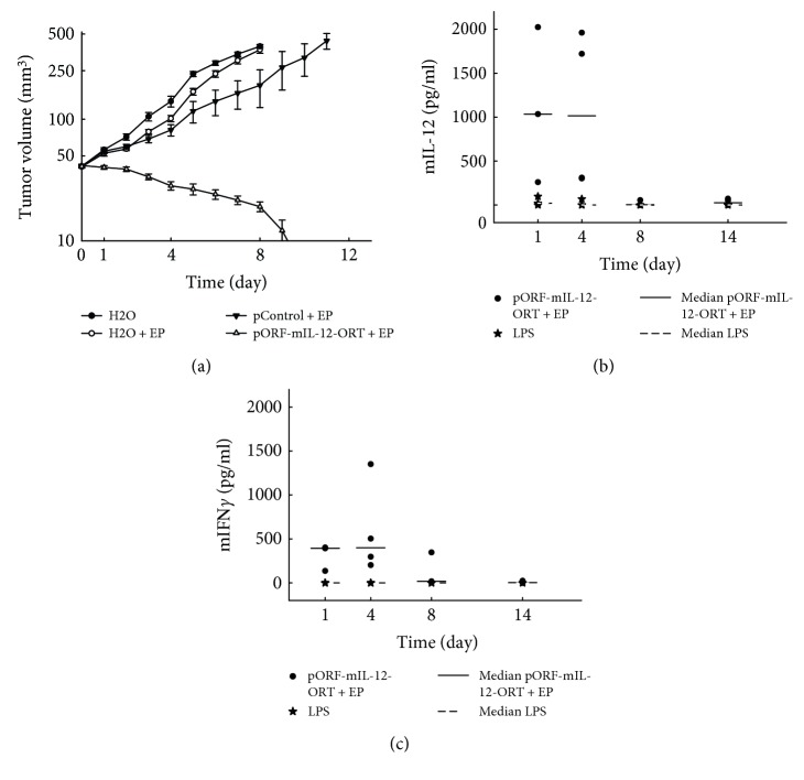
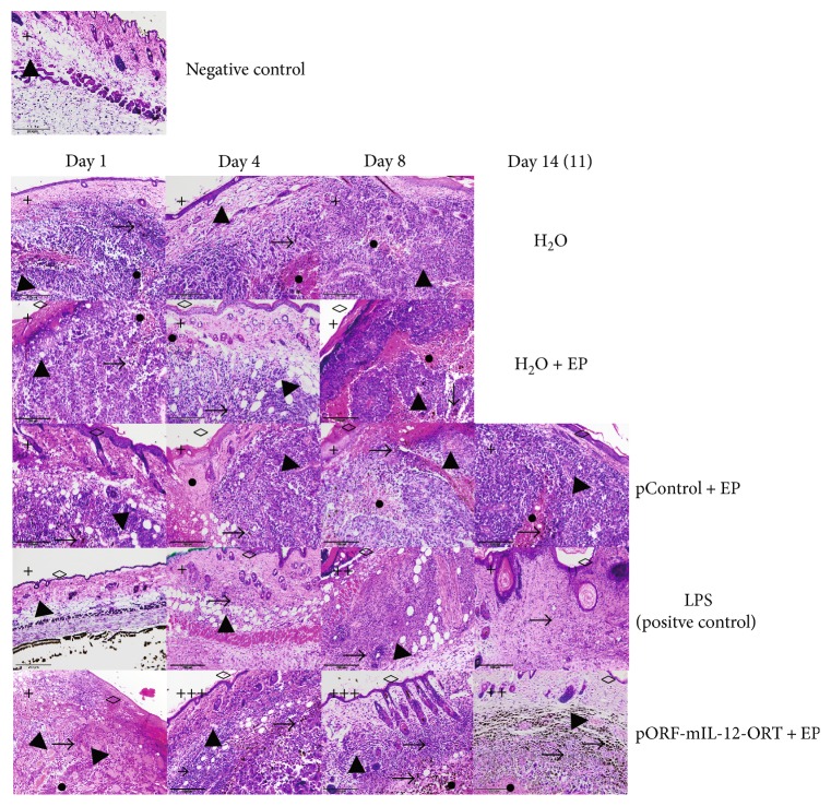
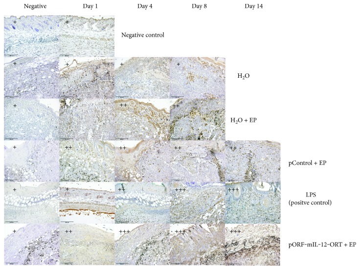
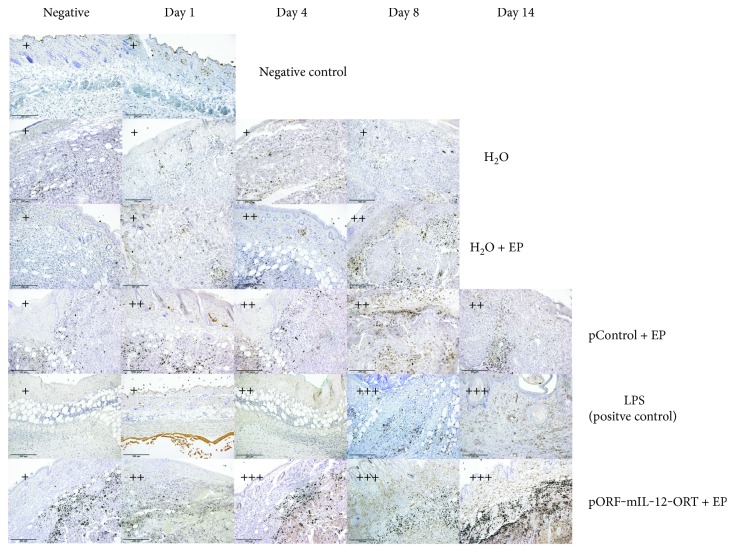
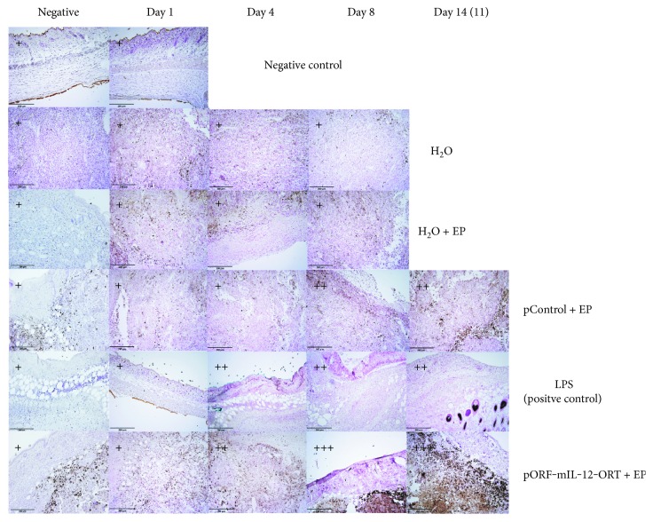
Cancer immunotherapy is currently one of the leading approaches in cancer treatment. Gene electrotransfer of plasmids encoding interleukin 12 (IL-12) into the cells leads to the production of IL-12, which drives immune cell polarization to an antitumoral response. One of the cell types that shows great promise in targeting tumor cells under the influence of IL-12 cytokine milieu is that of macrophages. Therefore, the aim of this study was to evaluate gene electrotransfer of antibiotic resistance-free plasmid DNA-encoding murine IL-12 (mIL-12) in mice bearing aggressive B16F10 murine melanoma. IL-12 electrotransfer resulted in the complete long-term eradication of the tumors. Serum mIL-12 and murine interferon γ (mIFNγ) were increased after IL-12 gene electrotransfer. Further on, hematoxylin and eosin (HE) staining showed increased infiltration of immune cells that lasted from day 4 until day 14. Immunohistochemistry (IHC) staining of F4/80, MHCII, and CD11c showed higher positive staining in the IL-12 gene electrotransfer group than in the control groups. Immune cell infiltration into the tumors and the high density of MHCII- and CD11c-positive cells suggest an antitumor polarization of macrophages and the presence of antigen-presenting cells that contributes to the important antitumor effectiveness of IL-12.
Figures
Similar articles
-
Radiosensitizing effect of intratumoral interleukin-12 gene electrotransfer in murine sarcoma.BMC Cancer. 2013 Jan 29;13:38. doi: 10.1186/1471-2407-13-38. BMC Cancer. 2013. PMID: 23360213 Free PMC article.
-
Gene electrotransfer of IL-2 and IL-12 plasmids effectively eradicated murine B16.F10 melanoma.Bioelectrochemistry. 2021 Oct;141:107843. doi: 10.1016/j.bioelechem.2021.107843. Epub 2021 May 18. Bioelectrochemistry. 2021. PMID: 34139572
-
Electrotransfer of plasmid DNA radiosensitizes B16F10 tumors through activation of immune response.Radiol Oncol. 2017 Feb 22;51(1):30-39. doi: 10.1515/raon-2017-0011. eCollection 2017 Mar 1. Radiol Oncol. 2017. PMID: 28265230 Free PMC article.
-
Antitumor effect of antibiotic resistance gene-free plasmids encoding interleukin-12 in canine melanoma model.Cancer Gene Ther. 2018 Oct;25(9-10):260-273. doi: 10.1038/s41417-018-0014-5. Epub 2018 Mar 29. Cancer Gene Ther. 2018. PMID: 29593358
-
Antitumor Activities of Interleukin-12 in Melanoma.Cancers (Basel). 2022 Nov 14;14(22):5592. doi: 10.3390/cancers14225592. Cancers (Basel). 2022. PMID: 36428682 Free PMC article. Review.
Cited by
-
Non-clinical evaluation of pmIL12 gene therapy for approval of the phase I clinical study.Sci Rep. 2024 Sep 27;14(1):22288. doi: 10.1038/s41598-024-73314-x. Sci Rep. 2024. PMID: 39333733 Free PMC article.
-
Clinical Applications and Immunological Aspects of Electroporation-Based Therapies.Vaccines (Basel). 2021 Jul 2;9(7):727. doi: 10.3390/vaccines9070727. Vaccines (Basel). 2021. PMID: 34358144 Free PMC article. Review.
-
Development of an Interleukin-12 Fusion Protein That Is Activated by Cleavage with Matrix Metalloproteinase 9.J Interferon Cytokine Res. 2019 Apr;39(4):233-245. doi: 10.1089/jir.2018.0129. Epub 2019 Mar 8. J Interferon Cytokine Res. 2019. PMID: 30848689 Free PMC article.
-
A virus based vaccine combined with IL12 gene therapy eradicates aggressive melanoma.Sci Rep. 2025 May 29;15(1):18786. doi: 10.1038/s41598-025-04026-z. Sci Rep. 2025. PMID: 40442309 Free PMC article.
-
Antitumor in situ vaccination effect of TNFα and IL-12 plasmid DNA electrotransfer in a murine melanoma model.Cancer Immunol Immunother. 2018 May;67(5):785-795. doi: 10.1007/s00262-018-2133-0. Epub 2018 Feb 21. Cancer Immunol Immunother. 2018. PMID: 29468364 Free PMC article.
References
MeSH terms
Substances
LinkOut - more resources
Full Text Sources
Other Literature Sources
Medical
Research Materials
