

Role of AXL in invasion and drug resistance of colon and breast cancer cells and its association with p53 alterations
- PMID: 28596680
- PMCID: PMC5442080
- DOI: 10.3748/wjg.v23.i19.3440
Role of AXL in invasion and drug resistance of colon and breast cancer cells and its association with p53 alterations
Abstract
Aim: To characterize AXL receptor tyrosine kinase (AXL) expression in relationship to tumor protein P53 (TP53 gene, p53 protein) and its role in tumor invasion and response to therapy.
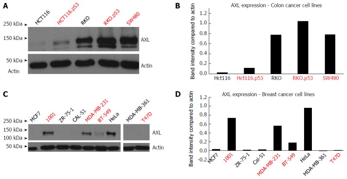
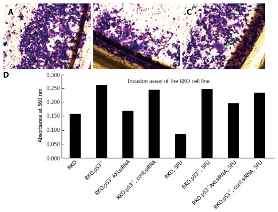
Methods: We used 14 cell lines, including 3 isogenic pairs carrying mutant/knockout p53, to gain insight into the relationship between AXL and TP53. These included HCT116, HCT116.p53 mutant, RKO, and RKO.p53-/- lines (all from colon cancers) as well as breast cancer cell lines MCF7 and 1001 (MCF7-p53 mutant clone). HeLa cell line was used as a positive control for epithelial to mesenchymal transition (EMT). AXL expression was determined by Western blotting using rabbit monoclonal antibody clone C89E7. AXL siRNA silencing was performed and followed by collagen invasion assay. Cell viability analysis using the sulforhodamine B assay and the invasion assay were performed after exposure to chemotherapeutic agents (doxorubicin for breast cancer cells; 5FU or irinotecan for colon cancer cells).
Results: We showed that the introduction of p53 mutations or knockout increased expression levels of AXL in isogenic cells compared to the matching p53 wild-type parental cells. Overall, we found a trend for correlation between the potential EMT candidate AXL, p53 alterations, and EMT markers in colorectal and breast cancers. The expression of AXL in RKO cells, a rare colon cancer cell line with inactive Wnt signaling, suggests that the AXL oncogene might provide an alternative genetic pathway for colorectal carcinogenesis in the absence of Wnt signaling activation and TP53 mutation. AXL silencing in the TP53 mutant isogenic cell lines 1001, HCT116.p53 mutant and RKO.P53-/- was > 95% efficient and the silenced cells were less invasive compared to the parental TP53 wild-type cells. AXL silencing showed a subtle trend to restore colon cancer cell sensitivity to 5FU or irinotecan. Importantly, AXL expressing cells developed more invasive potential after exposure to chemotherapy compared to the AXL-silenced cells.
Conclusion: AXL is influenced by p53 status and could cause the emergence of aggressive clones after exposure to chemotherapy. These findings could have applications in cancer management.
Keywords: AXL; Breast cancer; Chemotherapy; Colon cancer; Invasion.
Conflict of interest statement
Conflict-of-interest statement: The authors have no conflicts of interest to declare.
Figures
Similar articles
-
Silencing AXL by covalent siRNA-gelatin-antibody nanoconjugate inactivates mTOR/EMT pathway and stimulates p53 for TKI sensitization in NSCLC.Nanomedicine. 2019 Aug;20:102007. doi: 10.1016/j.nano.2019.04.010. Epub 2019 May 12. Nanomedicine. 2019. PMID: 31085346
-
Myeloid zinc finger 1 induces migration, invasion, and in vivo metastasis through Axl gene expression in solid cancer.Mol Cancer Res. 2010 Feb;8(2):159-69. doi: 10.1158/1541-7786.MCR-09-0326. Epub 2010 Feb 9. Mol Cancer Res. 2010. PMID: 20145042
-
Gas6/Axl Axis Contributes to Chemoresistance and Metastasis in Breast Cancer through Akt/GSK-3β/β-catenin Signaling.Theranostics. 2016 May 24;6(8):1205-19. doi: 10.7150/thno.15083. eCollection 2016. Theranostics. 2016. PMID: 27279912 Free PMC article.
-
AXL-Driven EMT State as a Targetable Conduit in Cancer.Cancer Res. 2017 Jul 15;77(14):3725-3732. doi: 10.1158/0008-5472.CAN-17-0392. Epub 2017 Jun 30. Cancer Res. 2017. PMID: 28667075 Review.
-
Axl inhibitors as novel cancer therapeutic agents.Life Sci. 2018 Apr 1;198:99-111. doi: 10.1016/j.lfs.2018.02.033. Epub 2018 Feb 27. Life Sci. 2018. PMID: 29496493 Review.
Cited by
-
Patient-derived xenografts and in vitro model show rationale for imatinib mesylate repurposing in HEY1-NCoA2-driven mesenchymal chondrosarcoma.Lab Invest. 2022 Sep;102(9):1038-1049. doi: 10.1038/s41374-021-00704-4. Epub 2021 Nov 26. Lab Invest. 2022. PMID: 34837064
-
Oncogenic Potential of Bisphenol A and Common Environmental Contaminants in Human Mammary Epithelial Cells.Int J Mol Sci. 2020 May 25;21(10):3735. doi: 10.3390/ijms21103735. Int J Mol Sci. 2020. PMID: 32466334 Free PMC article.
-
AXL Inactivation Inhibits Mesothelioma Growth and Migration via Regulation of p53 Expression.Cancers (Basel). 2020 Sep 25;12(10):2757. doi: 10.3390/cancers12102757. Cancers (Basel). 2020. PMID: 32992696 Free PMC article.
-
The Endocrine Disruptor Bisphenol A (BPA) Exerts a Wide Range of Effects in Carcinogenesis and Response to Therapy.Curr Mol Pharmacol. 2019;12(3):230-238. doi: 10.2174/1874467212666190306164507. Curr Mol Pharmacol. 2019. PMID: 30848227 Free PMC article. Review.
-
PEG-coated nanoparticles detachable in acidic microenvironments for the tumor-directed delivery of chemo- and gene therapies for head and neck cancer.Theranostics. 2020 May 17;10(15):6695-6714. doi: 10.7150/thno.45164. eCollection 2020. Theranostics. 2020. PMID: 32550898 Free PMC article.
References
-
- Ferlay J, Soerjomataram I, Ervik M, Dikshit R, Eser S, Mathers C, Rebelo M, Parkin DM, Forman D, Bray F. GLOBOCAN 2012 v1.0, Cancer Incidence and Mortality Worldwide: IARC CancerBase No. 11 [Internet]. Lyon, France: International Agency for Research on Cancer; 2013. Available from: http://globocan.iarc.fr, 2013.
-
- Nieminen TT, Shoman S, Eissa S, Peltomäki P, Abdel-Rahman WM. Distinct genetic and epigenetic signatures of colorectal cancers according to ethnic origin. Cancer Epidemiol Biomarkers Prev. 2012;21:202–211. - PubMed
-
- Kalia M. Biomarkers for personalized oncology: recent advances and future challenges. Metabolism. 2015;64:S16–S21. - PubMed
MeSH terms
Substances
LinkOut - more resources
Full Text Sources
Other Literature Sources
Medical
Research Materials
Miscellaneous
