

Tumor-associated autoantibodies are useful biomarkers in immunodiagnosis of α-fetoprotein-negative hepatocellular carcinoma
- PMID: 28596685
- PMCID: PMC5442085
- DOI: 10.3748/wjg.v23.i19.3496
Tumor-associated autoantibodies are useful biomarkers in immunodiagnosis of α-fetoprotein-negative hepatocellular carcinoma
Abstract
Aim: To determine the prevalence and diagnostic value of autoantibodies in α-fetoprotein (AFP)-negative hepatocellular carcinoma (HCC).
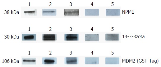
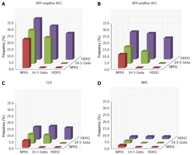
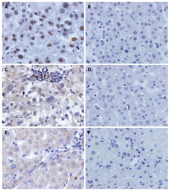
Methods: Fifty-six serum samples from AFP-negative HCC cases, 86 from AFP-positive HCC cases, 168 from chronic liver disease cases, and 59 from normal human controls were included in this study. Autoantibodies to nucleophosmin (NPM)1, 14-3-3zeta and mouse double minute 2 homolog (MDM2) proteins in AFP-negative HCC serum were evaluated by enzyme-linked immunosorbent assay. Partially positive sera were further evaluated by western blotting. Immunohistochemistry was used to detect the expression of three tumor-associated antigens (TAAs) in AFP-negative HCC and normal control tissues.
Results: The frequency of autoantibodies to the three TAAs in AFP-negative HCC sera was 21.4%, 19.6% and 19.6%, which was significantly higher than in the chronic liver disease cases and normal human controls (P < 0.01) as well as AFP-positive HCC cases. The sensitivity of the three autoantibodies for diagnosis of AFP-negative HCC ranged from 19.6% to 21.4%, and the specificity was approximately 95%. When the three autoantibodies were combined, the sensitivity reached 30.4% and the specificity reached 91.6%.
Conclusion: Autoantibodies to NPM1, 14-3-3zeta and MDM2 may be useful biomarkers for immunodiagnosis of AFP-negative HCC.
Keywords: 14-3-3zeta; Autoantibody; Hepatocellular carcinoma; Immunodiagnosis; Mouse double minute 2 homolog; Nucleophosmin 1; α-fetoprotein.
Conflict of interest statement
Conflict-of-interest statement: The authors declared that there is no conflict of interest related to this study.
Figures
Similar articles
-
Humoral autoimmune response to nucleophosmin in the immunodiagnosis of hepatocellular carcinoma.Oncol Rep. 2015 May;33(5):2245-52. doi: 10.3892/or.2015.3854. Epub 2015 Mar 16. Oncol Rep. 2015. PMID: 25779011 Free PMC article.
-
A cancer-related protein 14-3-3ζ is a potential tumor-associated antigen in immunodiagnosis of hepatocellular carcinoma.Tumour Biol. 2014 May;35(5):4247-56. doi: 10.1007/s13277-013-1555-8. Epub 2014 Jan 4. Tumour Biol. 2014. PMID: 24390614 Free PMC article.
-
A novel immunodiagnosis panel for hepatocellular carcinoma based on bioinformatics and the autoantibody-antigen system.Cancer Sci. 2022 Feb;113(2):411-422. doi: 10.1111/cas.15217. Epub 2021 Dec 14. Cancer Sci. 2022. PMID: 34821436 Free PMC article.
-
The threshold of alpha-fetoprotein (AFP) for the diagnosis of hepatocellular carcinoma: A systematic review and meta-analysis.PLoS One. 2020 Feb 13;15(2):e0228857. doi: 10.1371/journal.pone.0228857. eCollection 2020. PLoS One. 2020. PMID: 32053643 Free PMC article.
-
Current Status and Perspective Biomarkers in AFP Negative HCC: Towards Screening for and Diagnosing Hepatocellular Carcinoma at an Earlier Stage.Pathol Oncol Res. 2020 Apr;26(2):599-603. doi: 10.1007/s12253-019-00585-5. Epub 2019 Jan 19. Pathol Oncol Res. 2020. PMID: 30661224 Review.
Cited by
-
Autoantibody signature in hepatocellular carcinoma using seromics.J Hematol Oncol. 2020 Jul 2;13(1):85. doi: 10.1186/s13045-020-00918-x. J Hematol Oncol. 2020. PMID: 32616055 Free PMC article.
-
Value of anti-p53 antibody as a biomarker for hepatocellular carcinoma: Evidence from a meta-analysis.Medicine (Baltimore). 2020 Aug 21;99(34):e21887. doi: 10.1097/MD.0000000000021887. Medicine (Baltimore). 2020. PMID: 32846849 Free PMC article.
-
New Blood Biomarkers for the Diagnosis of AFP-Negative Hepatocellular Carcinoma.Front Oncol. 2020 Aug 14;10:1316. doi: 10.3389/fonc.2020.01316. eCollection 2020. Front Oncol. 2020. PMID: 32923383 Free PMC article. Review.
-
An Autoantigen-ome from HS-Sultan B-Lymphoblasts Offers a Molecular Map for Investigating Autoimmune Sequelae of COVID-19.bioRxiv [Preprint]. 2021 Apr 6:2021.04.05.438500. doi: 10.1101/2021.04.05.438500. bioRxiv. 2021. PMID: 33851168 Free PMC article. Updated. Preprint.
-
An Autoantigen Atlas From Human Lung HFL1 Cells Offers Clues to Neurological and Diverse Autoimmune Manifestations of COVID-19.Front Immunol. 2022 Mar 24;13:831849. doi: 10.3389/fimmu.2022.831849. eCollection 2022. Front Immunol. 2022. PMID: 35401574 Free PMC article.
References
-
- Bertino G, Ardiri A, Malaguarnera M, Malaguarnera G, Bertino N, Calvagno GS. Hepatocellualar carcinoma serum markers. Semin Oncol. 2012;39:410–433. - PubMed
-
- Poon D, Anderson BO, Chen LT, Tanaka K, Lau WY, Van Cutsem E, Singh H, Chow WC, Ooi LL, Chow P, et al. Management of hepatocellular carcinoma in Asia: consensus statement from the Asian Oncology Summit 2009. Lancet Oncol. 2009;10:1111–1118. - PubMed
-
- Lee JM, Yoon JH, Kim KW. Diagnosis of hepatocellular carcinoma: newer radiological tools. Semin Oncol. 2012;39:399–409. - PubMed
-
- Farinati F, Marino D, De Giorgio M, Baldan A, Cantarini M, Cursaro C, Rapaccini G, Del Poggio P, Di Nolfo MA, Benvegnù L, et al. Diagnostic and prognostic role of alpha-fetoprotein in hepatocellular carcinoma: both or neither? Am J Gastroenterol. 2006;101:524–532. - PubMed
-
- Ertle JM, Heider D, Wichert M, Keller B, Kueper R, Hilgard P, Gerken G, Schlaak JF. A combination of α-fetoprotein and des-γ-carboxy prothrombin is superior in detection of hepatocellular carcinoma. Digestion. 2013;87:121–131. - PubMed
MeSH terms
Substances
LinkOut - more resources
Full Text Sources
Other Literature Sources
Medical
Research Materials
