Review
. 2017 Jun 7:14:11.
doi: 10.1186/s12950-017-0159-2. eCollection 2017.

Regulating STING in health and disease

Affiliations
Review

Regulating STING in health and disease

Yang Li et al. J Inflamm (Lond). .

Abstract

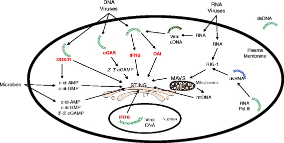
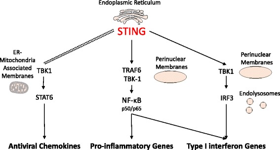
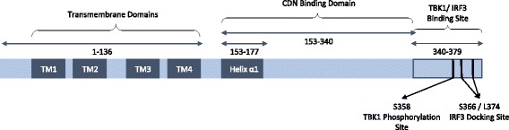
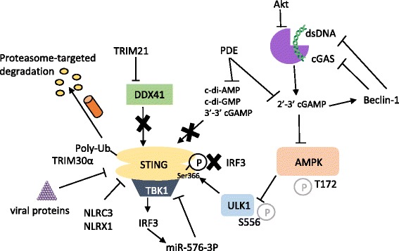
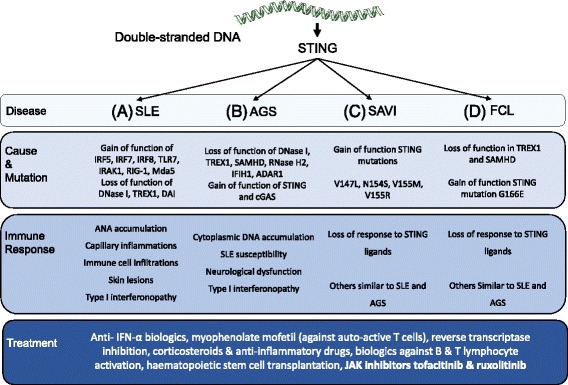
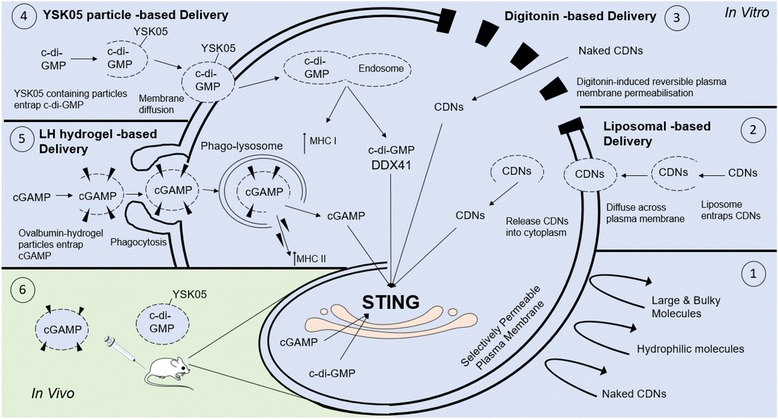
The presence of cytosolic double-stranded DNA molecules can trigger multiple innate immune signalling pathways which converge on the activation of an ER-resident innate immune adaptor named "STimulator of INterferon Genes (STING)". STING has been found to mediate type I interferon response downstream of cyclic dinucleotides and a number of DNA and RNA inducing signalling pathway. In addition to its physiological function, a rapidly increasing body of literature highlights the role for STING in human disease where variants of the STING proteins, as well as dysregulated STING signalling, have been implicated in a number of inflammatory diseases. This review will summarise the recent structural and functional findings of STING, and discuss how STING research has promoted the development of novel therapeutic approaches and experimental tools to improve treatment of tumour and autoimmune diseases.

Keywords: Cyclic dinucleotide; Double-stranded DNA sensor; Stimulator of Interferon Genes (STING); cGAS.


Figures

Fig. 1
Fig. 1
STING activation pathways. The endoplasmic reticulum (ER) adaptor STING is activated via recognition of bacteria-secreted 3′-5′ bond cyclic dinucleotides or DNA sensor cGAS-catalyzed 2′-5′ cGAMP. Cytoplasmic DNA, released from DNA viruses or reverse transcribed from the RNA viral genome, can induce direct interaction between STING and DNA sensors (in red) such as DDX41, IFI16, and DAI. Alternatively, RNA viruses also induce the RIG-I dependent MAVS activation which alters mitochondrial dynamics and indirectly induce STING activation. Mitochondrial stress can result in the release of mitochondrial DNA (mtDNA) which also induces DNA sensor activation (not shown) and STING-mediated signalling. RNA polymerase III (RNA Pol III) can convert dsDNA into dsRNA which activates RIG-I/ MAVS axis which has been shown to induce STING activation
Fig. 2
Fig. 2
STING activated signalling pathways. STING activation leads to translocation from ER membranes to the perinuclear vesicles where it induces the signalling of two major pathways: the NF-κB -dependent proinflammatory response and the IRF3-dependent type I interferon response. The activation of mitochondrial antiviral adaptor MAVS also results in the activation of STING and recruitment of TBK1, which upregulates the transcription of antiviral chemokines via STAT6
Fig. 3
Fig. 3
The domain structure of human STING protein. Human STING is a 379 amino-acid long ER-resident protein. The N-terminal contains 5 membrane-embedded domains (dark blue) including 4 transmembrane domains and Helix α1 responsible for ligand sensing and protein dimerisation. The C-terminal is mainly cytoplasmic (pale blue). It contains the cyclic dinucleotide (CDN) binding domain and interaction sites for TBK1 and IRF3 at the tail. Numbers above STING sequence indicate the amino acids comprising the functional domain
Fig. 4
Fig. 4
Negative regulation of STING-mediated response. STING-mediated signalling can be negatively regulated via multiple mechanisms, including E3 ubiquitin ligase TRIM30α- and TRIM21- mediated degradation of STING and its upstream DNA sensor DDX41, respectively. Certain phosphodiesterases (PDEs) also specifically hydrolyse bacterial cyclic dinucleotides to prevent them being sensed by STING. Akt kinase is also capable of inhibiting cGAS detection of cytoplasmic DNA. Activated cGAS produces 2′-3′ cGAMP to release AMPK-mediated inhibition of ULK1, which in turn blocks IRF3 recruitment downstream of STING activation. 2′-3′ cGAMP produced by cGAS can also activate Beclin-1 which can sequester cGAS as well as induce degradation of dsDNA. In a negative feedback loop, the product of the IRF3-dependent antiviral response, microRNA-576-3P (miR-576-3P), can prevent further STING activation. Some viruses can encode proteases or protein inhibitors to interfere in STING signalling, while others may enhance the activity of inflammasome complexes NLRC3 and NLRX1 to block STING/ TBK1 interaction
Fig. 5
Fig. 5
Cells and cytokines involved in STING-associated autoimmune diseases. Unresolved accumulation of cytoplasmic DNA can potentially trigger chronic inflammatory responses which result in autoimmune diseases, including a systemic lupus erythematosus (SLE) and b Aicardi-Goutières Syndrome (AGS). Both diseases are strongly associated with persistently enhanced type I interferon upregulation named type I interferonopathy and subsequent B and T lymphocyte activation that potentiates systemic tissue and organ damage. Though STING dysregulation has been suggested to play an essential role in the development of these diseases, current treatment of SLE and AGS still relies heavily on anti-inflammatory therapies and DNA resolving methods to ameliorate symptoms. Gain of function mutations of STING can cause two autoimmune diseases named c STING-associated vasculopathy with on-set in infancy (SAVI) and d familial chilblain lupus (FCL). Both diseases show similar manifestations to SLE and AGS and are much less responsive to STING ligands than other immune stimuli. Treatments for SAVI and FCL are limited but JAK inhibitors have been shown to ameliorate symptoms in patients with these two diseases
Fig. 6
Fig. 6
In vitro and in vivo delivery of STING agonists. The plasma membrane is a selectively permeable barrier that prevents cytoplasmic entry of large or hydrophilic molecules, including naked cyclic dinucleotides (CDNs) (1). In vitro (blue background) delivery of dinucleotide compounds could be achieved by the liposomal delivery system (2), or via reversible permeabilisation of plasma membrane to allow diffusion of naked CDNs into the cytoplasm (3). Recently designed YSK05-containing liposomes (4) could carry c-di-GMP across plasma membranes to induce DDX41-mediated STING activation as well as enhance the expression of MHC class I molecules and T cell co-stimulatory receptors (not demonstrated), and thus it is considered to be a potential adjuvant for cancer immunotherapy. In addition, the polyethyleneimine/ hyaluronic acid (LH) hydrogel-based vesicles use phagocytosis to deliver both STING ligands and antibody-stimulating agents such as ovalbumin (dark triangles) to cells (5), and enhance both STING-dependent innate immunity and MHC class II-activated adaptive immunity to suppress cancer growth. Both YSK05 particles and LH hydrogel-based particles have been tested in vivo (green background) to stall tumour progression in mice (6)

Similar articles

Cited by

References

    1. Holm CK, Paludan SR, Fitzgerald KA. DNA recognition in immunity and disease. Curr Opin Immunol. 2013;25:13–18. doi: 10.1016/j.coi.2012.12.006. - DOI - PMC - PubMed
    1. Zheng M, Zheng M, Xie L, Xie L, Liang Y, Wu S, et al. Recognition of cytosolic DNA attenuates glucose metabolism and induces AMPK mediated energy stress response. Int J Biol Sci. 2015;11:587–594. doi: 10.7150/ijbs.10945. - DOI - PMC - PubMed
    1. Fiszer-Kierzkowska A, Vydra N, Wysocka-Wycisk A, Kronekova Z, Jarząb M, Lisowska KM, et al. Liposome-based DNA carriers may induce cellular stress response and change gene expression pattern in transfected cells. BMC Mol Biol. 2011;12:27. doi: 10.1186/1471-2199-12-27. - DOI - PMC - PubMed
    1. Atianand MK, Fitzgerald KA. Long non-coding RNAs and control of gene expression in the immune system. Trends Mol Med. 2014;20:623–631. doi: 10.1016/j.molmed.2014.09.002. - DOI - PMC - PubMed
    1. Hornung V, Latz E. Intracellular DNA recognition. Nat Rev Immunol. 2010;10:123–130. doi: 10.1038/nri2690. - DOI - PubMed