. 2017 May 24:8:338.
doi: 10.3389/fphys.2017.00338. eCollection 2017.

RANK/RANKL/OPG Signalization Implication in Periodontitis: New Evidence from a RANK Transgenic Mouse Model

Affiliations

RANK/RANKL/OPG Signalization Implication in Periodontitis: New Evidence from a RANK Transgenic Mouse Model

Bouchra Sojod et al. Front Physiol. .

Abstract

Periodontitis is based on a complex inflammatory over-response combined with possible genetic predisposition factors. The RANKL/RANK/OPG signaling pathway is implicated in bone resorption through its key function in osteoclast differentiation and activation, as well as in the inflammatory response. This central element of osteo-immunology has been suggested to be perturbed in several diseases, including periodontitis, as it is a predisposing factor for this disease. The aim of the present study was to validate this hypothesis using a transgenic mouse line, which over-expresses RANK (RTg) and develops a periodontitis-like phenotype at 5 months of age. RTg mice exhibited severe alveolar bone loss, an increased number of TRAP positive cells, and disorganization of periodontal ligaments. This phenotype was more pronounced in females. We also observed dental root resorption lacunas. Hyperplasia of the gingival epithelium, including Malassez epithelial rests, was visible as early as 25 days, preceding any other symptoms. These results demonstrate that perturbations of the RANKL/RANK/OPG system constitute a core element of periodontitis, and more globally, osteo-immune diseases.

Keywords: RANK; alveolar bone; gingival epithelium; malassez epithelial rests (MER); osteoclasts; periodontitis; root resorption.


Figures

Figure 1
Figure 1
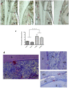
Micro-Computed-Tomography (micro-CT) analysis of alveolar bone height in 5-month-old mice. Distances between the cervical enamel and alveolar bone crest of the mandible were measured for each molar in eight positions (black lines for WT mice, and red lines for RTg mice). We observed a loss in height in RTg mice relative to WT mice, in the interproximal and inter-radicular areas (a,b), as well as the buccal (B) and lingual (L) alveolar crests in frontal sections (c,d). The mean distances between alveolar bone crest and cervical enamel (μm) were measured in eight different areas (e). Statistical analyses showed significant differences (p < 0.01), with greater bone loss in RTg mice (n = 10), which was greater in transgenic females (n = 5).
Figure 2
Figure 2
TRAP positive cells in 5-month-old mice. TRAP histo-enzymology performed on frontal sections (a,b) enabled visualization of a large increase in the number of TRAP positive cells (red staining) on both buccal (B) and lingual (L) surfaces of the alveolar bone of RTg mice (n = 10) relative to that of WT mice (n = 10). Magnifications of the lingual cervical dentin region (dashed rectangles in a,b) showed the presence of numerous TRAP positive cells on the dentin surface, almost exclusively in RTg mice. Quantification of TRAP-positive cells (C) on alveolar bone surfaces of WT and RTg females (in green) and males (blue) showed a significantly (***p < 0.001) higher number of cells respectively in both male (M) and female (F) RTg mice than in WT mice. Similar numbers of TRAP positive cells were visible at the cementum surface and in resorption lacunas through the cementum and dentin (d) in the root area of RTg mice. AB, alveolar bone; D, dentin; C, cementum; PL, periodontal ligament. Scale bars correspond to 10 μm in a,b, 20 μm in the enlarged a,b and right photograph in d, and 50 μm in the left photograph in d.
Figure 3
Figure 3
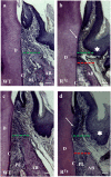
Hematoxylin-Eosin staining of alveolar bone and gingival epithelium. Frontal sections were cut in the molar plane 5 and 6-month-old WT (a,c) and RTg (b,d) mice, and a gradual reduction of the alveolar bone height was detected (green and red lines). This reduction was associated with the detachment and disruption of the epithelial junction (arrows in b,d). We also observed thickening of the gingival epithelium in RTg mice (stars in b,d). AB, alveolar bone; D, dentin; C, cementum; PL, periodontal ligament. Scale bars correspond to 20 μm.
Figure 4
Figure 4
Histological analysis of gingival epithelium and periodontal ligaments at 25 days. At this age, an important thickening of gingival epithelium was already clearly visible in RTg mice relative to WT mice (a) whereas we observed no difference in the structure of the periodontal ligament (b). Masson trichrome staining showed no difference regarding periodontal tissues or ligaments between WT and RTg mice. AB, alveolar bone; D, dentin; C, cementum; PL, periodontal ligament. Scale bars correspond to 50 μm in a, 10 μm in b and 50 μm in enlarged b.
Figure 5
Figure 5
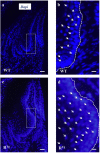
Evidence of gingival epithelium hyper-cellularity in RTg mice based on numbering of DAPI stained cell nucleus. Enlargement view of similar region of the gingival epithelium (rectangles in a,c) evidenced a two times augmentation of the number of cells (arrows) in the gingival epithelium of RTg mice comparatively to WT mice (b,d). Scale bars in A,C correspond to 100 μm and in b,d to 25 μm.
Figure 6
Figure 6
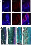
Immuno-histochemistry analyses of RANK and PCNA expressions comparatively in WT and RTg mice. The RANK expressing gingival epithelium thickness was already increased in 5 day-old transgenic mice comparatively to WT (a,b). PNCA staining performed on sections of at 25 day-old WT and RTg evidenced that gingival epithelium thickness increase is associated to an increase of proliferative cells number (c,d). Measurement of the epithelium thickness performed on these sections evidenced a significant increase (**p < 0.01) in the RTg mice (e). ge, gingival epithelium. Scale bars in a,b correspond to 50 μm and in C,D to 20 μm.
Figure 7
Figure 7
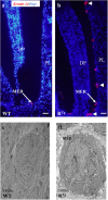
Histological (Keratin 14 immunohistochemistry) and structural (TEM) analyses of Malassez epithelial rests (MER) in 25-day-old mice. A higher number of MER was observed in RTg than WT mice (red staining in a,b) with most appearing to be hyperplasic and/or hypertrophic (arrow-heads in b). TEM analysis (c,d) showed that the hypertrophic appearance of MER was associated with an increased number of cells rather than a higher cell volume. DP, dental papilla; PL, periodontal ligament. Scale bars in a,b correspond to 50 μm. TEM magnification is 2400 X.
Figure 8
Figure 8
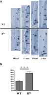
Longitudinal analyses from days 14 to 25 of the number of Malassez epithelial rests (MER) based on Keratin 14 immunohistochemistry comparatively in RTg and WT mice. Keratin 14 positive MER were numbered in equivalent areas of the mandible first molar roots of WT and RTg mice at 14, 17, 21, and 25 days-old (arrows in a). Obtained values were combined in a single statistic analysis in order to represent the median global variation of the MER number during the whole root elongation (b) showing a significant (***p < 0.001) increase in the RTg mice. P, pulp; D, dentin; C, cementum; PL, periodontal ligament; HES, Hertwig epithelial root sheath. Scale bars correspond to 10 μm.

Similar articles

Cited by

References

    1. Albandar J. M. (2005). Epidemiology and risk factors of periodontal diseases. Dent. Clin. North Am. 49, 517–532, v–vi. 10.1016/j.cden.2005.03.003 - DOI - PubMed
    1. Aminoshariae A., Aminoshariae A., Valiathan M., Kulild J. C. (2016). Association of genetic polymorphism and external apical root resorption. Angle Orthod. 86, 1042–1049. 10.2319/011916-50.1 - DOI - PMC - PubMed
    1. Bhuvaneswarri J., Gita B., Chandrasekaran S. C. (2014). Detection of rankl positive cells in gingival tissue in healthy & chronic periodontal disease patients-a comparative study. J. Clin. Diagn. Res. 8, ZC31–ZC34. 10.7860/jcdr/2014/9876.5125 - DOI - PMC - PubMed
    1. Bimstein E., Lustmann J., Sela M. N., Neriah Z. B., Soskolne W. A. (1990). Periodontitis associated with Papillon-Lefèvre syndrome. J. Periodontol. 61, 373–377. 10.1902/jop.1990.61.6.373 - DOI - PubMed
    1. Bord S., Ireland D. C., Beavan S. R., Compston J. E. (2003). The effects of estrogen on osteoprotegerin, RANKL, and estrogen receptor expression in human osteoblasts. Bone 32, 136–141. 10.1016/S8756-3282(02)00953-5 - DOI - PubMed