. 2016 Dec 23;5(1):e00282.
doi: 10.1002/prp2.282. eCollection 2017 Feb.

Acute cocaine exposure elicits rises in calcium in arousal-related laterodorsal tegmental neurons

Affiliations

Acute cocaine exposure elicits rises in calcium in arousal-related laterodorsal tegmental neurons

Mads Ødum Lambert et al. Pharmacol Res Perspect. .

Abstract

Cocaine has strong reinforcing properties, which underlie its high addiction potential. Reinforcement of use of addictive drugs is associated with rises in dopamine (DA) in mesoaccumbal circuitry. Excitatory afferent input to mesoaccumbal circuitry sources from the laterodorsal tegmental nucleus (LDT). Chronic, systemic cocaine exposure has been shown to have cellular effects on LDT cells, but acute actions of local application have never been demonstrated. Using calcium imaging, we show that acute application of cocaine to mouse brain slices induces calcium spiking in cells of the LDT. Spiking was attenuated by tetrodotoxin (TTX) and low calcium solutions, and abolished by prior exhaustion of intracellular calcium stores. Further, DA receptor antagonists reduced these transients, whereas DA induced rises with similar spiking kinetics. Amphetamine, which also results in elevated levels of synaptic DA, but via a different pharmacological action than cocaine, induced calcium spiking with similar profiles. Although large differences in spiking were not noted in an animal model associated with a heightened proclivity of acquiring addiction-related behavior, the prenatal nicotine exposed mouse (PNE), subtle differences in cocaine's effect on calcium spiking were noted, indicative of a reduction in action of cocaine in the LDT associated with exposure to nicotine during gestation. When taken together, our data indicate that acute actions of cocaine do include effects on LDT cells. Considering the role of intracellular calcium in cellular excitability, and of the LDT in addiction circuitry, our data suggest that cocaine effects in this nucleus may contribute to the high addiction potential of this drug.

Keywords: Arousal; REM sleep; cholinergic; in vitro; mouse.


Figures

Figure 1
Figure 1
Cocaine (5 μmol/L, 3 mL) induced changes in fluorescence (dF/F) indicative of rises in intracellular calcium in nearly half of the Fura‐2AM loaded cells examined in the laterodorsal tegmental nucleus (LDT). (A) LDT cells loaded with the calcium indicator dye, Fura‐2, are visible under fluorescent illumination with an excitation wavelength of 380 nm. Changes in fluorescence induced by applied drugs were measured within regions of interest drawn around each calcium indicator‐loaded cell. (B) Although the LDT was chosen by anatomical landmarks during recordings, bNOS immunohistochemistry was used post hoc to confirm that recordings sourced from brain slices containing the LDT. The perimeter of bNOS‐positive cells has been reported as a reliable method by which to define the boundaries of the LDT in mouse (Veleanu et al. 2016), and as shown in the top image in (B), which is a slice used in this study, at 4× magnification under fluorescent optics (wavelength excitation: 488 nm), bilateral clusters of bNOS‐positive cells can be seen ventral to the aqueduct (Aq). Recordings were conducted in these clusters. The bottom fluorescent image is from the same slice, at a higher magnification (20X), which allows for heightened visualization of the cellular details of the bNOS‐positive cells. (C) The most common response type induced by cocaine was categorized as a spiking response (Spikers), which exhibited a rapid rise and fall of fluorescence. Spikers could be further divided into three subtypes based on latency of occurrence of spiking (early, middle and late). The middle latency spiking response type was the most commonly‐occurring of the three types of spiking behavior, which is reflected in the histogram in C, that illustrates the proportion of each of the three types of spiking response occurring in a population of cells (n = 178 spikers/375 tested LDT cells). (E1, 2, 3) Examples of the most common spiking response are shown from three different representative cells responding to cocaine with changes in fluorescence categorized as a middle latency type of response. The two other types of spiker responses are shown in D and F, and designated as early and late, respectively. (G) In some cases, cells were exhibiting ongoing changes in fluorescence prior to drug application indicating spontaneous fluctuations in calcium activity. This ongoing behavior could be silenced by cocaine, but was often enhanced in amplitude and frequency by cocaine, as shown in this representative LDT cell. LDT, laterodorsal tegmental nucleus.
Figure 2
Figure 2
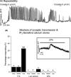
Cocaine responses were not repeatable within the first and second application intervals examined (20–80 min). (A) As shown in this dF/F recording taken from one representative LDT cell, even if spiking induced by a first application of cocaine began to wane, robust repeat responses could not be elicited by a second application of the drug. Often, responses to first applications failed to extinguish even at 1 h post application. Accordingly, determination of effects of antagonists or low calcium external solutions had to be conducted by comparing responses between two different populations of cells. (B) The proportions of cells responding with the early and middle spiking responses were significantly lower in the presence of tetrodotoxin (TTX), which blocks voltage‐dependent sodium channels, indicating reliance of these response types on intact synaptic transmission. The early and middle spiking response was eliminated by low calcium solutions. The late spiking response was also reduced in frequency in these conditions, however, this spiking profile was still elicited in a small proportion of cells (7%) raising the possibility that this response type was not entirely dependent upon calcium flux across the membrane. However, cocaine failed to elicit any of the three types of spiking behavior following dumping of the IP 3‐sensitive intracellular calcium stores. (B, inset) Success of cyclopiazonic acid in dumping these stores was monitored by fluorescent imaging conducted while this drug washed into the bath, and transient increases in dF/F were indicative of successful dumping of this store. When taken together, these data suggest that mechanisms occurring presynaptic to the postsynaptic imaged cells are involved in the cocaine‐induced changes in dF/F resulting in the early and middle latency spiking response, whereas intracellular calcium stores are involved in all three kinetic responses to this stimulant. Asterisks in this, and subsequent figures, denote statistical significance with alpha set lower than 0.05.
Figure 3
Figure 3
Application of DA (30 μmol/L) and of amphetamine (10 μmol/L) resulted in spiking behavior in Fura‐2AM loaded LDT neurons that was qualitatively very similar to that induced by cocaine, and DA receptor blockers attenuated cocaine‐induced spiking behavior. (A1) Representative LDT cell, in which a spiking change in dF/F is evoked by DA. (A2) Responses from the population of LDT cells examined revealed that the proportion of spiking behavior elicited by DA was not significantly different than the numbers of cells responding to cocaine, and the proportions of the three spiking response types induced by DA were not different from those elicited by cocaine. (A3) However, at the concentrations utilized, the amplitudes of two of the spiking behaviors (early and middle) elicited by DA were significantly smaller than those induced by cocaine, whereas, the amplitude of the late spiking response was not significantly different. (B1) Amphetamine induced spiking behavior that was also qualitatively similar to that induced by cocaine and to that induced by DA, as shown in this representative example of spiking behavior elicited by amphetamine in one LDT cell in this study. (B2) Population data show that the distribution of response types was not significantly different between cocaine and amphetamine. (B3) However, interestingly, at the concentrations of stimulants used, the average amplitudes of the early and middle spiking behavior induced by amphetamine were significantly smaller than that induced by cocaine. The amplitude of the late latency response was not significantly different between the two compounds. Experiments with DA and amphetamine were interleaved with control recordings and the same control population in which cocaine was applied was used to test significance across both data sets. (C1) Presence of the DA receptor antagonists, SCH‐23390 and raclopride (10–20 μmol/L), resulted in cocaine‐induced rises in dF/F which were significantly smaller in the population of cells test (DARA) than the amplitudes induced in the absence of these antagonists in another population (Con). (B2) Changes in dF/F induced by cocaine in the absence of DA receptor blockers (top recording, Cocaine Control) and in another cell (bottom recording, Cocaine/DARA), in the presence of the blockers showing a much smaller amplitude of spiking induced by cocaine when the blockers are present. DA, dopamine; LDT, laterodorsal tegmental nucleus.
Figure 4
Figure 4
Antagonists of ionotropic glutamate receptors reduced the amplitude of cocaine‐induced spiking, however, in a model of prenatal drug exposure shown to influence calcium arising subsequent to activation of glutamate mechanisms, the prenatal nicotine model (PNE), responses to cocaine were only subtly altered. (A1) Population data showing that the amplitude of the early and middle latency spiking responses were significantly attenuated when cocaine was applied in the presence of DNQX (10 μmol/L) and AP5 (50 μmol/L), antagonists of the AMPA and NMDA glutamate receptor, respectively, and noted as GlutA in histograms. However, the amplitude of the late spiking response was not significantly different when these receptor antagonists were present. (A2) Detail of one example of spiking behavior induced by cocaine in control conditions, and as shown in another cell, the typically much smaller amplitude change in dF/F induced by cocaine in the presence of the glutamate receptor blockers. (B) Cocaine‐induced changes in dF/F were monitored in LDT slices taken from animals prenatally exposed to nicotine (PNE) and compared to responses induced in saccharine‐exposed controls (PSE). There were no significant differences in the amplitude of the early, middle, and late spiking responses between PNE and PSE treatments groups (B1), which was confirmed using the K–S statistic as illustrated by the distribution of amplitudes of dF/F as shown here for the early latency response type (B2). However, the proportion of cells responding with the earliest latency spiking response was significantly lower in the LDT of PNE animals, than the number of cells exhibiting this spiking response type in the LDT of PSE animals. When taken together with our findings of cocaine responses during blockade of synaptic transmission, these data indicate that glutamate receptors, likely located in a presynaptic location are involved in the early and middle latency spiking behavior evoked by cocaine, whereas the late spiking response type is independent of this involvement. In addition, while differences in cocaine‐elicited spiking behaviors between the PNE and control LDT cells were slight, our data support the conclusion that calcium resulting from glutamate mechanisms is altered by gestational exposure to nicotine. LDT, laterodorsal tegmental nucleus; PNE, prenatal nicotine exposure; PSE, prenatal saccharine exposure.

Similar articles

Cited by

References

    1. Abreu‐Villaca Y, Seidler FJ, Slotkin TA (2004a). Does prenatal nicotine exposure sensitize the brain to nicotine‐induced neurotoxicity in adolescence? Neuropsychopharmacology 29: 1440–1450. - PubMed
    1. Abreu‐Villaca Y, Seidler FJ, Tate CA, Cousins MM, Slotkin TA (2004b). Prenatal nicotine exposure alters the response to nicotine administration in adolescence: effects on cholinergic systems during exposure and withdrawal. Neuropsychopharmacology 29: 879–890. - PubMed
    1. Baghdoyan HA, Rodrigo‐Angulo ML, McCarley RW, Hobson JA (1984). Site‐specific enhancement and suppression of desynchronized sleep signs following cholinergic stimulation of three brainstem regions. Brain Res 306: 39–52. - PubMed
    1. Boucetta S, Jones BE (2009). Activity profiles of cholinergic and intermingled GABAergic and putative glutamatergic neurons in the pontomesencephalic tegmentum of urethane‐anesthetized rats. J Neurosci 29: 4664–4674. - PMC - PubMed
    1. Boucetta S, Cisse Y, Mainville L, Morales M, Jones BE (2014). Discharge profiles across the sleep‐waking cycle of identified cholinergic, GABAergic, and glutamatergic neurons in the pontomesencephalic tegmentum of the rat. J Neurosci 34: 4708–4727. - PMC - PubMed

LinkOut - more resources