. 2017:2017:2180723.
doi: 10.1155/2017/2180723. Epub 2017 May 9.

Assessment of Vascular Change Using Swept-Source Optical Coherence Tomography Angiography: A New Theory Explains Central Visual Loss in Behcet's Disease

Affiliations

Assessment of Vascular Change Using Swept-Source Optical Coherence Tomography Angiography: A New Theory Explains Central Visual Loss in Behcet's Disease

Thanapong Somkijrungroj et al. J Ophthalmol. 2017.

Abstract

Objective: To evaluate retinal vascular structural change in ocular Behcet's using optical coherence tomography angiography (OCTA) and fluorescein angiography (FA).

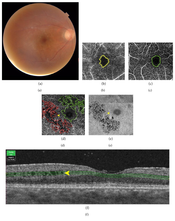
Methods: An analytic cross-sectional study of 37 eyes of 21 Behcet's uveitic patients was performed. Foveal retinal thickness (FRT), perifoveal hypoperfusion areas in superficial capillary plexus (SCP), and deep capillary plexus (DCP) were measured with swept-source optical coherence tomography and OCTA. FA images were used for assessing the vascular features and correlation.

Results: Twenty-one patients were enrolled (52.4% males). The average age at onset was 36.7 ± 12.93 years. The median of disease duration was 5 years (1-25). FRT was 118.1 ± 52.35 μm, which correlated with visual acuity (95% CI -60.47, -13.92). Using OCTA, the area of hypoperfusion in SCP (0.47 ± 0.17 mm2) was smaller than that in DCP (1.94 ± 3.87 mm2) (p < 0.001). Superficial to deep capillary plexus nonperfusion (SCP : DCP) ratio was 0.57 ± 0.27 which had the positive coefficient correlation with visual acuity (95% CI -0.644, -0.015).

Conclusions: OCTA is an alternative noninvasive method to monitor macular ischemia in Behcet. Behcet's uveitis affects DCP more than SCP. Decreasing SCP : DCP ratio and decrease FRT correlates with poor visual acuity. Macular ischemia and DCP loss can be found early and can explain vision loss in Behcet.


Figures

Figure 1
Figure 1
Multimodality imaging in the Behcet uveitis eye with 20/20 visual acuity complain about central visual loss. (a) Fundus photography shows generalized retinal artery attenuation. (b) Fluorescein angiography (FA) demonstrates hypoperfusion area measured in the early phase of FA (yellow area). (c) Superficial capillary plexus (SCP) slap in the optical coherence tomography angiography (OCTA) demonstrates hypoperfusion area in SCP (green area). (d) Deep capillary plexus (DCP) slap in the OCTA demonstrates hypoperfusion area in DCP (green area), and segmentation defect/artifact (red area) mimics hypoperfusion area caused by intraretinal cystoid change (arrow head) which corresponded with (e) structural en-face image and (f) B-scan OCT image. (e) Structural en-face image of DCP slap demonstrates intraretinal cystoid change.

Similar articles

Cited by

References

    1. Evereklioglu C. Current concepts in the etiology and treatment of Behcet disease. Survey of Ophthalmology. 2005;50(4):297–350. doi: 10.1016/j.survophthal.2005.04.009. - DOI - PubMed
    1. An L., Shen T. T., Wang R. K. Using ultrahigh sensitive optical microangiography to achieve comprehensive depth resolved microvasculature mapping for human retina. Journal of Biomedical Optics. 2011;16(10):p. 106013. doi: 10.1117/1.3642638. - DOI - PMC - PubMed
    1. de Carlo T. E., Romano A., Waheed N. K., Duker J. S. A review of optical coherence tomography angiography (OCTA) International Journal Retina Vitreous. 2015;1:p. 5. doi: 10.1186/s40942-015-0005-8. - DOI - PMC - PubMed
    1. Kam J., Zhang Q., Lin J., Liu J., Wang R. K., Rezaei K. Optical coherence tomography based microangiography findings in hydroxychloroquine toxicity. Quantitative Imaging in Medicine and Surgery. 2016;6(2):178–183. doi: 10.21037/qims.2016.01.01. - DOI - PMC - PubMed
    1. Agrawal R., Xin W., Keane P. A., Chhablani J., Agarwal A. Optical coherence tomography angiography: a non-invasive tool to image end-arterial system. Expert Review of Medical Devices. 2016;13(6):519–521. doi: 10.1080/17434440.2016.1186540. - DOI - PubMed

LinkOut - more resources