Review
. 2017:2017:3232848.
doi: 10.1155/2017/3232848. Epub 2017 May 17.

Soluble Urokinase Receptor and the Kidney Response in Diabetes Mellitus

Affiliations
Review

Soluble Urokinase Receptor and the Kidney Response in Diabetes Mellitus

Ranadheer R Dande et al. J Diabetes Res. 2017.

Abstract

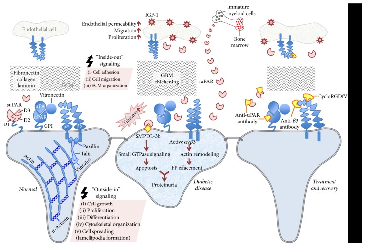
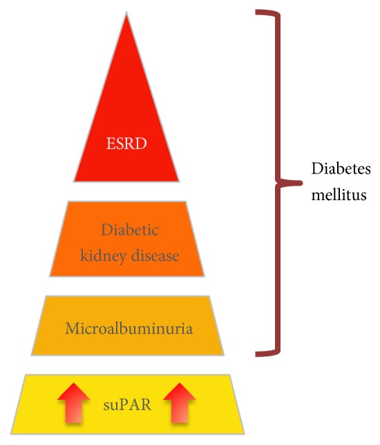
Diabetic nephropathy (DN) is the leading cause of end-stage renal disease (ESRD) worldwide. DN typically manifests by glomerular hyperfiltration and microalbuminuria; then, the disease progresses to impaired glomerular filtration rate, which leads to ESRD. Treatment options for DN include the strict control of blood glucose levels and pressure (e.g., intraglomerular hypertension). However, the search for novel therapeutic strategies is ongoing. These include seeking specific molecules that contribute to the development and progression of DN to potentially interfere with these "molecular targets" as well as with the cellular targets within the kidney such as podocytes, which play a major role in the pathogenesis of DN. Recently, podocyte membrane protein urokinase receptor (uPAR) and its circulating form (suPAR) are found to be significantly induced in glomeruli and sera of DN patients, respectively, and elevated suPAR levels predicted diabetic kidney disease years before the occurrence of microalbuminuria. The intent of this review is to summarize the emerging evidence of uPAR and suPAR in the clinical manifestations of DN. The identification of specific pathways that govern DN will help us build a more comprehensive molecular model for the pathogenesis of the disease that can inform new opportunities for treatment.


Figures

Figure 1
Figure 1
Schematic depiction of uPAR/suPAR-αvβ3 integrin signaling at the glomerular filtration level in health and diabetic disease. In resting podocytes, uPAR interacts with uPA and anchored to the outer plasma membrane with GPI. This complex is connected to αvβ3 integrin through vitronectin, a β3 integrin ligand. This leads to the initiation of “outside-in” signaling events, which requires the recruitment of linker proteins (paxillin, talin, and vinculin) by integrins for actin involvement. This signaling pathway is responsible for proper actin cytoskeleton assembly, lamellipodia formation, growth, proliferation, differentiation, and cell survival. ECM proteins such as fibronectin, collagen, and laminin are also involved in many cellular activities including ECM organization, cell adhesion and migration. Three homologous domains of uPAR are denoted by D1, D2, and D3, respectively (left panel). In the hyperglycemic state, αvβ3 integrin activity increases causing altered adhesion, migration, and proliferation. These intracellular changes might initiate an “inside-out” signaling affecting integrin's binding affinity. Soluble uPAR also increases in circulation and probably contributes to the pathology of the diabetic kidney disease, which can be characterized as impaired cytoskeletal organization and podocyte FP effacement. The pathogenic suPAR is mainly generated by bone marrow-immature myeloid cells. Podocyte-specific expression of SMPDL-3b, which is elevated during the course of diabetic kidney disease, prevents αvβ3 integrin activation by interacting with suPAR. This eventually increases RhoA activity and podocyte susceptibility to apoptosis. αvβ3 integrin receptors are also expressed in glomerular endothelium and exposure of endothelial cells to hyperglycemia leads to pathologic outcomes in these cells such as endothelial permeability, migration, and proliferation in response to the ligand occupancy of αvβ3 and concomitant stimulation of IGF-1 (middle panel). Targeting uPAR and suPAR with an uPAR-specific monoclonal antibody can attenuate the adverse effects of uPAR/suPAR-dependent integrin signaling. Using antibodies that bind preferentially to the activated and/or ligand-occupied forms of β3 integrin and β3 integrin small molecule inhibitor, cycloRGDfV, offer alternative ways to disentangle its interactions with uPAR/suPAR. Blocking the ligand occupancy of αvβ3 inhibits the pathogenic mechanisms stimulated by IGF-1 (right panel).
Figure 2
Figure 2
Schematic depiction of suPAR as a predictor for future diabetic kidney disease.

Similar articles

Cited by

References

    1. Satirapoj B., Adler S. G. Prevalence and management of diabetic nephropathy in Western countries. Kidney Disease (Basel) 2015;1(1):61–70. doi: 10.1159/000382028. - DOI - PMC - PubMed
    1. Chen L., Magliano D. J., Zimmet P. Z. The worldwide epidemiology of type 2 diabetes mellitus - present and future perspectives. Nature Reviews. Endocrinology. 2012;8(4):228–236. doi: 10.1038/nrendo.2011.183. - DOI - PubMed
    1. Mogensen C. E. Microalbuminuria as a predictor of clinical diabetic nephropathy. Kidney International. 1987;31(2):673–689. doi: 10.1038/ki.1987.50. - DOI - PubMed
    1. Eknoyan G., Hostetter T., Bakris G. L., et al. Proteinuria and other markers of chronic kidney disease: a position statement of the national kidney foundation (NKF) and the national institute of diabetes and digestive and kidney diseases (NIDDK) American Journal of Kidney Diseases. 2003;42(4):617–622. doi: 10.1016/S0272-6386(03)00826-6. - DOI - PubMed
    1. Kramer H. J., Nguyen Q. D., Curhan G., Hsu C. Y. Renal insufficiency in the absence of albuminuria and retinopathy among adults with type 2 diabetes mellitus. JAMA. 2003;289(24):3273–3277. doi: 10.1001/jama.289.24.3273. - DOI - PubMed

Substances

LinkOut - more resources