. 2017 Mar;3(1):15-31.
doi: 10.1007/s40883-016-0022-7. Epub 2017 Jan 30.

Biodegradable Polyphosphazene-Based Blends for Regenerative Engineering

Affiliations

Biodegradable Polyphosphazene-Based Blends for Regenerative Engineering

Kenneth S Ogueri et al. Regen Eng Transl Med. 2017 Mar.

Abstract

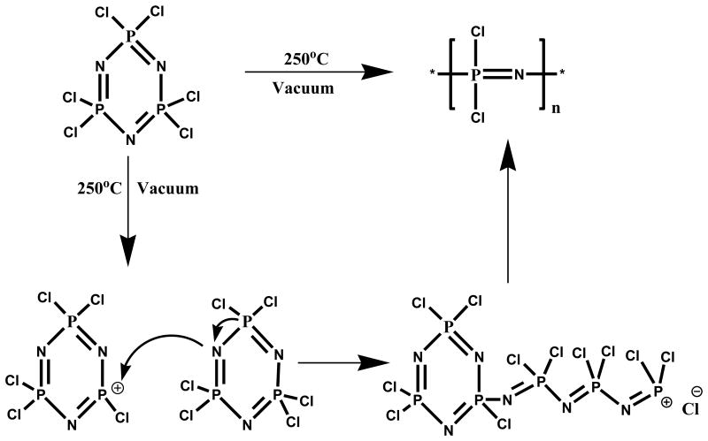
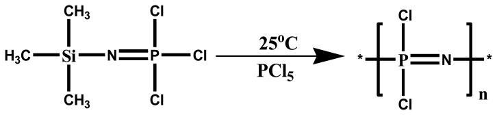
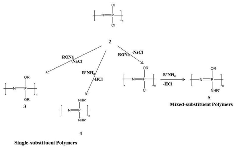
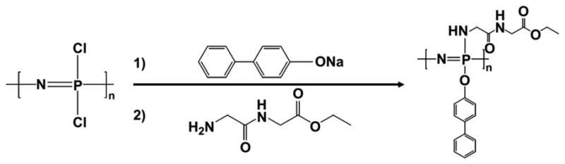
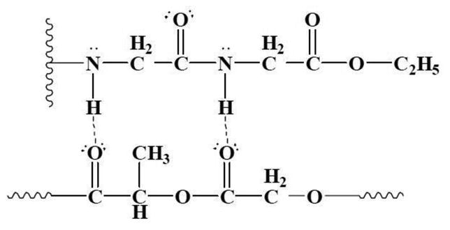
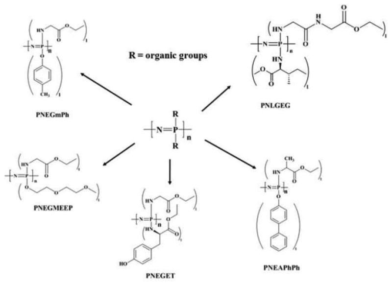
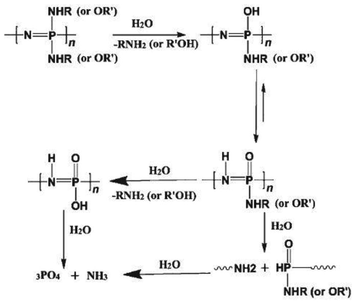
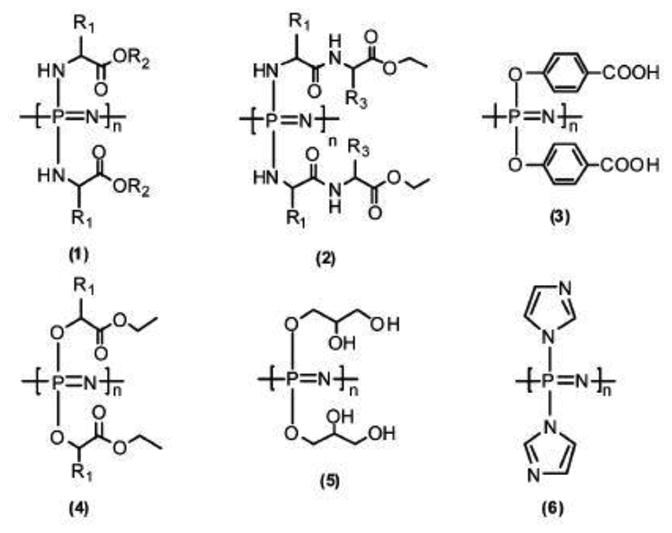
The occurrence of musculoskeletal tissue injury or disease and the subsequent functional impairment is at an alarming rate. It continues to be one of the most challenging problems in the human health care. Regenerative engineering offers a promising transdisciplinary strategy for tissues regeneration based on the convergence of tissue engineering, advanced materials science, stem cell science, developmental biology and clinical translation. Biomaterials are emerging as extracellular-mimicking matrices designed to provide instructive cues to control cell behavior and ultimately, be applied as therapies to regenerate damaged tissues. Biodegradable polymers constitute an attractive class of biomaterials for the development of scaffolds due to their flexibility in chemistry and the ability to be excreted or resorbed by the body. Herein, the focus will be on biodegradable polyphosphazene-based blend systems. The synthetic flexibility of polyphosphazene, combined with the unique inorganic backbone, has provided a springboard for more research and subsequent development of numerous novel materials that are capable of forming miscible blends with poly (lactide-co-glycolide) (PLAGA). Laurencin and co-workers has demonstrated the exploitation of the synthetic flexibility of Polyphosphazene that will allow the design of novel polymers, which can form miscible blends with PLAGA for biomedical applications. These novel blends, due to their well-tuned biodegradability, and mechanical and biological properties coupled with the buffering capacity of the degradation products, constitute ideal materials for regeneration of various musculoskeletal tissues.

Lay summary: Regenerative engineering aims to regenerate complex tissues to address the clinical challenge of organ damage. Tissue engineering has largely focused on the restoration and repair of individual tissues and organs, but over the past 25 years, scientific, engineering, and medical advances have led to the introduction of this new approach which involves the regeneration of complex tissues and biological systems such as a knee or a whole limb. While a number of excellent advanced biomaterials have been developed, the choice of biomaterials, however, has increased over the past years to include polymers that can be designed with a range of mechanical properties, degradation rates, and chemical functionality. The polyphosphazenes are one good example. Their chemical versatility and hydrogen bonding capability encourages blending with other biologically relevant polymers. The further development of Polyphosphazene-based blends will present a wide spectrum of advanced biomaterials that can be used as scaffolds for regenerative engineering and as well as other biomedical applications.

Keywords: Biodegradable polymers; Dipeptide-based Polyphosphazene; Musculoskeletal; Polyphosphazene Blends; Regenerative engineering.


Figures

Fig. 1
Fig. 1. Synthesis of poly (dichlorophospazene): Synthetic Schematics of ring opening polymerization [31]
Fig. 2
Fig. 2. Alternative route to the synthesis of poly(dichlorophospazene): living cationic polymerization of a phosphoranamine monomer[32]
Fig. 3
Fig. 3. Macromolecular substitution of PDCP with a wide array of organic substituent groups forming single-substituent polymers (simultaneous replacement of chlorine atoms) or mixed-substituent polymers (sequential substitution of chlorine atoms)[8, 32]
Fig. 4
Fig. 4. Schematic illustrations of a mixed-substituent biodegradable polyphosphazene poly((glycine ethyl glycinato)1(phenyl phenoxy)1phosphazene) (PNGEGPhPh)[18]
Fig. 5
Fig. 5. Illustration of intermolecular hydrogen bonding network between PLAGA and PNGEGPhPh through the glycylglycine dipeptides [18]
Fig. 6
Fig. 6. Structures of mixed-substituent PPHOS for coordination with PLAGA. The primary selection criterion for side groups was based on its capacity to form miscible blends with PLAGA through intermolecular hydrogen bonding interactions[15]
Fig. 7
Fig. 7. Pathway to the degradation of biodegradable Polyphosphazenes [61]
Fig. 8
Fig. 8. Structures of bio-erodible Polyphosphazene[5]
Fig. 9
Fig. 9
Time dependent topographical imaging of the polymeric blends immersed in aqueous media for 12 weeks at 37 °C. Top row: SEM topography of Matrix1 at 0, 4, 7, and 12 weeks of time intervals of in vitro degradation. Spots (e) and (g) show the detailed 3D Spherical structures. Bottom row: SEM topography of Matrix2 under vitro degradation at 0, 4, 7, and 12 weeks. An assemblage of microspheres with interconnected porous structures was evident due to the unique polymer erosion of the blend system[51]
Fig. 10
Fig. 10
Graphs showing a 12-week study of in vitro degradation of the blend systems in pH 7.4 aqueous media at 37°C(a) Comparison of the molecular weight percentages of PLAGA in pristine PLAGA and the two blend systems after degradation (b) Mass of the residual systems expressed in percentage; (c) Comparison of the molecular weight percentage of polyphosphazene in the residual blends matrices (d) pH profiles of the media during degradation of pristine PLAGA and blend matrices. Slower degradation rate was evident in the blend systems than the pristine PLAGA. The trends for molecular weight changes of the PLAGA and phosphazene reasserted the similarities in the degradations of the two components [51].
Fig. 11
Fig. 11
Histological examination reveals the formation of pore system that stems from the arrangement of newly formed polymer spheres. This is capable of accommodating cell infiltration and tissue in-growth within the blend systems. (a, b, H&E): The arrows shows the formation of polymer sphere within Matrix1 and Matrix2 after 7 weeks of implantation, respectively; (c, d, H&E): The arrows show the formation of polymer sphere within Matrix1 and Matrix2 after 12 weeks of implantation, respectively. After 12 weeks of implantation, strong collagen tissue infiltration via in situ formed pores within the matrix was apparent in (C) and (D) with TRI. This reaffirms the availability and enhancement of cell infiltration and collagen tissue in-growth through the formation of in situ 3D interconnected porous structure [51]
Fig. 12
Fig. 12
(a) Electrospun polyphosphazene nanofibrous scaffolds; (b) SEM-SE image showing the topographical fixtures of 3D biomimetic scaffolds seeded with cells after 28 days of culture. (c) Immunohistochemical staining for osteopontin (OPN), an important component of the mineralized ECM, showing a homogenous ECM distribution throughout the scaffold architecture at day 28. * indicates interlamellar space, whereas ** indicates central cavity[61].

Similar articles

Cited by

References

    1. Laurencin CT, Khan Y. Regenerative engineering. Science translational medicine. 2012;4(160):160ed169–160ed169. - PubMed
    1. Laurencin CT, Nair LS. Regenerative Engineering: Approaches to Limb Regeneration and Other Grand Challenges. Regenerative Engineering and Translational Medicine. 2015;1(1):1–3. - PMC - PubMed
    1. Reichert WM, Ratner BD, Anderson J, Coury A, Hoffman AS, Laurencin CT, Tirrell D. 2010 Panel on the biomaterials grand challenges. J Biomed Mater Res A. 2011;96(2):275–287. - PMC - PubMed
    1. Polyphosphazenes for Biomedical Applications (1) Hoboken, US: Wiley; 2009.
    1. Allcock HR, Morozowich NL. Bioerodible polyphosphazenes and their medical potential. Polymer Chemistry. 2012;3(3):578–590.

LinkOut - more resources