

Magnetic Resonance Imaging of Cerebral Malaria Patients Reveals Distinct Pathogenetic Processes in Different Parts of the Brain
- PMID: 28596990
- PMCID: PMC5463026
- DOI: 10.1128/mSphere.00193-17
Magnetic Resonance Imaging of Cerebral Malaria Patients Reveals Distinct Pathogenetic Processes in Different Parts of the Brain
Abstract
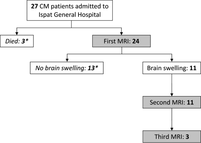
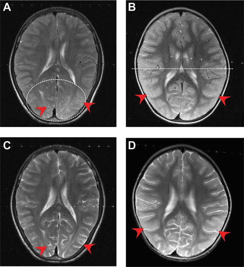
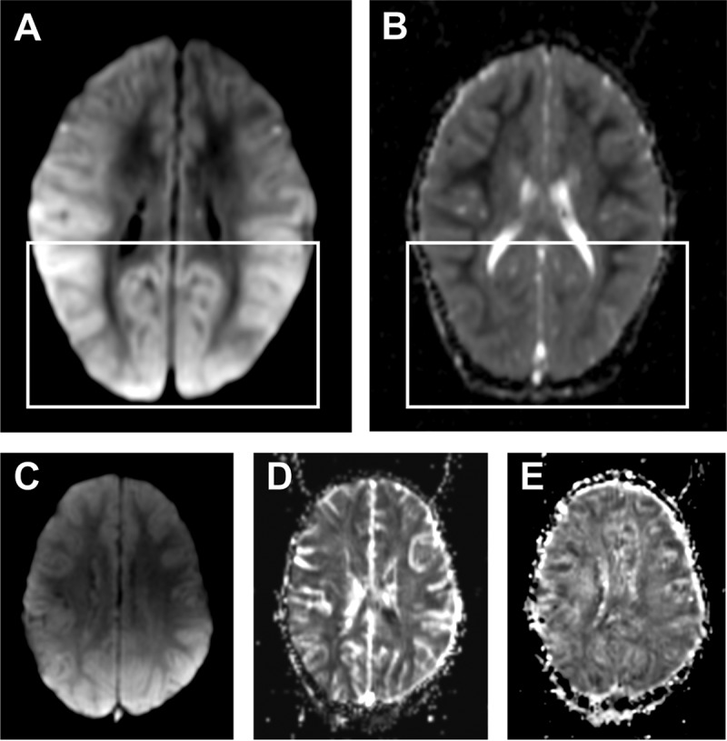
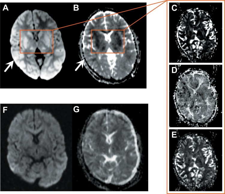
The mechanisms underlying the rapidly reversible brain swelling described in patients with cerebral malaria (CM) are unknown. Using a 1.5-Tesla (T) magnetic resonance imaging (MRI) scanner, we undertook an observational study in Rourkela, India, of 11 Indian patients hospitalized with CM and increased brain volume. Among the 11 cases, there were 5 adults and 6 children. All patients had reduced consciousness and various degrees of cortical swelling at baseline. The latter was predominately posterior in distribution. The findings on diffusion-weighted imaging (DWI) and apparent diffusion coefficient (ADC) maps were consistent with vasogenic edema in all cases. Reversibility after 48 to 72 h was observed in >90% of cases. DWI/ADC mismatch suggested the additional presence of cytotoxic edema in the basal nuclei of 5 patients; all of these had perfusion parameters consistent with vascular engorgement and not with ischemic infarcts. Our results suggest that an impairment of the blood-brain barrier is responsible for the brain swelling in CM. In 5 cases, vasogenic edema occurred in conjunction with changes in the basal nuclei consistent with venous congestion, likely to be caused by the sequestration of Plasmodium falciparum-infected erythrocytes. While both mechanisms have been individually postulated to play an important role in the development of CM, this is the first demonstration of their concurrent involvement in different parts of the brain. The clinical and radiological characteristics observed in the majority of our patients are consistent with posterior reversible encephalopathy syndrome (PRES), and we show for the first time a high frequency of PRES in the context of CM. IMPORTANCE The pathophysiology and molecular mechanisms underlying cerebral malaria (CM) are still poorly understood. Recent neuroimaging studies demonstrated that brain swelling is a common feature in CM and a major contributor to death in pediatric patients. Consequently, determining the precise mechanisms responsible for this swelling could open new adjunct therapeutic avenues in CM patients. Using an MRI scanner with a higher resolution than the ones used in previous reports, we identified two distinct origins of brain swelling in both adult and pediatric patients from India, occurring in distinct parts of the brain. Our results support the hypothesis that both endothelial dysfunction and microvascular obstruction by Plasmodium falciparum-infected erythrocytes make independent contributions to the pathogenesis of CM, providing opportunities for novel therapeutic interventions.
Keywords: MRI; PRES; Plasmodium falciparum; cerebral malaria; vasogenic edema.
Figures
References
-
- Seydel KB, Kampondeni SD, Valim C, Potchen MJ, Milner DA, Muwalo FW, Birbeck GL, Bradley WG, Fox LL, Glover SJ, Hammond CA, Heyderman RS, Chilingulo CA, Molyneux ME, Taylor TE. 2015. Brain swelling and death in children with cerebral malaria. N Engl J Med 372:1126–1137. doi: 10.1056/NEJMoa1400116. - DOI - PMC - PubMed
Grants and funding
LinkOut - more resources
Full Text Sources
Other Literature Sources
