

Long Persistence of a Streptococcus pneumoniae 23F Clone in a Cystic Fibrosis Patient
- PMID: 28596991
- PMCID: PMC5463027
- DOI: 10.1128/mSphere.00201-17
Long Persistence of a Streptococcus pneumoniae 23F Clone in a Cystic Fibrosis Patient
Abstract
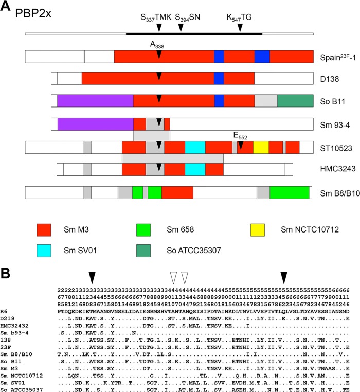
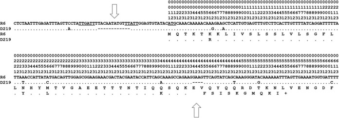
Streptococcus pneumoniae isolates of serotype 23F with intermediate penicillin resistance were recovered on seven occasions over a period of 37 months from a cystic fibrosis patient in Berlin. All isolates expressed the same multilocus sequence type (ST), ST10523. The genome sequences of the first and last isolates, D122 and D141, revealed the absence of two phage-related gene clusters compared to the genome of another ST10523 strain, D219, isolated earlier at a different place in Germany. Genomes of all three strains carried the same novel mosaic penicillin-binding protein (PBP) genes, pbp2x, pbp2b, and pbp1a; these genes were distinct from those of other penicillin-resistant S. pneumoniae strains except for pbp1a of a Romanian S. pneumoniae isolate. All PBPs contained mutations that have been associated with the penicillin resistance phenotype. Most interestingly, a mosaic block identical to an internal pbp2x sequence of ST10523 was present in pbp2x of Streptococcus mitis strain B93-4, which was isolated from the same patient. This suggests interspecies gene transfer from S. pneumoniae to S. mitis within the host. Nearly all genes expressing surface proteins, which represent major virulence factors of S. pneumoniae and are typical for this species, were present in the genome of ST10523. One exception was the hyaluronidase gene hlyA, which contained a 12-nucleotide deletion within the promoter region and an internal stop codon. The lack of a functional hyaluronidase might contribute to the ability to persist in the host for an unusually long period of time. IMPORTANCEStreptococcus pneumoniae is a common resident in the human nasopharynx. However, carriage can result in severe diseases due to a unique repertoire of pathogenicity factors that are rare in closely related commensal streptococci. We investigated a penicillin-resistant S. pneumoniae clone of serotype 23F isolated from a cystic fibrosis patient on multiple occasions over an unusually long period of over 3 years that was present without causing disease. Genome comparisons revealed an apparent nonfunctional pneumococcus-specific gene encoding a hyaluronidase, supporting the view that this enzyme adds to the virulence potential of the bacterium. The 23F clone harbored unique mosaic genes encoding penicillin resistance determinants, the product of horizontal gene transfer involving the commensal S. mitis as donor species. Sequences identical to one such mosaic gene were identified in an S. mitis strain from the same patient, suggesting that in this case S. pneumoniae played the role of donor.
Keywords: 23F clone; Streptococcus pneumoniae; cystic fibrosis; hyaluronidase; penicillin-binding proteins; persistence.
Figures
Similar articles
-
[Evaluation of penicillin-binding protein genotypes in penicillin susceptible and resistant Streptococcus pneumoniae isolates].Mikrobiyol Bul. 2012 Apr;46(2):190-201. Mikrobiyol Bul. 2012. PMID: 22639308 Turkish.
-
Highly variable penicillin resistance determinants PBP 2x, PBP 2b, and PBP 1a in isolates of two Streptococcus pneumoniae clonal groups, Poland 23F-16 and Poland 6B-20.Antimicrob Agents Chemother. 2008 Mar;52(3):1021-7. doi: 10.1128/AAC.01082-07. Epub 2007 Dec 26. Antimicrob Agents Chemother. 2008. PMID: 18160523 Free PMC article.
-
Insight into the Diversity of Penicillin-Binding Protein 2x Alleles and Mutations in Viridans Streptococci.Antimicrob Agents Chemother. 2017 Apr 24;61(5):e02646-16. doi: 10.1128/AAC.02646-16. Print 2017 May. Antimicrob Agents Chemother. 2017. PMID: 28193649 Free PMC article.
-
Diversity of Mosaic pbp2x Families in Penicillin-Resistant Streptococcus pneumoniae from Iran and Romania.Antimicrob Agents Chemother. 2017 Nov 22;61(12):e01535-17. doi: 10.1128/AAC.01535-17. Print 2017 Dec. Antimicrob Agents Chemother. 2017. PMID: 28971878 Free PMC article.
-
Horizontal genetic exchange, evolution, and spread of antibiotic resistance in bacteria.Clin Infect Dis. 1998 Aug;27 Suppl 1:S12-20. doi: 10.1086/514917. Clin Infect Dis. 1998. PMID: 9710667 Review.
Cited by
-
Competence in Streptococcus pneumoniae and Close Commensal Relatives: Mechanisms and Implications.Front Cell Infect Microbiol. 2019 Apr 3;9:94. doi: 10.3389/fcimb.2019.00094. eCollection 2019. Front Cell Infect Microbiol. 2019. PMID: 31001492 Free PMC article. Review.
-
Ceftriaxone-resistant viridans streptococci bacteraemia among patients treated at a large comprehensive cancer care centre: a retrospective eighteen-year study.JAC Antimicrob Resist. 2024 Aug 5;6(4):dlae126. doi: 10.1093/jacamr/dlae126. eCollection 2024 Aug. JAC Antimicrob Resist. 2024. PMID: 39104770 Free PMC article.
-
How Streptococcus suis escapes antibiotic treatments.Vet Res. 2022 Nov 12;53(1):91. doi: 10.1186/s13567-022-01111-3. Vet Res. 2022. PMID: 36371221 Free PMC article. Review.
-
Puzzling Over the Pneumococcal Pangenome.Front Microbiol. 2018 Oct 30;9:2580. doi: 10.3389/fmicb.2018.02580. eCollection 2018. Front Microbiol. 2018. PMID: 30425695 Free PMC article. Review.
References
-
- Short KR, Diavatopoulos DA. 2015. Nasopharyngeal colonization with Streptococcus pneumoniae, p 279–291. In Brown J, Hammerschmidt S, Orihuela C (ed), Streptococcus pneumoniae molecular mechanisms of host-pathogen interactions. Academic Press, London, United Kingdom.
-
- Nunes S, Sá-Leão R, Carriço J, Alves CR, Mato R, Avô AB, Saldanha J, Almeida JS, Sanches IS, de Lencastre H. 2005. Trends in drug resistance, serotypes, and molecular types of Streptococcus pneumoniae colonizing preschool-age children attending day care centers in Lisbon, Portugal: a summary of 4 years of annual surveillance. J Clin Microbiol 43:1285–1293. doi:10.1128/JCM.43.3.1285-1293.2005. - DOI - PMC - PubMed
-
- Ercibengoa M, Arostegi N, Marimón JM, Alonso M, Pérez-Trallero E. 2012. Dynamics of pneumococcal nasopharyngeal carriage in healthy children attending a day care center in northern Spain. Influence of detection techniques on the results. BMC Infect Dis 12:69. doi:10.1186/1471-2334-12-69. - DOI - PMC - PubMed
LinkOut - more resources
Full Text Sources
Other Literature Sources
Research Materials
Miscellaneous
