Multicenter Study
. 2017 Apr;3(4):393-402.
doi: 10.1016/j.jacep.2016.10.006.

Recurrent Post-Ablation Paroxysmal Atrial Fibrillation Shares Substrates With Persistent Atrial Fibrillation : An 11-Center Study

Affiliations
Multicenter Study

Recurrent Post-Ablation Paroxysmal Atrial Fibrillation Shares Substrates With Persistent Atrial Fibrillation : An 11-Center Study

Junaid A B Zaman et al. JACC Clin Electrophysiol. 2017 Apr.

Abstract

Introduction: The role of atrial fibrillation (AF) substrates is unclear in patients with paroxysmal AF (PAF) that recurs after pulmonary vein isolation (PVI). We hypothesized that patients with recurrent post-ablation (redo) PAF despite PVI have electrical substrates marked by rotors and focal sources, and structural substrates that resemble persistent AF more than patients with (de novo) PAF at first ablation.

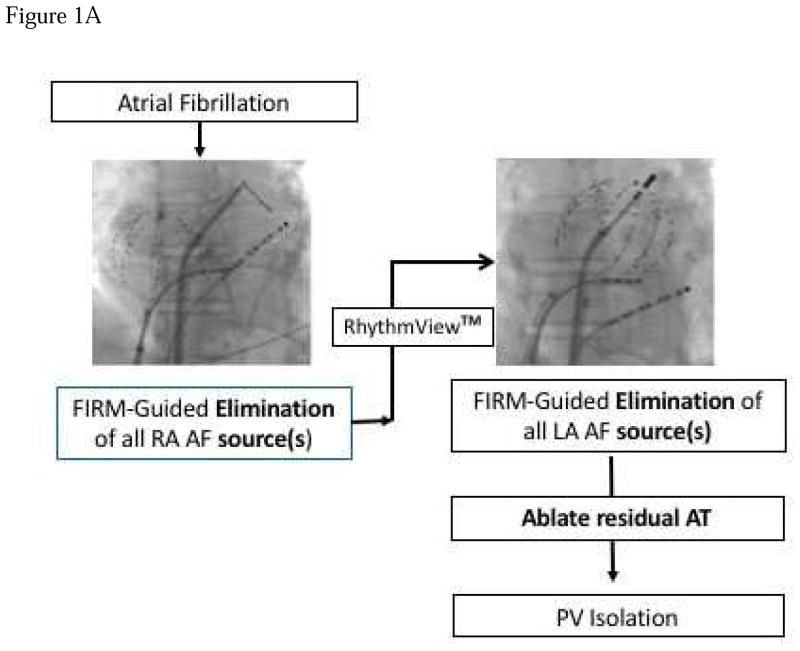
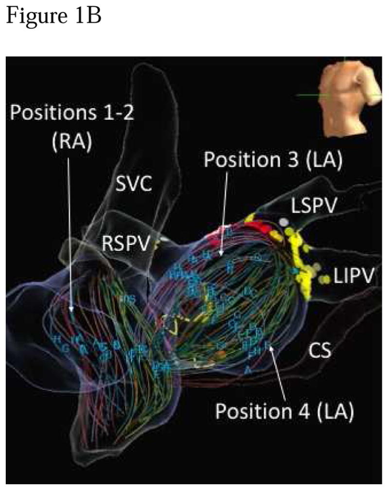
Methods: In 175 patients at 11 centers, we compared AF substrates in both atria using 64 pole-basket catheters and phase mapping, and indices of anatomical remodeling between patients with de novo or redo PAF and first ablation for persistent AF.

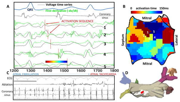
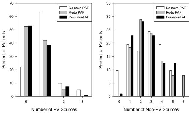
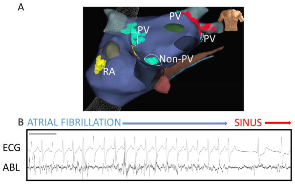
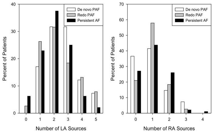
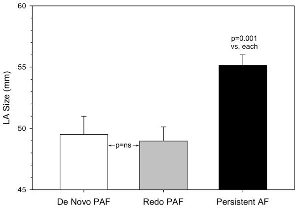
Results: Sources were seen in all patients. More patients with de novo PAF (78.0%) had sources near PVs than patients with redo PAF (47.4%, p=0.005) or persistent AF (46.9%, p=0.001). The total number of sources per patient (p=0.444), and number of non-PV sources (p=0.701) were similar between groups, indicating that redo PAF patients had residual non-PV sources after elimination of PV sources by prior PVI. Structurally, left atrial size did not separate de novo from redo PAF (49.5±9.5 vs. 49.0±7.1mm, p=0.956) but was larger in patients with persistent AF (55.2±8.4mm, p=0.001).

Conclusions: Patients with paroxysmal AF despite prior PVI show electrical substrates that resemble persistent AF more closely than patients with paroxysmal AF at first ablation. Notably, these subgroups of paroxysmal AF are indistinguishable by structural indices. These data motivate studies of trigger versus substrate mechanisms for patients with recurrent paroxysmal AF after PVI.

Keywords: ablation; atrial fibrillation; sources.


Figures

Figure 1
Figure 1. Technique for FIRM Guided Ablation
A) Typical atrial basket placement is indicated in the fluoroscopic images. Sequential atrial basket recordings were taken with source elimination prior to PVI. B) Multiple basket positions used per FIRM-mapping epoch to minimize unmapped atrial regions. Positions 1–2 are in the right atrium, positions 3–4 are in the LA. SVC – superior vena cava, RSPV – right superior pulmonary vein, RIPV right inferior pulmonary vein, LSPV – left superior pulmonary vein, LIPV – left inferior pulmonary vein.
Figure 2
Figure 2. Rotational Activity at FIRM-mapped Rotor, where Ablation terminated AF to Atrial Tachycardia
In this 61 year old man with persistent AF, unipolar AF electrograms confirm rotation around rotor core. A) Electrograms (unipoles) and dV/dt (first derivative, green) shown, with one cycle annotated (red lines). B) Left atrial shell with activation map from the annotated cycle, demonstrating earliest-latest interaction in a rotational pattern. C) Termination panel shows abrupt termination of AF to organized AT with ablation at this site, prior to any PVI. Black bar represents 1000ms. D) Location on LA posterior wall of termination site (red) during ablation of rotor area (white).
Figure 3
Figure 3. Higher number of PV sources in de novo PAF
(Left) The bar chart shows a higher percentage of patients with PV rotors/sources in de novo PAF compared to redo PAF and persistent AF (p=0.002). Differences in the number of non-PV sources (right) were not significant (p=0.701).
Figure 4
Figure 4. Ablation of AF sources adjacent and remote to PVs causing acute termination to sinus rhythm
A) Antero-posterior bi-atrial NavX map with RA, non-PV and PV LA sources labelled in a patient at redo ablation for PAF. B) AF signals on ablation catheter at FIRM-mapped rotor adjacent to prior left superior PVI lesion set, where AF was terminated by FIRM-guided ablation. Black bar represents 1000ms. ABL – ablation catheter.
Figure 5
Figure 5. Non-significant differences in RA and LA sources
There was a trend towards a difference among the groups in the number of LA sources (p=0.070) which was somewhat higher in de novo PAF than in the other groups. There was no group difference in the number of RA sources (p=0.541).
Figure 6
Figure 6. LA size did not separate de novo from redo PAF
LA size was higher only in the persistent AF group, despite the electrophysiological differences in this study. (Mean + SEM).

Comment in

References

    1. Verma A, Jiang C, Betts TR, et al. Approaches to Catheter Ablation for Persistent Atrial Fibrillation. N Engl J Med. 2015;372(19):1812–22. doi: 10.1056/NEJMoa1408288. - DOI - PubMed
    1. Natale A, Reddy VY, Monir G, et al. Paroxysmal AF catheter ablation with a contact force sensing catheter: results of the prospective, multicenter SMART-AF trial. J Am Coll Cardiol. 2014;64(7):647–56. doi: 10.1016/j.jacc.2014.04.072. - DOI - PubMed
    1. Calkins H. Demonstrating the Value of Contact Force Sensing More Difficult Than Meets the Eye. Circulation. 2015:901–4. doi: 10.1161/CIRCULATIONAHA.115.018354. - DOI - PubMed
    1. Dukkipati SR, Cuoco F, Kutinsky I, et al. Pulmonary Vein Isolation Using the Visually Guided Laser Balloon. J Am Coll Cardiol. 2015;66(12):1350–60. doi: 10.1016/j.jacc.2015.07.036. - DOI - PubMed
    1. Jiang R-H, Po SS, Tung R, et al. Incidence of pulmonary vein conduction recovery in patients without clinical recurrence after ablation of paroxysmal atrial fibrillation: mechanistic implications. Heart Rhythm. 2014;11(6):969–76. doi: 10.1016/j.hrthm.2014.03.015. - DOI - PubMed

Publication types