. 2017 Dec;39(12):1317-1322.
doi: 10.1007/s00276-017-1885-0. Epub 2017 Jun 8.

Ultrasound visualization of an underestimated structure: the bicipital aponeurosis

Affiliations

Ultrasound visualization of an underestimated structure: the bicipital aponeurosis

M Konschake et al. Surg Radiol Anat. 2017 Dec.

Abstract

Purpose: We established a detailed sonographic approach to the bicipital aponeurosis (BA), because different pathologies of this, sometimes underestimated, structure are associated with vascular, neural and muscular lesions; emphasizing its further implementation in routine clinical examinations.

Methods: The BA of 100 volunteers, in sitting position with the elbow lying on a suitable table, was investigated. Patients were aged between 18 and 28 with no history of distal biceps injury. Examination was performed using an 18-6 MHz linear transducer (LA435; system MyLab25 by Esaote, Genoa, Italy) utilizing the highest frequency, scanned in two planes (longitudinal and transverse view). In each proband, scanning was done with and without isometric contraction of the biceps brachii muscle.

Results: The BA was characterized by two clearly distinguishable white lines enveloping a hypoechoic band. In all longitudinal images (plane 1), the lacertus fibrosus was clearly seen arising from the biceps muscle belly, the biceps tendon or the myotendinous junction, respectively. In transverse images (plane 2) the BA spanned the brachial artery and the median nerve in all subjects. In almost all probands (97/100), the BA was best distinguishable during isometric contraction of the biceps muscle.

Conclusion: With the described sonographic approach, it should be feasible to detect alterations and unusual ruptures of the BA. Therefore, we suggest additional BA scanning during clinical examinations of several pathologies, not only for BA augmentation procedures in distal biceps tendon tears.

Keywords: Biceps brachii muscle; Bicipital aponeurosis; Lacertus fibrosus; Ultrasonography.


Conflict of interest statement

The authors declare that they have no conflict of interest.

Figures
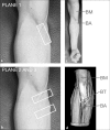
Fig. 1
Fig. 1
a Probe placement for plane 1, longitudinal view: the transducer was placed in line with the assumed aponeurosis’ main bundle, illustrated by the white rectangle. b Probe placement for plane 2 and 3, transverse views: the probe was turned 90° at two different levels as shown by the two white rectangles. c Isometric contraction during examination, showing the biceps brachii muscle (BM) and the bicipital aponeurosis (BA). d Specimen showing the biceps brachii muscle (BM), the biceps tendon (BT) and the bicipital aponeurosis (BA)
Fig. 2
Fig. 2
Bicipital aponeurosis (BA) longitudinal view. The BA (white arrowheads) is seen as double contour emerging from the myotendineous junction of biceps brachii muscle (orange arrowhead), bridging the brachial artery (red dashed oval) and connecting to the antebrachial fascia that covers the pronator teres muscle (grey arrowhead). Note that the BA is clearly distinguishable from the subcutis (asterisks)! BR brachialis muscle (color figure online)
Fig. 3
Fig. 3
Bicipital aponeurosis (BA) transverse view. The BA (white arrowheads) is seen as double contour bridging the brachial artery (red dashed oval) and the median nerve (yellow arrowhead) before it connects to the antebrachial fascia that covers the pronator teres muscle (orange arrowhead). Note that, due to anisotropy, the biceps tendon is not delineated here in contrast to Fig. 4. BR brachialis muscle (color figure online)
Fig. 4
Fig. 4
Bicipital aponeurosis (BA) transverse view more proximal compared to Fig. 3. The BA (white arrowheads) is seen as double contour bridging the brachial artery (colored mainly blue) and the median nerve (yellow arrowhead). Note that the BA appears slightly arched; the biceps tendon (blue arrowhead) is partially seen. BR brachialis muscle (color figure online)

References

    1. Bassett FH, 3rd, Nunley JA. Compression of the musculocutaneous nerve at the elbow. J Bone Jt Surg Am. 1982;64:1050–1052. doi: 10.2106/00004623-198264070-00013. - DOI - PubMed
    1. Bassett FH, 3rd, Spinner RJ, Schroeter TA. Brachial artery compression by the lacertus fibrosus. Clin Orthop Relat Res. 1994;307:110–116. - PubMed
    1. Benjamin M, Kaiser E, Milz S. Structure-function relationships in tendons: a review. J Anat. 2008;212:211–228. doi: 10.1111/j.1469-7580.2008.00864.x. - DOI - PMC - PubMed
    1. Biemans RG. Brachial artery entrapment syndrome. Intermittent arterial compression as a result of muscular hypertrophy. J Cardiovasc Surg (Torino) 1977;18:367–371. - PubMed
    1. Blasi M, de la Fuente J, Martinoli C, Blasi J, Perez-Bellmunt A, Domingo T, Miguel-Perez M. Multidisciplinary approach to the persistent double distal tendon of the biceps brachii. Surg Radiol Anat. 2014;36:17–24. doi: 10.1007/s00276-013-1136-y. - DOI - PubMed

LinkOut - more resources