

Multiple Desmoplastic Cellular Neurothekeomas in Child: Report of the First Oral Case and Review of the Literature
- PMID: 28597210
- PMCID: PMC5873483
- DOI: 10.1007/s12105-017-0828-8
Multiple Desmoplastic Cellular Neurothekeomas in Child: Report of the First Oral Case and Review of the Literature
Abstract
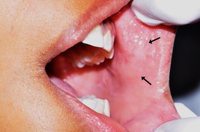
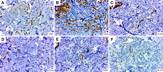
Cellular neurothekeomas (CNs) are distinctive benign tumors of uncertain histogenesis, with predilection for the skin of the head and neck region. We describe the first case of multiple desmoplastic CNs (DCNs) affecting the oral cavity in a 9-year-old girl. Histopathologic evaluation showed a proliferation of spindle and epithelioid cells, forming nests and bundles, supported by exuberant fibrous stroma, as well as scattered multinucleated floret-like giant cells. The tumor cells were immunopositive for vimentin, CD63, CD56, whereas AE1/AE3, S100, CD34, α-SMA, GFAP, EMA, CD57 and NSE were negative. Ki-67 was <2%. Multiple DCNs should be considered in the differential diagnosis of oral nodular lesions.
Keywords: Cellular neurothekeoma; Child; Desmoplastic cellular neurothekeoma; Immunohistochemistry; Oral cavity.
Figures

Similar articles
-
Multiple desmoplastic cellular neurothekeomas localized to the face of a 16-year-old boy.Am J Dermatopathol. 2010 Jul;32(5):509-13. doi: 10.1097/DAD.0b013e3181c98f89. Am J Dermatopathol. 2010. PMID: 20571347
-
Multiple desmoplastic cellular neurothekeomas localized to the face of a 16-year-old boy.Am J Dermatopathol. 2010 Dec;32(8):841-5. Am J Dermatopathol. 2010. PMID: 21137111
-
Multiple cellular neurothekeomas--a case report and review on the role of immunohistochemistry as a histologic adjunct.J Cutan Pathol. 2006 Jan;33(1):51-6. doi: 10.1111/j.0303-6987.2006.00400.x. J Cutan Pathol. 2006. PMID: 16441413
-
Neurothekeoma in the middle cranial fossa as a rare location: Case report and literature review.Neurochirurgie. 2016 Dec;62(6):336-338. doi: 10.1016/j.neuchi.2016.07.003. Epub 2016 Nov 2. Neurochirurgie. 2016. PMID: 27816187 Review.
-
Atypical cellular neurothekeoma of the oral mucosa: A rare case report and literature review.J Cutan Pathol. 2024 Dec;51(12):930-937. doi: 10.1111/cup.14693. Epub 2024 Jul 18. J Cutan Pathol. 2024. PMID: 39021336 Review.
Cited by
-
Oral neural tumors: Clinicopathologic analysis of 157 cases and review of the literature.J Clin Exp Dent. 2019 Aug 1;11(8):e721-e731. doi: 10.4317/jced.55944. eCollection 2019 Aug. J Clin Exp Dent. 2019. PMID: 31598201 Free PMC article.
-
Nerve sheath myxoma masqueraded as intramuscular myxoma: an extremely rare tumor with unusual location - a case report and literature review.Ann Med Surg (Lond). 2023 Mar 16;85(6):2953-2957. doi: 10.1097/MS9.0000000000000133. eCollection 2023 Jun. Ann Med Surg (Lond). 2023. PMID: 37363581 Free PMC article.
-
Neurothekeoma Presenting as a Rare Post Aural Swelling in an Adolescent Male with Review of Literature of Benign Spindle Cell Lesions.Indian J Otolaryngol Head Neck Surg. 2025 Apr;77(4):1831-1836. doi: 10.1007/s12070-025-05417-4. Epub 2025 Mar 25. Indian J Otolaryngol Head Neck Surg. 2025. PMID: 40226276
References
-
- Fetsch JF, Laskin WB, Miettinen M. Nerve sheath myxoma: a clinicopathologic and immunohistochemical analysis of 57 morphologically distinctive, S-100 protein- and GFAP-positive, myxoid peripheral nerve sheath tumors with a predilection for the extremities and a high local recurrence rate. Am J Surg Pathol. 2005;29(12):1615–1624. doi: 10.1097/01.pas.0000173025.87476.a4. - DOI - PubMed
Publication types
MeSH terms
Substances
LinkOut - more resources
Full Text Sources
Other Literature Sources
Medical
Research Materials
Miscellaneous
