Review
. 2017 Jun;5(3):10.1128/microbiolspec.tbtb2-0014-2016.
doi: 10.1128/microbiolspec.TBTB2-0014-2016.

Oxidative Phosphorylation as a Target Space for Tuberculosis: Success, Caution, and Future Directions

Affiliations
Review

Oxidative Phosphorylation as a Target Space for Tuberculosis: Success, Caution, and Future Directions

Gregory M Cook et al. Microbiol Spectr. 2017 Jun.

Abstract

The emergence and spread of drug-resistant pathogens, and our inability to develop new antimicrobials to combat resistance, have inspired scientists to seek out new targets for drug development. The Mycobacterium tuberculosis complex is a group of obligately aerobic bacteria that have specialized for inhabiting a wide range of intracellular and extracellular environments. Two fundamental features in this adaptation are the flexible utilization of energy sources and continued metabolism in the absence of growth. M. tuberculosis is an obligately aerobic heterotroph that depends on oxidative phosphorylation for growth and survival. However, several studies are redefining the metabolic breadth of the genus. Alternative electron donors and acceptors may provide the maintenance energy for the pathogen to maintain viability in hypoxic, nonreplicating states relevant to latent infection. This hidden metabolic flexibility may ultimately decrease the efficacy of drugs targeted against primary dehydrogenases and terminal oxidases. However, it may also open up opportunities to develop novel antimycobacterials targeting persister cells. In this review, we discuss the progress in understanding the role of energetic targets in mycobacterial physiology and pathogenesis and the opportunities for drug discovery.


Figures

FIGURE 1
FIGURE 1
Generalized schematic overview of relevant electron transfer components of M. tuberculosis. Complexes indicated in blue oxidize various substrates to reduce quinones. The resulting (mena)quinol molecules (orange) can be oxidized to result in reduction of various terminal electron acceptors, mediated by the complexes shown in purple.
FIGURE 2
FIGURE 2
Mechanisms by which a proton motive (membrane potential [Δψ] + transmembrane pH gradient [ZΔpH]) force can be generated in mycobacteria. (1) Cotransport of protons driven by solute (succinate) symport into the periplasm. (2) Redox-loop separation of charge; (mena)quinol oxidation results in proton release into the periplasm by virtue of (mena)quinol site proximity to the periplasm, while electrons are transferred to reduce a terminal electron acceptor (e.g., nitrate, fumarate) in the cytoplasm that results in neutralization of charge. (3) Proton translocation mediated by primary proton-pumping complexes (bc1-aa3 supercomplex).
FIGURE 3
FIGURE 3
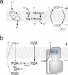
Traditional inhibitors of proton motive force generation. (a) Valinomycin is an ionophore, selective for potassium ions, which equilibrates the potassium gradient—dissipating the Δψ (electrogenic). Nigericin is a hydrophobic weak carboxylic acid which can traverse the membrane as its either protonated acid or neutral salt. It dissipates chemical gradients (i.e., ΔpH) but maintains the charge (one positive charge exchanged for one positive charge—electroneutral) (3). Carbonyl cyanide m-chlorophenyl hydrazine (CCCP) is an electrogenic protonophore. CCCP is driven to the periplasm by the Δψ, while CCCPH is driven to the cytoplasm by the ΔpH. It can equilibrate both Δψ and ΔpH. (b) Model for uncoupling by either pyrazinamide (PZA) or BDQ. (Left side) PZA diffuses into the cell and is converted to pyrazinoic acid (POA) by PncA (pyrazinamidase). Anionic POA could effectively inhibit growth through anion accumulation in the neutral pH of the cytoplasm and/or efflux from the cells to become protonated in the acidic extracellular environment (POA-H). POA-H would then diffuse back into the cell driven by the ΔpH gradient and dissociate in the cytoplasm (neutral pH), leading to intracellular acidification and cell death. (Right side) In a typical mycobacterial cell, the majority of ATP synthesis is respiratory, driven by the PMF. The binding of BDQ to the c-ring most likely perturbs the a-c subunit interface, causing an uncontrolled proton leak uncoupled from ATP synthesis and resulting in a futile proton cycle. Compensation by the exchange of other cations (i.e., K+) would allow the process to remain electroneutral.
FIGURE 4
FIGURE 4
Inhibitors of the electron transport chain and F1F0-ATP synthase of M. tuberculosis. Selected inhibitors of these complexes are indicated with flathead arrows and do not reflect the binding site of the inhibitors. Abbreviations: QPs, quinolinyl pyrimidines; TPZ, trifluoperazine; CFZ, clofazimine; 3-NP, 3-nitropropionate; SQ109, N-adamantan-2-yl-N′-((E)-3,7-dimethyl-octa-2,6-dienyl)-ethane-1,2-diamine; LPZ, lansoprazole; Q203, imidazopyridine amide; BDQ, bedaquiline.
FIGURE 5
FIGURE 5
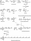
Proposed menaquinone biosynthesis pathway in mycobacteria based on the known pathway in E. coli. In this scheme the product of MenA is depicted as the quinone rather than the quinol. This is consistent with the majority of the menaquinone literature (167), which indicates that the oxidation from quinol to quinone is spontaneous but differs from ubiquinone synthesis. The arrows indicate C2 and C3 of menaquinone-9(II-H2). Abbreviations: DHNA, 1,4-dihydroxy-2-naphthoate; DHNA-CoA, 1,4-dihydroxy-2-naphthoyl-CoA; OSB, o-succinylbenzoate; OSB-CoA, o-succinylbenzoyl-CoA; SEPHCHC, 2-succinyl-5-enolpyruvyl-6-hydroxy-3-cyclohexadiene-1-carboxylate; SHCHC, 2-succinyl-6-hydroxy-2,4-cyclohexadiene-1-carboxylate.

References

    1. Brodie AF, Gutnik DL (ed). 1972. Electron Transport and Oxidative Phosphorylation in Microbial Systems. Marcel Dekker Inc., New York, NY.
    1. Rao M, Streur TL, Aldwell FE, Cook GM. 2001. Intracellular pH regulation by Mycobacterium smegmatis and Mycobacterium bovis BCG. Microbiology 147:1017–1024 10.1099/00221287-147-4-1017. - DOI - PubMed
    1. Rao SP, Alonso S, Rand L, Dick T, Pethe K. 2008. The protonmotive force is required for maintaining ATP homeostasis and viability of hypoxic, nonreplicating Mycobacterium tuberculosis. Proc Natl Acad Sci USA 105:11945–11950 10.1073/pnas.0711697105. - DOI - PMC - PubMed
    1. Jormakka M, Byrne B, Iwata S. 2003. Protonmotive force generation by a redox loop mechanism. FEBS Lett 545:25–30 10.1016/S0014-5793(03)00389-2. - DOI - PubMed
    1. Dimroth P, Cook GM. 2004. Bacterial Na+ - or H+ -coupled ATP synthases operating at low electrochemical potential. Adv Microb Physiol 49:175–218 10.1016/S0065-2911(04)49004-3. - DOI - PubMed

Publication types

MeSH terms

LinkOut - more resources