. 2017 Jun 9:8:15775.
doi: 10.1038/ncomms15775.

Centromeres are maintained by fastening CENP-A to DNA and directing an arginine anchor-dependent nucleosome transition

Affiliations

Centromeres are maintained by fastening CENP-A to DNA and directing an arginine anchor-dependent nucleosome transition

Lucie Y Guo et al. Nat Commun. .

Abstract

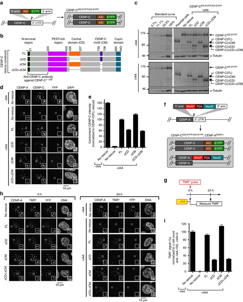
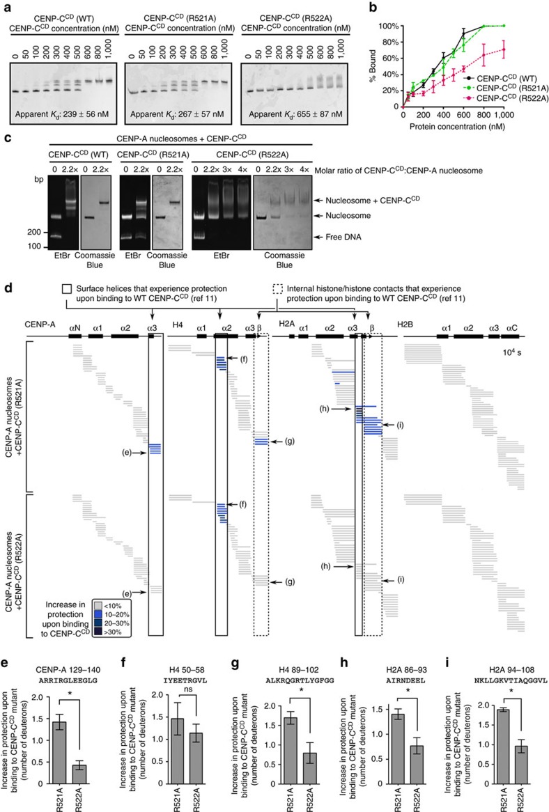
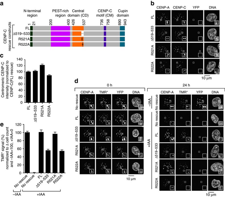
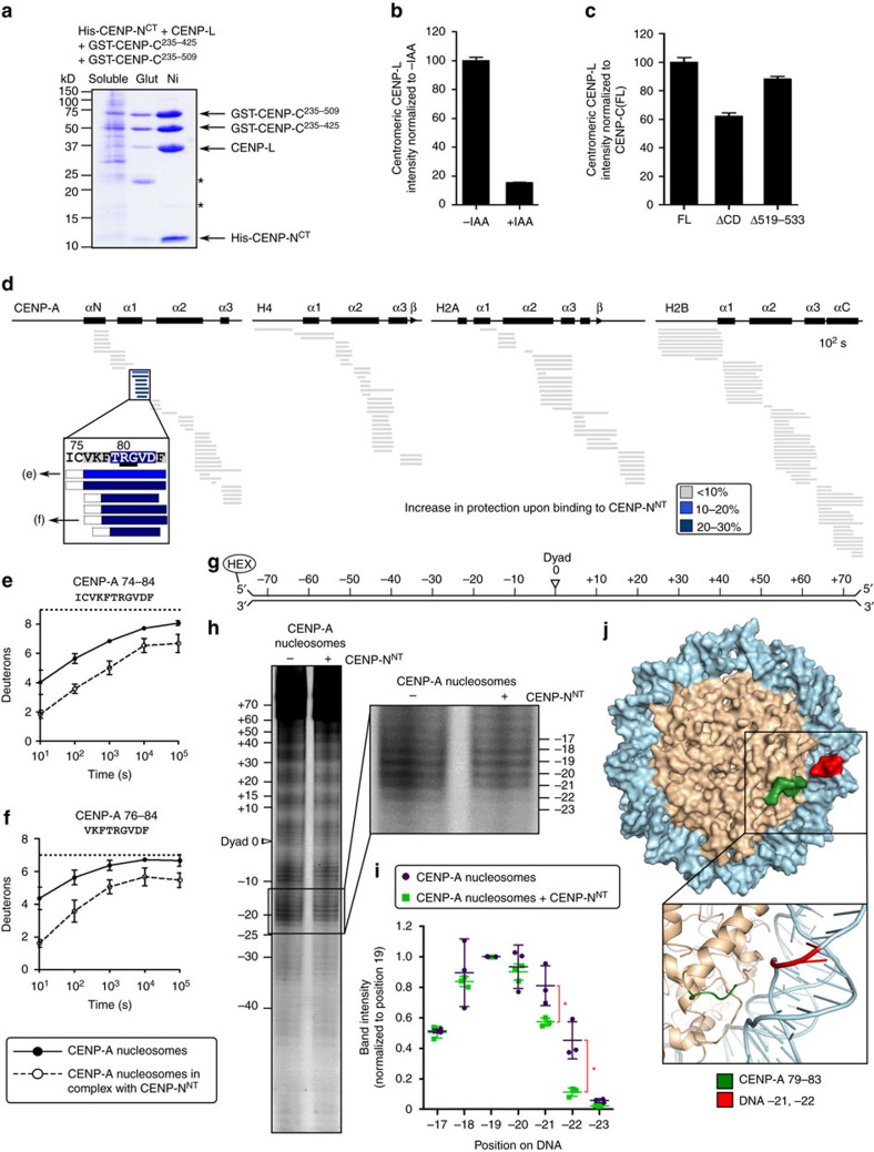
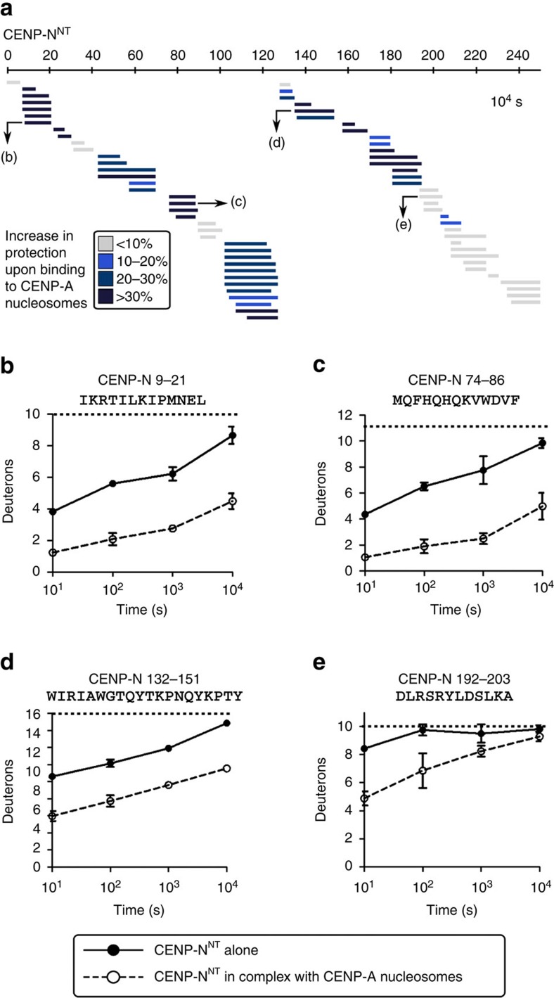
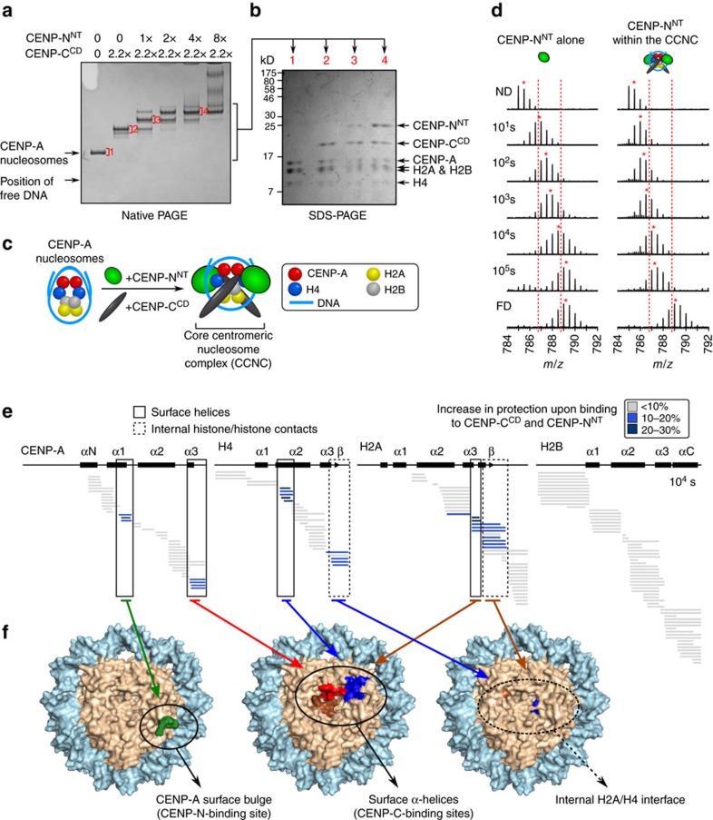
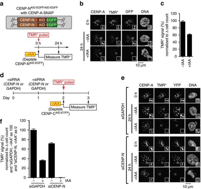
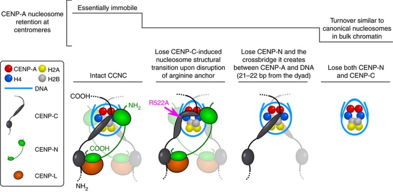
Maintaining centromere identity relies upon the persistence of the epigenetic mark provided by the histone H3 variant, centromere protein A (CENP-A), but the molecular mechanisms that underlie its remarkable stability remain unclear. Here, we define the contributions of each of the three candidate CENP-A nucleosome-binding domains (two on CENP-C and one on CENP-N) to CENP-A stability using gene replacement and rapid protein degradation. Surprisingly, the most conserved domain, the CENP-C motif, is dispensable. Instead, the stability is conferred by the unfolded central domain of CENP-C and the folded N-terminal domain of CENP-N that becomes rigidified 1,000-fold upon crossbridging CENP-A and its adjacent nucleosomal DNA. Disrupting the 'arginine anchor' on CENP-C for the nucleosomal acidic patch disrupts the CENP-A nucleosome structural transition and removes CENP-A nucleosomes from centromeres. CENP-A nucleosome retention at centromeres requires a core centromeric nucleosome complex where CENP-C clamps down a stable nucleosome conformation and CENP-N fastens CENP-A to the DNA.


Conflict of interest statement

The authors declare no competing financial interests.

Figures

Figure 1
Figure 1. CENP-CCD is the only nucleosome-binding domain of CENP-C required for retention of CENP-A nucleosomes.
(a) Schematic representation of CENP-CAID-EYFP/AID-EYFP cells. (b) Rescue constructs constitutively expressed at unique FRT site. FL, full length. (c) Immunoblot of CENP-CAID-EYFP/AID-EYFP cells (with and without 4 h of auxin-induced CENP-C depletion), using an antibody generated against CENP-C (a.a. 1–198). See Supplementary Fig. 9 for uncropped blot. (d) Representative images, in which the loss of YFP signal verifies depletion of CENP-C-AID-EYFP after 24 h of IAA, and CENP-C antibody then exclusively detects rescue constructs. Scale bar, 10 μm. (e) Quantification of d. (f) Schematic representation for SNAP-tagging CENP-A at its endogenous locus. (g) Schematic representation for pulse-chase experiment, in which CENP-CAID-EYFP/AID-EYFP cells expressing rescue constructs of either CENP-C(FL) or CENP-C domain deletion mutants were pulse-labelled with TMR* and assessed for retention of the existing pool of CENP-A molecules. (h) Representative images from experiment diagrammed in g. Scale bar, 10 μm. (i) Quantification of h. See also Supplementary Fig. 1k–m. All graphs are shown as mean±95% confidence interval (n>2,000 centromeres in all cases).
Figure 2
Figure 2. The arginine anchor of CENP-CCD is critical for the CENP-A nucleosome structural transition.
(a) Representative native PAGE analysis of CENP-A NCPs harbouring Cy5-labelled histone H2B that have been incubated with the indicated concentrations of CENP-CCD (WT or the indicated mutants). Each reaction contains 200 nM nucleosomes. Cy5 fluorescence was detected on a Typhoon phosphorimager, and CENP-C binding retards the mobility. Both WT and R521A show crisp shifts to bands with one or two copies of CENP-C bound to the nucleosome. R522A exhibits a more smeary appearance when bound to the CENP-A nucleosome (see also c), and the species with a single molecule of CENP-CCD(R522A) was not clearly resolved. Listed on the graphs are apparent Kd values for these binding experiments (values shown are mean±s.d.; n=3). (b) Quantification of three independent experiments (values shown are mean±s.d.) performed as in a. Note that for some data points, the error bars are too small to be visible in the graph. (c) CENP-A NCPs in complex with WT or mutant CENP-CCD, as assessed by native PAGE stained with ethidium bromide (EtBr) and then Coomassie Blue. (d) HXMS of all histone subunits of the CENP-A NCP from a single timepoint (104 s), showing regions that exhibit additional protection from HX upon binding of CENP-CCD(R521A) or CENP-CCD(R522A). Each horizontal bar represents an individual peptide, placed beneath the schematics of secondary structural elements of the CENP-A nucleosome. When available, we present the data from all measurable charge states of each of the unique peptides (here and in the similarly formatted plots in the experiments presented in Figs 4, 5, 6). (ei) Representative peptides from various histone regions, comparing protection from exchange when the nucleosome is bound to CENP-CCD R521A versus R522A, showing faithful detection of differences between the two mutants across multiple replicate experiments (plotted as the mean±s.d.; n=3). Asterisks denotes differences that are statistically significant (P<0.05; Student's t-test).
Figure 3
Figure 3. The arginine anchor of CENP-CCD is required for CENP-A nucleosome stability at centromeres.
(a) Rescue constructs constitutively expressed at the unique FRT site in CENP-CAID-EYFP/AID-EYFP cells. FL, full length. (b) Representative images showing localization of CENP-C rescue constructs in CENP-CAID-EYFP/AID-EYFP cells after 24 h of IAA treatment. Scale bar, 10 μm. (c) Quantification of b. (d) Representative images showing CENP-A retention as measured by TMR* assay in cells, similar to schematic representation in Fig. 1g. Scale bar, 10 μm. (e) Quantification of d. All graphs are shown as mean±95% confidence interval (n>2000 centromeres in all cases).
Figure 4
Figure 4. CENP-NNT crossbridges CENP-A to DNA.
(a) Coomassie Blue-stained SDS–PAGE of co-purification with described protocol of CENP-L/His-CENP-NCT with GST-CENP-C235–509 and GST-CENP-C235–425 by glutathione-agarose (Glut) or Nickel-NTA-agarose (Ni). (b) Localization of CENP-L-N in CENP-CAID-EYFP/AID-EYFP cells before and after 24 h of IAA treatment, assessed using anti-CENP-L (Supplementary Fig. 3b for images). (c) Localization of CENP-L-N in CENP-CAID-EYFP/AID-EYFP cells constitutively expressing the rescue constructs CENP-C(FL), CENP-C(ΔCD) or CENP-C(Δ519–533), after 24 h of IAA treatment. (Supplementary Fig. 3c for images) All graphs are shown as mean±95% confidence interval (n>2,000 centromeres in all cases). (d) HXMS of all histone subunits of the CENP-A NCP from a single timepoint (102 s), showing protection at CENP-A(79–83) upon binding to CENP-NNT. The first two residues of each peptide are boxed in dashed black lines because exchange of the first two backbone amide protons cannot be measured. (e,f) Representative peptides spanning the CENP-A surface bulge over the timecourse. The maximum number of deuterons possible to measure by HXMS for each peptide is shown by the dotted line. All peptides are plotted at every timepoint as mean±s.d. from triplicate experiments. Note that for some data points, the error bars are too small to be visible in the graph. (g) Schematic representation of the 5′-fluorescently labelled 147 bp α-satellite DNA sequence used in footprinting experiments. (h) Representative hydroxyl radical footprinting experiment of CENP-A nucleosomes vs. CENP-A nucleosomes in complex with CENP-NNT, with inset showing magnification of positions −17 to −23. (i) Quantification of band intensities from three independent experiments, shown as mean±s.d. normalized to DNA position −19 (this position was chosen because it was expected to be very exposed for hydroxyl radical-mediated cleavage with and without CENP-NNT). Asterisks denotes differences that are statistically significant (P<0.05; Student's t-test). (j) A molecular model of the CENP-A nucleosome (PDB 3AN2), in which the DNA sequence was modified to that used in the footprinting experiment: CENP-A a.a. 79−83 is labelled in green, and DNA positions −21 and −22 are labelled in red.
Figure 5
Figure 5. CENP-NNT undergoes global stabilization upon binding to the CENP-A nucleosome.
(a) HXMS of CENP-NNT from a single timepoint (104 s), showing substantial protection from HX spanning the ∼200 a.a. domain upon binding to CENP-A NCP. (be) Representative peptides spanning CENP-NNT over the timecourse. All peptides are plotted at every timepoint as mean±s.d. from triplicate experiments. Note that for some data points, the error bars are too small to be visible in the graph.
Figure 6
Figure 6. CENP-CCD and CENP-NNT simultaneously bind to the same CENP-A NCP and generate internal and surface stability.
(a) Coomassie Blue-stained native PAGE of binding reactions with CENP-NNT and CENP-CCD and CENP-A NCPs. (b) Indicated bands from native PAGE excised and run on SDS–PAGE. (c) Schematic representation of formation of the CCNC. (d) A representative peptide of CENP-NNT (a.a. 9−21) that shows substantial protection upon binding to CENP-A nucleosomes. The peptide is shown from CENP-NNT alone (left) versus as part of the CCNC (right). Dotted red lines serve as guideposts to highlight the differences in m/z shifts between the two samples. A red asterisk denotes the centroid location of each peptide envelope, and the numerical value in blue indicates the centroid mass of the peptide envelope. It is important to note that this peptide exhibits clear EX2 behaviour at all timepoints when part of the CCNC (without any evidence of bimodal peaks), indicating that this complex is stable in solution even on timescales of 100,000 s (∼28 h). (e) HXMS of all histone subunits of the CENP-A NCP from a single timepoint (104 s). (f) Regions showing substantial protection from HX mapped onto the structure of the CENP-A NCP (PDB ID 3AN2). (left) The exposed CENP-A bulge, to which CENP-N binds. (middle) The surface helices to which CENP-C binds. (right) Internal histone–histone contacts that undergo stability upon CENP-C binding.
Figure 7
Figure 7. CENP-C and CENP-N collaborate to maintain CENP-A nucleosomes at centromeres.
(a) Schematic for experiment in which CENP-NAID-EGFP/AID-EGFP cells expressing CENP-A-SNAP at a unique FRT site were pulse-labelled with TMR* and assessed for retention of the existing pool of CENP-A molecules. (b) Representative images from experiment diagrammed in a. Scale bar, 10 μm. (c) Quantification of b. (d) Schematic representation of experiment, in which CENP-CAID-EYFP/AID-EYFP cells were treated with siCENP-N or siGAPDH and pulse-labelled with TMR*, and the relative CENP-A-SNAP signals were analysed after 24 h (with or without CENP-C depletion by IAA treatment). (e) Representative images from experiment described in d. Scale bar, 10 μm. (f) Quantification of e. The value of the ‘siGAPDH, −IAA' condition is normalized as 100%, and the value of the ‘siCENP-N, +IAA' condition is normalized as 0%. All graphs are shown as mean±95% confidence interval (n>2,000 centromeres in all cases).
Figure 8
Figure 8. Model of the physical basis for the stability of CENP-A nucleosomes within the CCNC.
See text for the details of our model of centromere maintenance. We note that CENP-CCD is shown as an elongated oval that represents a structured loop that has no conventional secondary structure, despite having been historically called a ‘domain'. Also, a flexible linker is shown between CENP-CCD and the CENP-C contact point with the CENP-L-N complex, in line with proposals that CENP-C largely exists as an extended and unfolded protein that may span >100 nm at mitotic kinetochores. In addition, it is also not known if there is a fixed or variable distance at centromeres from the CENP-C-L-N contact point to the CENP-A nucleosome. It is also unclear if this contact point on CENP-C with CENP-L-N is a folded domain or if it contacts one or both subunits of CENP-L-N.

Similar articles

Cited by

References

    1. Westhorpe F. G. & Straight A. F. The centromere: epigenetic control of chromosome segregation during mitosis. Cold Spring Harb. Perspect. Biol. 7, a015818 (2015). - PMC - PubMed
    1. McKinley K. L. & Cheeseman I. M. The molecular basis for centromere identity and function. Nat. Rev. Mol. Cell Biol. 17, 16–29 (2016). - PMC - PubMed
    1. Earnshaw W. C. & Rothfield N. Identification of a family of human centromere proteins using autoimmune sera from patients with scleroderma. Chromosoma 91, 313–321 (1985). - PubMed
    1. Palmer D. K. & Margolis R. L. Kinetochore components recognized by human autoantibodies are present on mononucleosomes. Mol. Cell. Biol. 5, 173–186 (1985). - PMC - PubMed
    1. Black B. E. & Cleveland D. W. Epigenetic centromere propagation and the nature of CENP-A nucleosomes. Cell 144, 471–479 (2011). - PMC - PubMed

Publication types

MeSH terms