Review
. 2017 Sep;246(9):641-656.
doi: 10.1002/dvdy.24530. Epub 2017 Jul 12.

Conserved signaling mechanisms in Drosophila heart development

Affiliations
Review

Conserved signaling mechanisms in Drosophila heart development

Shaad M Ahmad. Dev Dyn. 2017 Sep.

Abstract

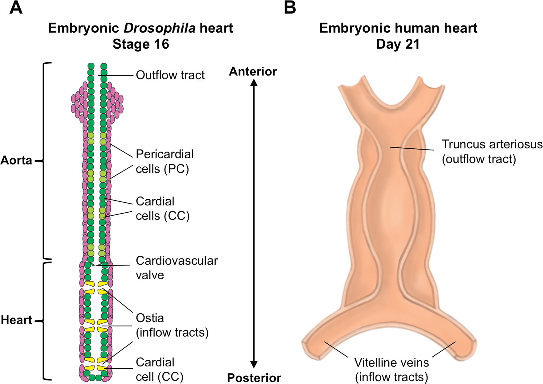
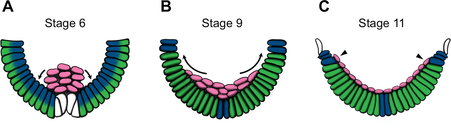
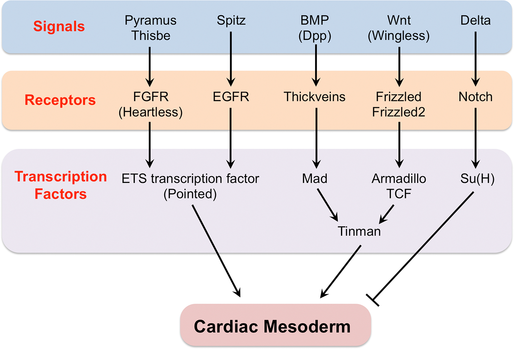
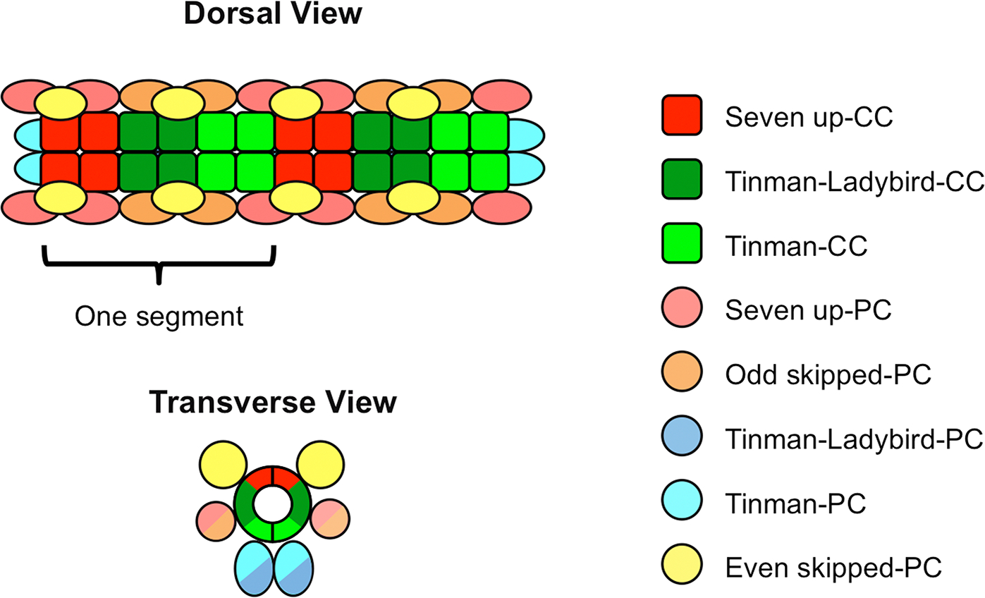
Signal transduction through multiple distinct pathways regulates and orchestrates the numerous biological processes comprising heart development. This review outlines the roles of the FGFR, EGFR, Wnt, BMP, Notch, Hedgehog, Slit/Robo, and other signaling pathways during four sequential phases of Drosophila cardiogenesis-mesoderm migration, cardiac mesoderm establishment, differentiation of the cardiac mesoderm into distinct cardiac cell types, and morphogenesis of the heart and its lumen based on the proper positioning and cell shape changes of these differentiated cardiac cells-and illustrates how these same cardiogenic roles are conserved in vertebrates. Mechanisms bringing about the regulation and combinatorial integration of these diverse signaling pathways in Drosophila are also described. This synopsis of our present state of knowledge of conserved signaling pathways in Drosophila cardiogenesis and the means by which it was acquired should facilitate our understanding of and investigations into related processes in vertebrates. Developmental Dynamics 246:641-656, 2017. © 2017 Wiley Periodicals, Inc.

Keywords: Dpp/BMP signaling; Drosophila cardiogenesis; EGFR signaling; FGF signaling; Hedgehog signaling; Integrin mediated signaling; Notch signaling; Pvr/VEGF signaling; Slit-Robo signaling; Wnt signaling; cardiac mesoderm specification; cardiac morphogenesis; cardiac valve formation; heart development; ostia formation; signaling pathways.


Figures

Fig. 1.
Fig. 1.. The embryonic heart in Drosophila and humans.
(A) The tubular heart of a stage 16 Drosophila embryo, divided into a narrow anterior structure termed the aorta and a wider posterior structure referred to as the heart proper. Both structures consist of an inner tube of contractile cardial cells (CCs; Tinman-Ladybird-CCs and Tinman-CCs are colored dark green; Seven up-CCs are colored light green and yellow) surrounded by an external sheath of non-muscle nephrocytic pericardial cells (PCs, pink), all arranged metamerically in repeated units. Ostia (inflow tracts) are present only in the posterior compartment, three on each side, with two morphologically distinct Seven up CCs (yellow) acting as a valve to regulate ingress through each ostium. (B) The tubular single-chambered heart of a 21-day human embryo, prior to the looping and septation which will produce four chambers. Note that both hearts are organized as tubes along an anterior-posterior axis, with inflow tracts entering the heart at the posterior part of the organ, and circulatory fluid (hemolymph in Drosophila, blood in humans) being pumped anteriorwards and exiting through the outflow tracts at the anterior.
Fig. 2.
Fig. 2.. Mesoderm migration in Drosophila cardiogenesis.
(A-C) Schematic cross-sections of Drosophila embryos at stages 6 (A), 9 (B), and 11 (C) showing the dorsolateral migration (arrows) of the FGFR Heartless-expressing mesodermal cells (pink) after invagination through the ventral furrow at gastrulation. Dynamic and broad expression patterns of the FGFs Pyramus (blue) and Thisbe (green) along the ectoderm direct this migration (cells which are half green and half blue express both FGFs). The dorsalmost cells at stage 11 (arrowheads) receive inductive signals to become specified as the cardiac mesoderm. Adapted from Kadam et al., 2009.
Fig. 3.
Fig. 3.. Regulatory network of signaling pathways responsible for establishing the Drosophila cardiac mesoderm.
Only the signaling ligands, their receptors, and the terminal transcription factors of these signal transduction pathways are shown.
Fig. 4
Fig. 4. Schematic diagram showing the stereotyped positions of the eight different cell types comprising the Drosophila heart.
An individual heart segment, composed of two contralateral hemisegments mirroring each other across the dorsal midline, is indicated.
Fig. 5.
Fig. 5.. Pericardial fate specification by the Notch signaling pathway.
Schematic showing the regulation of pericardial cell (PC)-specific genes. Binding sites for the Su(H) transcription factor are enriched along the enhancers of PC-specific genes. In cardial cells (CCs), the enhancers of PC-specific genes are repressed by the binding of the Su(H)-co-repressor complex. However, the Delta ligand expressed specifically by the CCs activates the Notch receptor in the neighboring cells, with the resulting cleaved Nicd fragment associating with Su(H) and displacing the co-repressor. The resulting elimination of the repressor complex is sufficient to initiate transcription of the PC-specific genes due to the presence of other local activators, leading these neighboring cells to adopt a pericardial fate. Transcription of the PC-specific genes is further enhanced by the Nicd-Su(H) complex acting as an activator. Adapted from Ahmad et al., 2014.
Fig. 6.
Fig. 6.. Schematic representation of two cardial cells illustrating the temporal sequence of events during heart tube and lumen formation.
(A) At an early stage in migration both cardial cells (CCs) are cuboidal. (B) As they approach one another, they become constricted along their dorsal domain. (C) Each CC makes initial contact with its contralateral counterpart across the midline along this constricted dorsal domain. (D) Each cell in these dorsally conjoined CC pairs then begins adopting a crescent shape. (E) Ultimately, this shape change also allows the ventral domains of the CCs to join and form a tube which encloses an internal central lumen. Localization of Slit and Robo is shown in green while that of E-cadherin and β-catenin is shown in red. Adapted from Medioni et al., 2008 and Santiago-Martinez et al., 2008.

Similar articles

Cited by

References

    1. Abu-Issa R, Smyth G, Smoak I, Yamamura K, Meyers EN. 2002. Fgf8 is required for pharyngeal arch and cardiovascular development in the mouse. Development 129:4613–4625. - PubMed
    1. Afouda BA, Martin J, Liu F, Ciau-Uitz A, Patient R, Hoppler S. 2008. GATA transcription factors integrate Wnt signalling during heart development. Development 135:3185–3190. - PubMed
    1. Ahmad SM, Bhattacharyya P, Jeffries N, Gisselbrecht SS, Michelson AM. 2016. Two Forkhead transcription factors regulate cardiac progenitor specification by controlling the expression of receptors of the fibroblast growth factor and Wnt signaling pathways. Development 143:306–317. - PMC - PubMed
    1. Ahmad SM, Busser BW, Huang D, Cozart EJ, Michaud S, Zhu X, Jeffries N, Aboukhalil A, Bulyk ML, Ovcharenko I, Michelson AM. 2014. Machine learning classification of cell-specific cardiac enhancers uncovers developmental subnetworks regulating progenitor cell division and cell fate specification. Development 141:878–888. - PMC - PubMed
    1. Ahmad SM, Tansey TR, Busser BW, Nolte MT, Jeffries N, Gisselbrecht SS, Rusan NM, Michelson AM. 2012. Two forkhead transcription factors regulate the division of cardiac progenitor cells by a Polo-dependent pathway. Dev Cell 23:97–111. - PMC - PubMed

MeSH terms

Substances

LinkOut - more resources