. 2017 Jun 9;12(6):e0179341.
doi: 10.1371/journal.pone.0179341. eCollection 2017.

Long-term cryopreservation of decellularised oesophagi for tissue engineering clinical application

Affiliations

Long-term cryopreservation of decellularised oesophagi for tissue engineering clinical application

Luca Urbani et al. PLoS One. .

Abstract

Oesophageal tissue engineering is a therapeutic alternative when oesophageal replacement is required. Decellularised scaffolds are ideal as they are derived from tissue-specific extracellular matrix and are non-immunogenic. However, appropriate preservation may significantly affect scaffold behaviour. Here we aim to prove that an effective method for short- and long-term preservation can be applied to tissue engineered products allowing their translation to clinical application. Rabbit oesophagi were decellularised using the detergent-enzymatic treatment (DET), a combination of deionised water, sodium deoxycholate and DNase-I. Samples were stored in phosphate-buffered saline solution at 4°C (4°C) or slow cooled in medium with 10% Me2SO at -1°C/min followed by storage in liquid nitrogen (SCM). Structural and functional analyses were performed prior to and after 2 and 4 weeks and 3 and 6 months of storage under each condition. Efficient decellularisation was achieved after 2 cycles of DET as determined with histology and DNA quantification, with preservation of the ECM. Only the SCM method, commonly used for cell storage, maintained the architecture and biomechanical properties of the scaffold up to 6 months. On the contrary, 4°C method was effective for short-term storage but led to a progressive distortion and degradation of the tissue architecture at the following time points. Efficient storage allows a timely use of decellularised oesophagi, essential for clinical translation. Here we describe that slow cooling with cryoprotectant solution in liquid nitrogen vapour leads to reliable long-term storage of decellularised oesophageal scaffolds for tissue engineering purposes.


Conflict of interest statement

Competing Interests: The authors have declared that no competing interests exist.

Figures

Fig 1
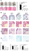
Fig 1. Decellularisation efficiency and scaffolds characterization.
(A) Efficient cell removal after 2 DET cycles was evident by macroscopic appearance, H&E staining and DNA quantification. (B) Immunohistochemistry for extracellular matrix composition. Masson’s Trichrome and Picrosirius Red staining demonstrated collagen preservation in both submucosa and among muscle fibers (inset). Elastin Van Gieson staining showed elastic fibers in the submucosa, around blood vessels and surrounding muscle fascicles both in the fresh and DET tissues. Muscular elastin staining was reduced after 3 DET cycles (inset). Alcian Blue staining indicated glycosaminoglycan preservation (bar = 100μm). (C) Extracellular component quantification demonstrated a gradual decrease in collagen after the first and second DET cycle. Elastin decreased after 3 DET cycles. glycosaminoglycan were partially reduced by the first DET cycle. DET = Detergent-Enzymatic Treatment, MT = Masson’s Trichrome, PR = Picrosirius Red, EVG = Elastin Van Gieson, AB = Alcian Blue. *p<0.05; **p<0.01.
Fig 2
Fig 2. Functional analysis of decellularised scaffolds.
(A,B) Synchrotron analysis after 2 DET cycles confirmed extracellular matrix preservation in the lamina propria, submucosa and muscularis. (C) An intact basement membrane was detected across the entire scaffold segment. (D) Blood vessels were seen converging towards the scaffold in a spoked wheel manner after 10 days from placement on the CAM as confirmed by blinded quantification of the vessels compared to the negative control (**p< 0.01). (E) The maximum tensile stress at which the samples broke and the elasticity modulus remained comparable to fresh after decellularisation. (F) Characteristic stress-strain curves showing that by increasing the number of DET cycles the tensile stress at which the samples break remains the same as seen in E. DET = Detergent-Enzymatic Treatment, CAM = Chicken chorioallantoic membrane assay.
Fig 3
Fig 3. Macro- and microscopic appearance of stored decellularised scaffolds at 6 months.
(A) Macroscopic appearances varied in the two protocols. (B) Scanning electron microscopy analysis. While SCM scaffolds demonstrated a good preservation of all oesophageal layers, in 4°C samples extracellular matrix was falling apart with signs of degradation. SCM = slow cooling medium, 4°C = 4°C in PBS.
Fig 4
Fig 4. Composition and mechanical properties of decellularised scaffolds after storage.
(A) Masson’s Trichrome staining demonstrated a progressive loss of architecture in 4°C-treated scaffolds. (B) Elastin staining showed maintenance of this protein in SCM scaffolds. Elastin was progressively lost in 4°C scaffolds. (C) Alcian Blue staining showed glycosaminoglycan maintenance in both storage methods. (D) Samples stored for 6 months with SCM maintained comparable ultimate tensile stress with 2 week-stored scaffolds despite a decreased Young's modulus with no impact on the maximum stress that the material could withstand. While 4°C samples at 2 weeks showed similar values to SCM, prolonged 4°C storage had a profound impact on the scaffold with a reduction of both values. *p<0.05 (bar = 100μm). SCM = slow cooling medium, 4°C = 4°C in PBS.

Similar articles

Cited by

References

    1. Mansbridge J. Commercial considerations in tissue engineering. J. Anat. 2006; 209: 527–532. doi: 10.1111/j.1469-7580.2006.00631.x - DOI - PMC - PubMed
    1. Ferlay J, Shin HR, Bray F, Forman D, Mathers C, Parkin DM. Estimates of worldwide burden of cancer in 2008: GLOBOCAN 2008. Int. J. Cancer. 2010; 127: 2893–2917. doi: 10.1002/ijc.25516 - DOI - PubMed
    1. Spitz L. Oesophageal atresia. Orphanet J. Rare Dis. 2007; 2: 24 doi: 10.1186/1750-1172-2-24 - DOI - PMC - PubMed
    1. Zani A, Pierro A, Elvassore N, De Coppi P. Tissue engineering: an option for esophageal replacement? Semin. Pediatr. Surg. 2009; 18: 57–62. doi: 10.1053/j.sempedsurg.2008.10.011 - DOI - PubMed
    1. Totonelli G, Maghsoudlou P, Garriboli M, Riegler J, Orlando G, Burns AJ, et al. A rat decellularized small bowel scaffold that preserves villus-crypt architecture for intestinal regeneration. Biomaterials. 2012; 33: 3401–3410. doi: 10.1016/j.biomaterials.2012.01.012 - DOI - PMC - PubMed

Substances

LinkOut - more resources