. 2017 Dec;177(6):1601-1611.
doi: 10.1111/bjd.15716. Epub 2017 Nov 16.

Coexistence of Eph receptor B1 and ephrin B2 in port-wine stain endothelial progenitor cells contributes to clinicopathological vasculature dilatation

Affiliations

Coexistence of Eph receptor B1 and ephrin B2 in port-wine stain endothelial progenitor cells contributes to clinicopathological vasculature dilatation

W Tan et al. Br J Dermatol. 2017 Dec.

Abstract

Background: Port-wine stain (PWS) is a vascular malformation characterized by progressive dilatation of postcapillary venules, but the molecular pathogenesis remains obscure.

Objectives: To illustrate that PWS endothelial cells (ECs) present a unique molecular phenotype that leads to pathoanatomical PWS vasculatures.

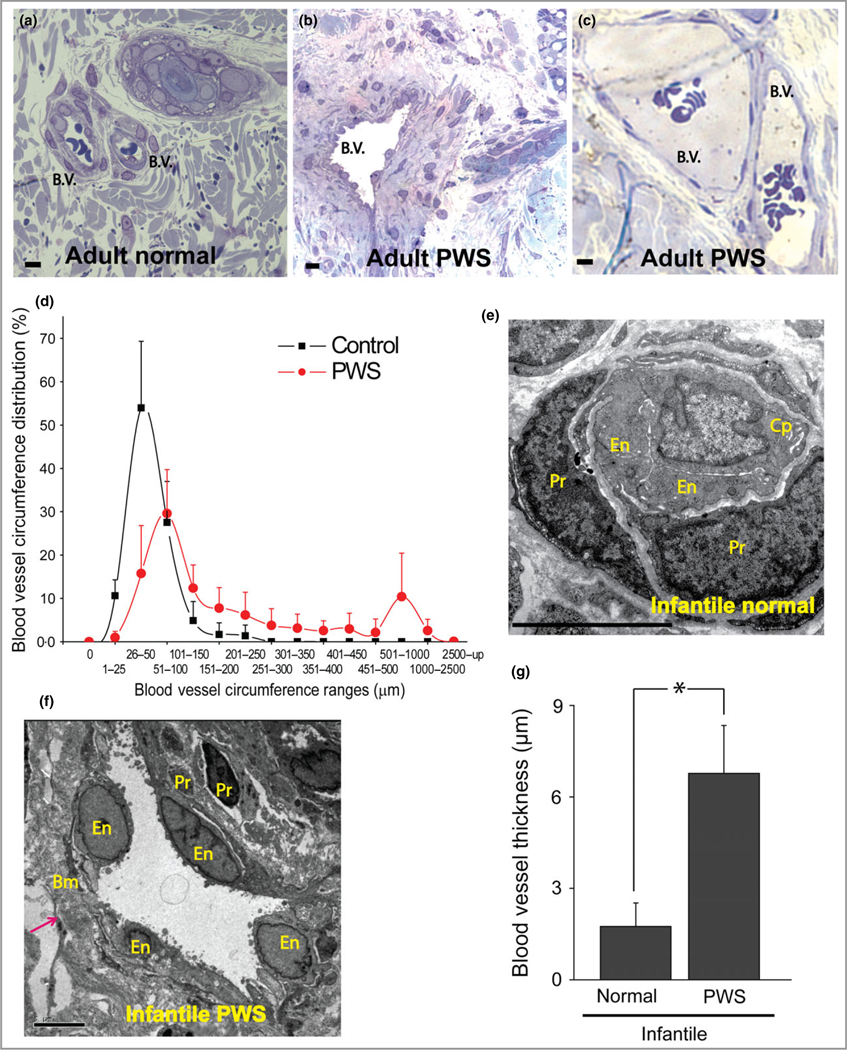
Methods: Immunohistochemistry and transmission electron microscopy were used to characterize the ultrastructure and molecular phenotypes of PWS blood vessels. Primary culture of human dermal microvascular endothelial cells and in vitro tube formation assay were used for confirmative functional studies.

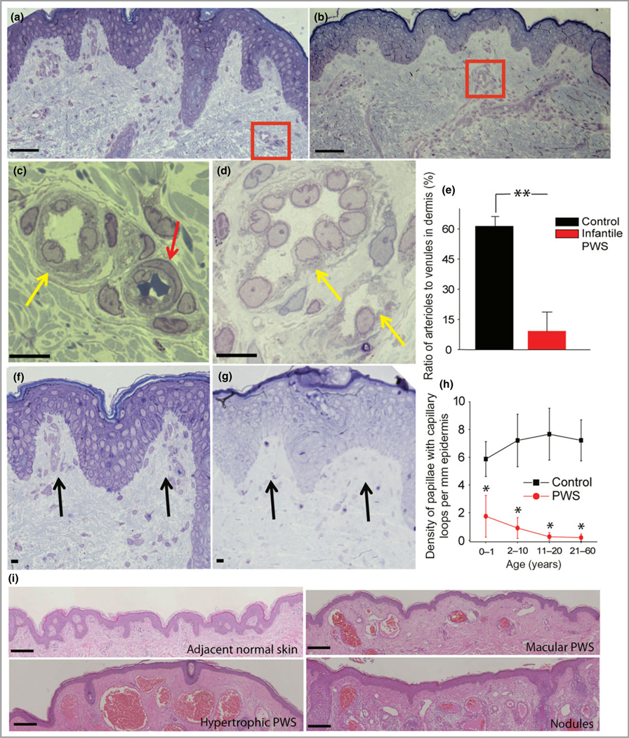
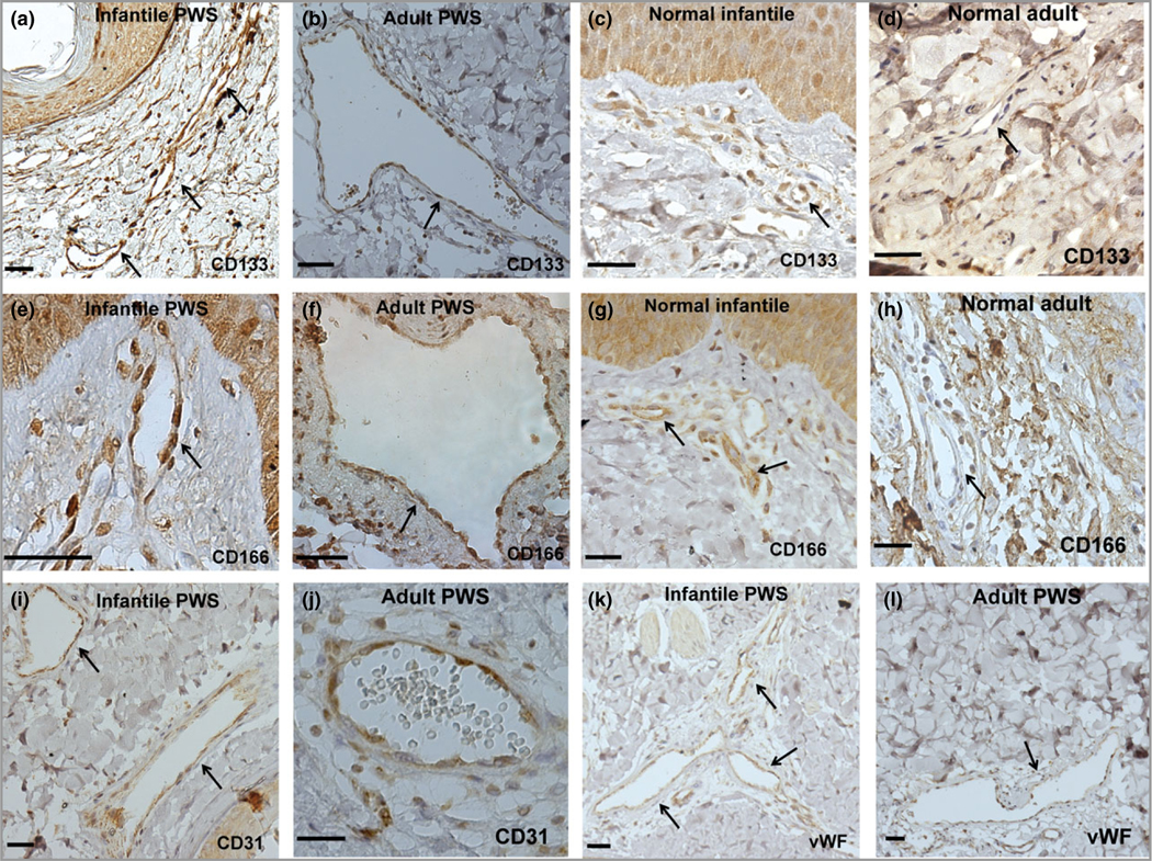
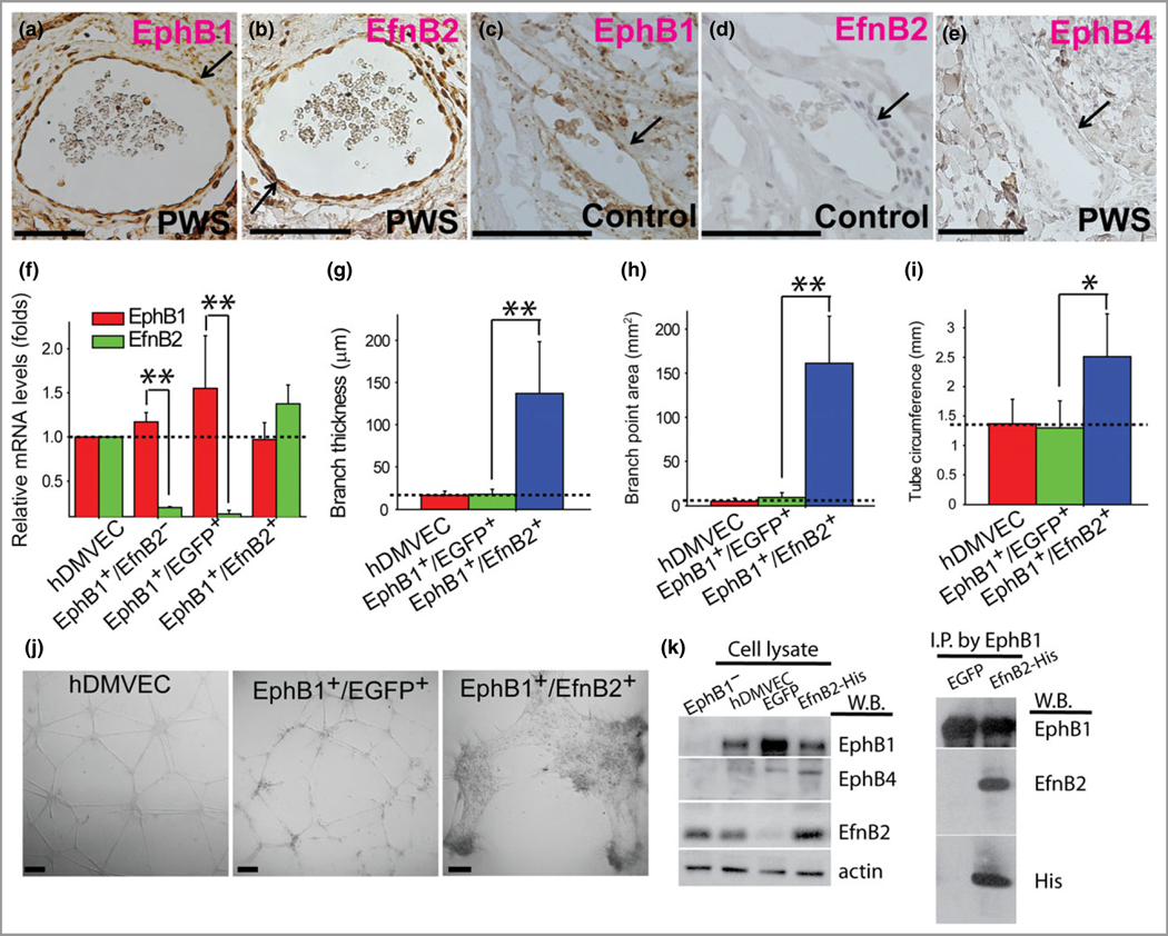
Results: Multiple clinicopathological features of PWS blood vessels during the development and progression of the disease were shown. There were no normal arterioles and venules observed phenotypically and morphologically in PWS skin; arterioles and venules both showed differentiation impairments, resulting in a reduction of arteriole-like vasculatures and defects in capillary loop formation in PWS lesions. PWS ECs showed stemness properties with expression of endothelial progenitor cell markers CD133 and CD166 in non-nodular lesions. They also expressed dual venous/arterial identities, Eph receptor B1 (EphB1) and ephrin B2 (EfnB2). Co-expression of EphB1 and EfnB2 in normal human dermal microvascular ECs led to the formation of PWS-like vasculatures in vitro, for example larger-diameter and thick-walled capillaries.

Conclusions: PWS ECs are differentiation-impaired, late-stage endothelial progenitor cells with a specific phenotype of CD133+ /CD166+ /EphB1+ /EfnB2+ , which form immature venule-like pathoanatomical vasculatures. The disruption of normal EC-EC interactions by coexistence of EphB1 and EfnB2 contributes to progressive dilatation of PWS vasculatures.


Conflict of interest statement

Conflicts of interest

None declared.

Figures

Fig 1.
Fig 1.
Thick- and thin-walled blood vessels in adult and infant port-wine stain (PWS) lesions. (a) Normal blood vessels (b.v.) in adjacent normal skin from an adult with PWS. (b, c) Thick- and thin-walled PWS blood vessels from adults with PWS. (d) PWS blood vessel circumference distribution vs. normal dermal vasculatures (four patients with PWS and four normal adult participants). (e) Electron microscopy (EM) showed a normal capillary in adjacent normal skin from an infant with PWS. En, endothelial cell; Pr, pericyte; Cp, capillary. (f) EM showed an ectatic, thick-walled blood vessel with replication of the basement membranes in PWS from the same subject as in (e). Bm, basement membrane. The arrow indicates the blood vessel wall. (g) Infantile PWS blood vessels showed a significantly thicker blood vessel wall compared with normal dermal blood vessels from the same subjects (n = 4). Scale bar = 5 μm. *P < 0·05 vs. control.
Fig 2.
Fig 2.
Multiple developmental impairments of infant port-wine stain (PWS) vasculatures. (a) Semi-thin section showed normal adjacent skin from an infant with PWS. (b) Semi-thin section showed PWS lesional skin from the same subject as in (a). Scale bar = 20 lm. (c) A normal venule (yellow arrow) and arteriole (red arrow) from the red-boxed area in (a). (d) PWS pathoanatomical venule-like vasculatures (yellow arrows) from the red-boxed area in (b). Scale bar = 5 μm. (e) The ratio of arteriole to venule-like vasculatures in infantile PWS lesions was significantly reduced compared with normal adjacent skin from the same subjects (n = 4). (f) Normal formation of capillary loop (black arrows) in adjacent normal skin from an infant with PWS. (g) Defects in capillary loop formation along with normal development of epidermal rete ridges in PWS from the same subject as in (d). Scale bar = 5 μm. (h) Quantitative analysis of the density of papillae containing capillary loops per mm epidermis in patients with PWS vs. normal subjects among groups of different ages. (i) Reduction of capillary loops and rete ridges in PWS flat reddish macular, protuberant hypertrophic areas and nodules from the same subject. Scale bar = 100 μm. **P < 0·01 and *P < 0·05 vs. the control groups in (e) and (h).
Fig 3.
Fig 3.
Port-wine stain (PWS) endothelial cells (ECs) presented stemness phenotypes of CD133+/CD166+ in non-nodular lesions. (a–h) Expression of CD133 and CD166 in infant and adult PWS and normal subjects. (i–l) PWS ECs expressed EC markers CD31 and von Willebrand factor (vWF). Positive stain is diaminobenzidine (DAB) (brown). Scale bar = 50 μm. Arrows indicate blood vessels.
Fig 4.
Fig 4.
Port-wine stain (PWS) endothelial cells (ECs) showed dual arterial and venous identities of co-expression of Eph receptor B1 (EphB1) and ephrin B2 (EfnB2). (a, b) PWS ECs were EphB1+ and EfnB2+. (c, d) A normal dermal venule showed expression of EphB1 but not EfnB2. (e) PWS ECs were EphB4. (f) Relative mRNA levels of EphB1 and EfnB2 in selected normal human dermal microvascular endothelial cell (hDMVEC) subtypes. (g–i) Forced co-expression of EphB1 and EfnB2 in normal hDMVECs showed a significant increase in (g) branch thickness, (h) branch point area and (i) tube circumference of the capillary tubes formed in vitro compared with controls. (j) PWS blood vessel-like phenotypes were observed in EphB1+/EfnB2+ but not in wild-type and EphB1+/EGFP+ control hDMVECs in an in vitro capillary tube formation assay at 12 h after cell plating. Positive stain is diaminobenzidine (DAB; brown). Scale bar = 100 μm. *P < 0·05, ** P < 0·01 vs. control. The arrows indicate blood vessels. (k) Left panel, detection of expression of EphB1, EphB4, β-actin and EfnB2 by Western blot (W.B.) in various hDMVEC subpopulations. hDMVEC, heterogeneous population prior to EfnB2–Fc selection; EphB1 hDMVEC, the remaining hDMVEC subpopulation after EfnB2–Fc selection; enhanced green fluorescent protein (EGFP) or EfnB2–His, overexpression of EGFP or EfnB2 in the EfnB2–Fc selected hDMVEC subpopulation. Right panel, an anti-EphB1 antibody was used to immunoprecipitate EphB1 from cell lysate and EfnB2 was detected from the immunoprecipitated protein complex using an anti-EfnB2 or anti-His antibody.

Comment in

Similar articles

Cited by

References

    1. Jacobs AH, Walton RG. The incidence of birthmarks in the neonate. Pediatrics 1976; 58:218–22. - PubMed
    1. Pratt AG. Birthmarks in infants. Arch Dermatol Syphilol 1953; 67:302–5. - PubMed
    1. Geronemus RG, Ashinoff R. The medical necessity of evaluation and treatment of port-wine stains. J Dermatol Surg Oncol 1991; 17:76–9. - PubMed
    1. Lever WF, Schaumburg-Lever G. Histopathology of the Skin, 7th edn. Philadelphia, PA: JB Lippincott, 1990.
    1. Kalick SM. Toward an interdisciplinary psychology of appearances. Psychiatry 1978; 41:243–53. - PubMed

MeSH terms