. 2017 Jul 18;8(29):47816-47830.
doi: 10.18632/oncotarget.18001.

Optimizing a machine learning based glioma grading system using multi-parametric MRI histogram and texture features

Affiliations

Optimizing a machine learning based glioma grading system using multi-parametric MRI histogram and texture features

Xin Zhang et al. Oncotarget. .

Abstract

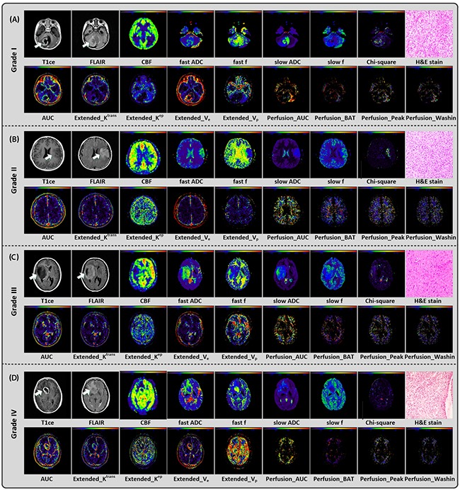
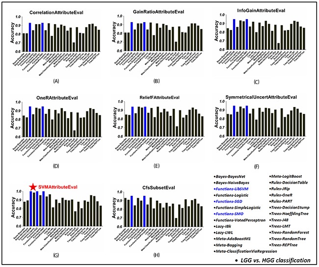
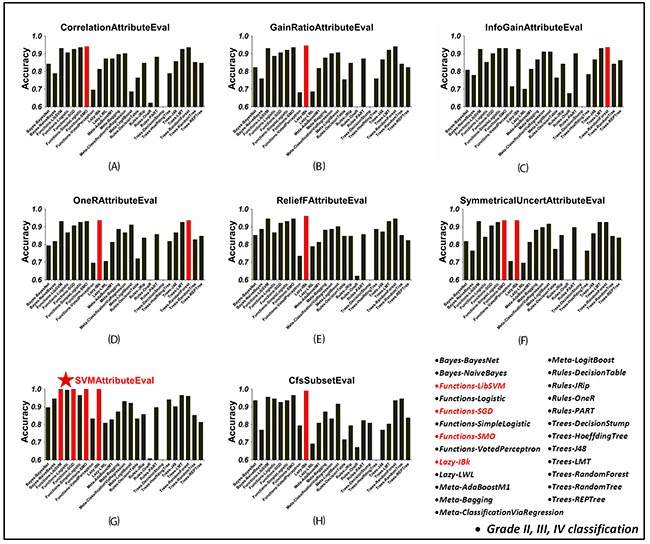
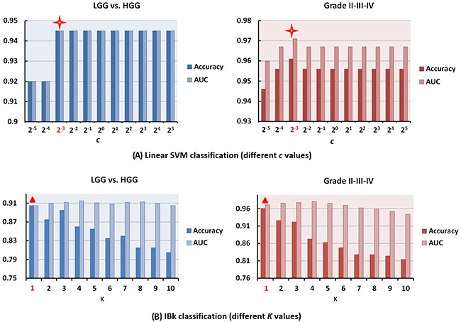
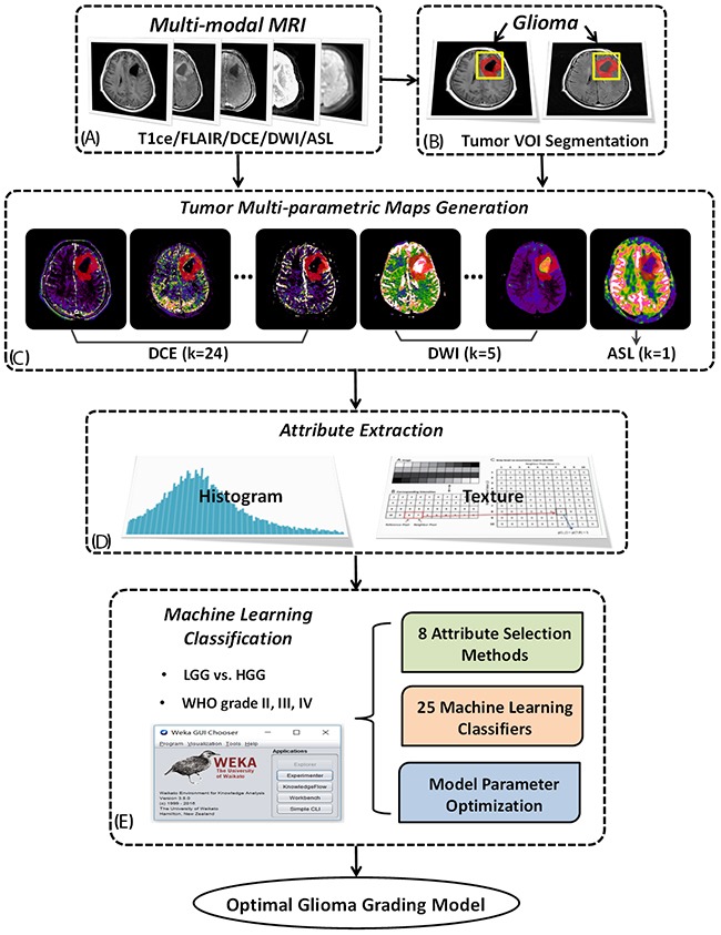
Current machine learning techniques provide the opportunity to develop noninvasive and automated glioma grading tools, by utilizing quantitative parameters derived from multi-modal magnetic resonance imaging (MRI) data. However, the efficacies of different machine learning methods in glioma grading have not been investigated.A comprehensive comparison of varied machine learning methods in differentiating low-grade gliomas (LGGs) and high-grade gliomas (HGGs) as well as WHO grade II, III and IV gliomas based on multi-parametric MRI images was proposed in the current study. The parametric histogram and image texture attributes of 120 glioma patients were extracted from the perfusion, diffusion and permeability parametric maps of preoperative MRI. Then, 25 commonly used machine learning classifiers combined with 8 independent attribute selection methods were applied and evaluated using leave-one-out cross validation (LOOCV) strategy. Besides, the influences of parameter selection on the classifying performances were investigated. We found that support vector machine (SVM) exhibited superior performance to other classifiers. By combining all tumor attributes with synthetic minority over-sampling technique (SMOTE), the highest classifying accuracy of 0.945 or 0.961 for LGG and HGG or grade II, III and IV gliomas was achieved. Application of Recursive Feature Elimination (RFE) attribute selection strategy further improved the classifying accuracies. Besides, the performances of LibSVM, SMO, IBk classifiers were influenced by some key parameters such as kernel type, c, gama, K, etc. SVM is a promising tool in developing automated preoperative glioma grading system, especially when being combined with RFE strategy. Model parameters should be considered in glioma grading model optimization.

Keywords: MRI; attribute selection; glioma grading; machine learning; support vector machine (SVM).


Conflict of interest statement

CONFLICTS OF INTEREST

No conflicts of interest to declare.

Figures

Figure 1
Figure 1
Conventional/multi-parametric MRI maps and H&E stain results of 4 individuals diagnosed as grade I (A), II (B), III (C), and IV (D) gliomas, respectively. For each individual, 1 parametric map derived from 3D ASL (i.e. CBF), 5 parametric maps derived from multi b-value DWI (i.e. fast ADC, fast f, slow ADC, slow f and Chi-square maps), part of parametric maps derived from DCE (9 out of 24, i.e. AUCAIF, Extended_Krans, Extended_Kep, Extended_Ve, Extended_Vp, Perfusion_AUCFP Perfusion_BAT, Perfusion_Peak, and Perfusion_Washin) and H&E stain (i.e. haematoxylin and eosin) result were shown.
Figure 2
Figure 2. The classification accuracy of 25 WEKA classifiers in LGG and HGG classification, using each attribute selection strategy
(A)–(G) Using ‘CorrelationAttributeEval’, ‘GainRatioAttributeEval’, ‘InfoGainAttributeEval’, ‘OneRAttributeEval’, ‘ReliefFAttributeEval’, ‘SymmetricalUncertAttributeEval’ and ‘SVMAttributeEval’ with ‘Ranker’ search method, respectively. (H) Using ‘CfsSubsetEval’ with ‘BestFirst’ search method. Under each attribute selection strategy, the highest accuracy among all the 25 WEK. In each figure, blue bars mean the highest classification accuracy across classifiers using the corresponding attribute selection method. The overall best result was achieved when using ‘SVMAttributeEval’ attribute slection method with LibSVM/SGD/SMO classifiers as shown in (G).
Figure 3
Figure 3. The classification accuracy of 25 WEKA classifiers in grade II, III and IV gliomas classification, using each attribute selection strategy
(A)~(G) Using ‘CorrelationAttributeEval’, ‘GainRatioAttributeEval’, ‘InfoGainAttributeEval’, ‘OneRAttributeEval’, ‘ReliefFAttributeEval’, ‘SymmetricalUncertAttributeEval’ and ‘SVMAttributeEval’ with ‘Ranker’ search method, respectively. (H) Using ‘CfsSubsetEval’ with ‘BestFirst’ search method. In each figure, red bars mean the highest classification accuracy across classifiers using the corresponding attribute selection method. The overall best result was achieved when using ‘SVMAttributeEval’ attribute slection method with LibSVM/SGD/SMO/IBk classifiers as shown in (G).
Figure 4
Figure 4. The influence of key model parameters for linear SVM and IBk classifiers
(A) The classification performance of LibSVM (linear) classifier using different c. When using c=2-3, the best classification performance was achieved for both LGG and HGG (Accuracy/AUC = 0.945/0.945) as well as grade II, III, and IV (Accuracy/AUC = 0.961/0.971) gliomas classification. (B) The classification accuracy and AUC values of IBk classifiers using different K in KNN for LGG and HGG as well as grade II, III, IV gliomas classification, respectively.
Figure 5
Figure 5. The flowchart of the current study
Based on multi-modal MRI data including DCE-MRI, multi-b DWI and 3D-ASL (A) and tumor volume of interest (VOI) manually drawn on resampled T1ce or FLAIR image (B), a group of permeability, diffusion and perfusion parametric images were derived and the corresponding parametric maps of the whole tumor region were extracted (C). Utilizing histogram analysis and texture analysis, a big collection of tumor parameter attributes was acquired for the following machine learning process (D). 25 commonly used classifiers and 8 attribute selection methods were implemented and compared using WEKA software with additional discussion on model parameters to construct the optimal glioma grading model (E).

References

    1. Wu CC, Guo WY, Chen MH, Ho DM, Hung AS, Chung HW. Direct measurement of the signal intensity of diffusion-weighted magnetic resonance imaging for preoperative grading and treatment guidance for brain gliomas. J Chin Med Assoc. 2012;75:581–8. doi: 10.1016/j.jcma.2012.08.019. - DOI - PubMed
    1. Arevalo-Perez J, Peck KK, Young RJ, Holodny AI, Karimi S, Lyo JK. Dynamic contrast-enhanced perfusion MRI and diffusion-weighted imaging in grading of gliomas. J Neuroimaging. 2015;25:792–8. doi: 10.1111/jon.12239. - DOI - PMC - PubMed
    1. Li X, Zhu Y, Kang H, Zhang Y, Liang H, Wang S, Zhang W. Glioma grading by microvascular permeability parameters derived from dynamic contrast-enhanced MRI and intratumoral susceptibility signal on susceptibility weighted imaging. Cancer Imaging. 2015;15:4. doi: 10.1186/s40644-015-0039-z. - DOI - PMC - PubMed
    1. Zacharaki EI, Kanas VG, Davatzikos C. Investigating machine learning techniques for MRI-based classification of brain neoplasms. Int J Comput Assist Radiol Surg. 2011;6:821–828. doi: 10.1007/s11548-011-0559-3. - DOI - PMC - PubMed
    1. Law M, Yang S, Wang H, Babb JS, Johnson G, Cha S, Knopp EA, Zagzag D. Glioma grading: sensitivity, specificity, and predictive values of perfusion MR imaging and proton MR spectroscopic imaging compared with conventional MR imaging. AJNR Am J Neuroradiol. 2003;24:1989–98. - PMC - PubMed