. 2017 Jun 9;5(1):46.
doi: 10.1186/s40478-017-0445-5.

Brain-derived exosomes from dementia with Lewy bodies propagate α-synuclein pathology

Affiliations

Brain-derived exosomes from dementia with Lewy bodies propagate α-synuclein pathology

Jennifer Ngolab et al. Acta Neuropathol Commun. .

Erratum in

Abstract

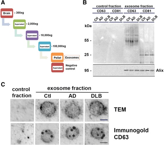
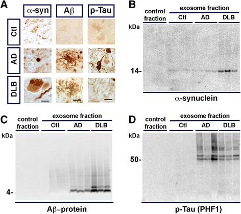
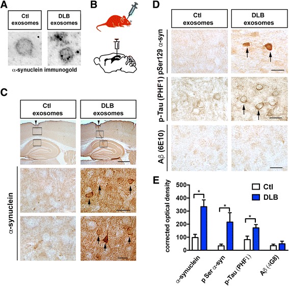
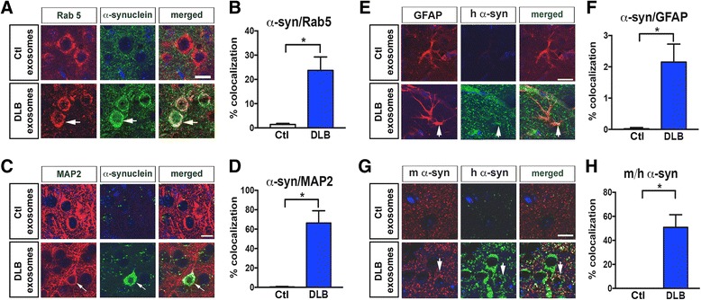
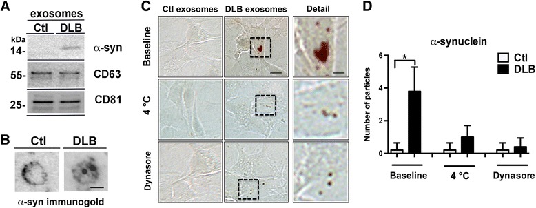
Proteins implicated in neurodegenerative conditions such as Alzheimer's disease (AD) and Dementia with Lewy Bodies (DLB) have been identified in bodily fluids encased in extracellular vesicles called exosomes. Whether exosomes found in DLB patients can transmit pathology is not clear. In this study, exosomes were successfully harvested through ultracentrifugation from brain tissue from DLB and AD patients as well as non-diseased brain tissue. Exosomes extracted from brains diagnosed with either AD or DLB contained aggregate-prone proteins. Furthermore, injection of brain-derived exosomes from DLB patients into the brains of wild type mice induced α-synuclein (α-syn) aggregation. As assessed through immunofluorescent double labeling, α-syn aggregation was observed in MAP2+, Rab5+ neurons. Using a neuronal cell line, we also identified intracellular α-syn aggregation mediated by exosomes is dependent on recipient cell endocytosis. Together, these data suggest that exosomes from DLB patients are sufficient for seeding and propagating α-syn aggregation in vivo.

Keywords: Alpha synuclein; Alzheimer’s disease; Amyloid beta; Dementia; Exosomes; Lewy body; Tau.


Figures

Fig. 1
Fig. 1
Isolation of exosomes from patient brain tissue. a Schematic of ultracentrifugation protocol used to isolate exosomes from tissue. Note that the pellet is the “exosome fraction”, and the supernatant is the “control fraction”. b Exosome identification markers present in the exosome fraction of Ctl, AD and DLB brain tissue. c Top Row: Representative EM micrographs from control fraction as well as Ctl, AD and DLB exosome fractions. Bottom Row: EM micrographs from control fraction and Ctl, AD and DLB exosome fractions stained with immunogold labeled anti-CD 63. Scale bar = 50 nm
Fig. 2
Fig. 2
Aggregate prone proteins associated with disease are present in exosome cargo. a Representative micrographs of Ctl, AD and DLB brain tissue, immunohistochemically stained for α-synuclein (α-syn), Amyloid Beta (Aβ) and phosphorylated tau (p-Tau). Scale bar for: α-syn = 5 μm, Aβ = 10 μm and p-Tau = 5 μm. b-d Western blots detecting the presence of α-syn (b), Aβ-protein (c) and p-Tau (d) contained within each fraction
Fig. 3
Fig. 3
Administration of DLB exosomes into mouse brains initiates intracellular accumulation of phosphorylated proteins. a Representative EM micrographs of exosomes from Ctl and DLB brain samples immunolabeled for α-syn. b Schematic of stereotaxic injection into the hippocampus of C57BL/6 N × DBA/2 F1 mice. c Representative brightfield micrographs from mouse brains injected with control or DLB exosomes. Row 1: Sagittal view of the hippocampal area. Needle entry site is highlighted by arrowhead. Boxes highlight region of interest depicted in micrographs below. Scale bar = 150 μm. Rows 2–3: High magnification view of highlighted areas. Arrowheads highlight immunolabeled cell bodies. Scale bar = 25 μm. d Micrographs of brain tissue stained with phosphorylated α-syn (pSer129), PHF1 and Aβ (6E10). Scale bar = 25 μm. e Quantification of immunohistochemical stains from (c) and (d) through optical density. * = P < 0.05. Error bars indicate SEM
Fig. 4
Fig. 4
Human α-syn internalization occurs in neurons and astrocytes. Representative immunofluoresent micrographs of mouse hippocampal brain slices injected with either Ctl or DLB exosomes, depicting human α-syn (Green), DAPI (Blue) and a Rab5, c MAP2, e GFAP or g mouse α-syn (Red). Scale bar = 25 μm. Percent colocalization of α-syn and b Rab5, d MAP2, f GFAP and h mouse α-syn. * = P < 0.05. Error bars indicate SEM
Fig. 5
Fig. 5
Inhibition of endocytosis inhibits exosomes-mediated α-syn accumulation. a Immunoblot and b electron microscopy confirm the presence of α-syn containing exosomes from DLB patient. Scale bar = 50 nm. c B103 neuronal cell lines incubated with either Ctl or DLB exosomes and were left alone (first row, Baseline), incubated in 4 °C (second row) or incubated with Dynasore (third row). Accumulation of α-syn was visualized using the Syn1 antibody. Detail column indicates the region of interest magnified. d Quantification of α-syn positive particles in 37 °C (baseline), incubation at 4 °C and at 37 °C with Dynasore. * = P < 0.05 (DLB compared to Ctl). Error bars indicate SEM

References

    1. Baker HF, Ridley RM, Duchen LW, Crow TJ, Bruton CJ (1994) Induction of β (A4)-amyloid in primates by injection of Alzheimer’s disease brain homogenate. Mol Neurobiol 8:25–39. doi:10.1007/BF02778005 - PubMed
    1. Bayer TA. Proteinopathies, a core concept for understanding and ultimately treating degenerative disorders? Eur Neuropsychopharmacol. 2015;25:713–724. doi: 10.1016/j.euroneuro.2013.03.007. - DOI - PubMed
    1. Bellingham SA, Guo BB, Coleman BM, Hill AF (2012) Exosomes: vehicles for the transfer of toxic proteins associated with neurodegenerative diseases? Front Physiol MAY 3:1–12. doi:10.3389/fphys.2012.00124 - PMC - PubMed
    1. Brettschneider J, Del Tredici K, M-Y Lee V, Trojanowski JQ (2015) Spreading of pathology in neurodegenerative diseases: a focus on human studies. Nat Publ Gr. doi:10.1038/nrn3887 - PMC - PubMed
    1. Bucci C, Parton RG, Mather IH, Stunnenberg H, Simons K, Hoflack B, et al. The small GTPase rab5 functions as a regulatory factor in the early endocytic pathway. Cell. 1992;70:715–728. doi: 10.1016/0092-8674(92)90306-W. - DOI - PubMed

Publication types

MeSH terms