. 2017 Aug 1;428(1):188-203.
doi: 10.1016/j.ydbio.2017.06.003. Epub 2017 Jun 7.

Planar cell polarity-dependent and independent functions in the emergence of tissue-scale hair follicle patterns

Affiliations

Planar cell polarity-dependent and independent functions in the emergence of tissue-scale hair follicle patterns

Maureen Cetera et al. Dev Biol. .

Abstract

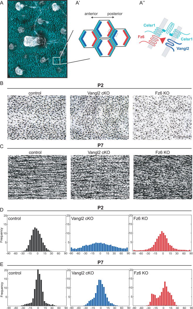
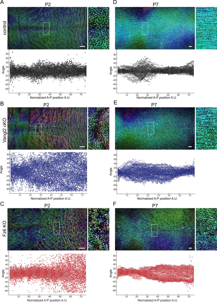
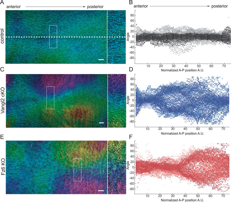
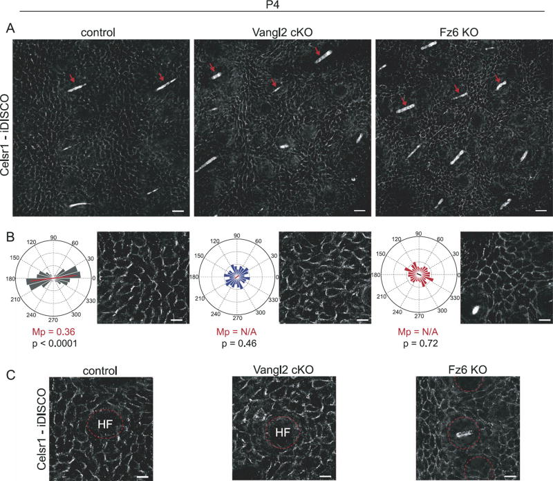
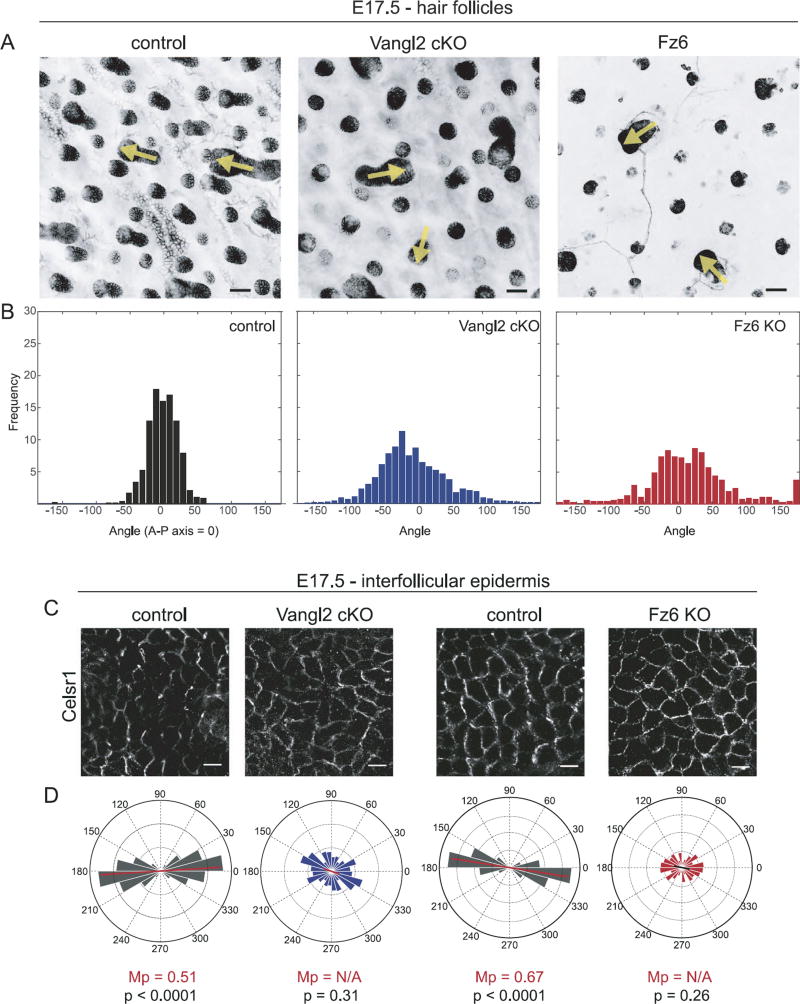
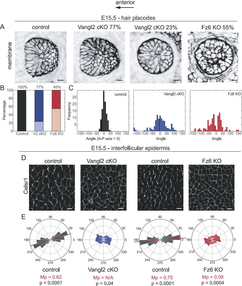
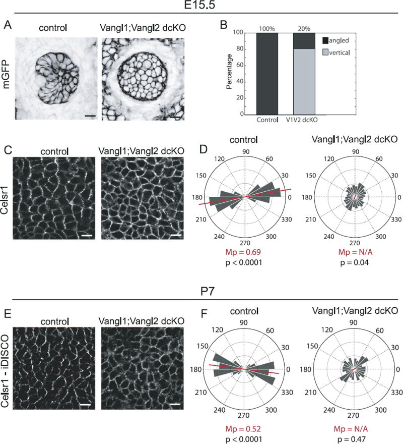
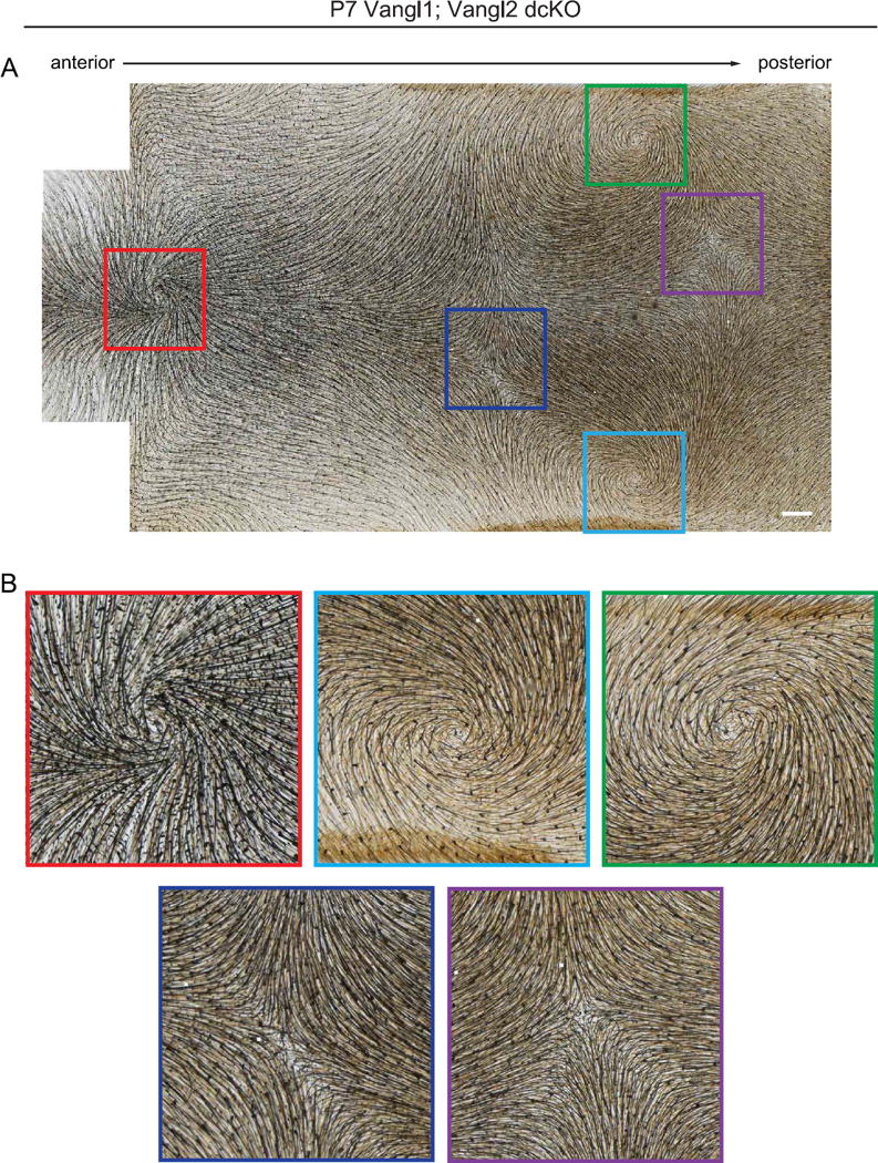
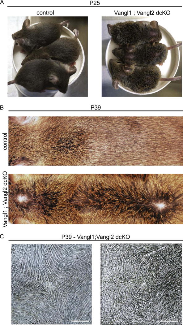
Hair follicles of the mammalian epidermis display local order and global alignment, a complex pattern instructed by the core planar cell polarity (PCP) pathway. Here we address the contributions of core PCP genes, Van Gogh-like and Frizzled, to the establishment, local refinement, and global order of embryonic and postnatal hair follicles. We find that, similar to Fz6 mutants, the disordered hair patterns of Vangl2 mutants are refined over time and eventually corrected. In both mutants, we find that tissue-level reorientation occurs through locally coordinated follicle rotation at stereotyped locations. Strikingly, Vangl2 and Fz6 mutant follicles collectively rotate with opposing directionalities, suggesting that redundant core PCP signals contribute to their directed realignment. Consistently, global follicle alignment is not restored upon conditional ablation of both Vangl1 and Vangl2 genes. Instead, spatially distinct patterns of whorls and crosses emerge and persist even after a complete cycle of hair follicle regeneration. Thus, local refinement of hair follicles into higher order patterns can occur independently of the core PCP system, however, their global alignment with the body axes requires PCP function throughout morphogenesis, growth and regeneration.

Keywords: Celsr1; Epidermis; Fz6; Hair follicles; Planar cell polarity; Vangl2.


Figures

Fig. 1
Fig. 1
Disordered hair patterns in Vangl2 and Fz6 mutants correct over time. (A) Schematic of planar cell polarity in the embryonic epidermis at the tissue (A), cellular (A′) and subcellular level (A″). (A, A′) PCP proteins are localized to AP junctions where they instruct the anterior tilt of the follicle. Celsr1 is shown in cyan and hair follicles are shown in white. (A′,A″) Celsr1, Vangl2 (cyan, blue) complexes localize at the anterior cell border while Celsr1, Fz6 (cyan, red) complexes localize at the posterior cell border. These complexes interact across AP borders. (B,C) Hair follicles from wild type control, Vangl2 cKO, and Fz6 KO dorsal epidermis at P2 (B) and P7 (C). See Table 1 for specific genotypes. Skins were cleared and hair follicle pigment was imaged by bright field microscopy. Anterior is to the left in all figures. (D,E) Quantification of hair follicle orientations at P2 (D) and P7 (E). See Methods. Anterior-posterior axis = 0 degrees. Angular frequencies at P2 are pooled from control (n=6 animals); Vangl2 cKO (n=5 animals); Fz6 KO (n=3 animals). P7: controls (n=4 animals), Vangl2 cKO (n=5 animals), Fz6 KO (n=3 animals). Scale bar, 500 µm.
Fig. 2
Fig. 2
Postnatal hair follicle refinement is independent of Vangl2 and Fz6. (A–C) Wild type control (A), Vangl2 cKO, (B) and Fz6 KO (C) dorsal skins at P2. Hair follicles are pseudo-colored according to their angle using Orientation J. Green-blue represents hair follicles oriented closest to the AP axis ~ 0° to +/− 45°. Pink-purple ~ −45° to −90°. Orange-yellow ~ +45° to +90°. The boxed regions are shown zoomed in on the right. Quantification of hair follicle angles plotted relative to their position along the anterior-posterior axis is shown below. Each dot represents the average orientation of follicles within one square window, tiled across the backskin. See Methods. Each color shade represents data from one animal. For control n=6, Vangl2 cKO n=5, and Fz6 KO n=3 animals. (D–F) The same analysis was carried out at P7. For control n=4, Vangl2 cKO n=5, and Fz6 KO n=3. Note that the disordered hair pattern at P2 is aligned with the anterior-posterior axis by P7. Scale bar, 1 mm.
Fig. 3
Fig. 3
Stereotyped hair patterns emerge during follicle refinement. (A) Hair follicles from wild type control, Vangl2 cKO, and Fz6 KO dorsal epidermis at postnatal day 4 (P4). Skins were cleared and hair follicle pigment was imaged by bright field microscopy. Zoomed regions (2x) are shown below. (B) Quantification of hair follicle orientations at P4. Anterior-posterior axis = 0°. Frequencies are pooled from control (n=5 animals); Vangl2 cKO (n=6 animals); Fz6 KO (n=6 animals). Scale bar, 1 mm.
Fig. 4
Fig. 4
Vangl2 cKO and Fz6 KO follicles refine in opposing directions. (A,C,E) Wild type control (A), Vangl2 cKO (C) and Fz6KO (E) dorsal skins at P4 (left). Hair follicles are pseudo-colored according to their angle using Orientation J. Green-blue represents hair follicles oriented closest to the AP axis ~ 0° to +/− 45°, anterior-posterior axis. Pink-purple ~ −45° to −90°. Orange-yellow ~ +45° to +90°. Boxed region is shown zoomed on the right. The dotted horizontal line indicates the midline. The position of the midline is consistent across all whole backskin images. Note that while anterior-most follicles have the correct orientation in all conditions, central and posterior follicles point inward, toward the midline, in Vangl2 cKO skin (C) and outward, away from the midline, in Fz6 KO skin (E). For additional examples, see Supplemental Fig. 1. (B,D,F) Quantification of hair follicle angles plotted relative to their position along the anterior-posterior axis. Each dot represents the average orientation of follicles within one square window, which were tiled across the entire backskin. See Section 2. Each color shade represents data from one animal. n=6 animals for control, Vangl2 cKO, and Fz6 KO. Scale bar, 1 mm.
Fig. 5
Fig. 5
During refinement, hair follicle orientation is uncoupled from Celsr1 asymmetry in the interfollicular epidermis. (A) Immunofluorescence images of wild type control, Vangl2 cKO, and Fz6 KO dorsal skin at P4 labeled with antibodies against Celsr1. Tissues were cleared and processed for immunofluorescence using the iDISCO technique. Images are oriented with hairs (red arrows) aligned in the same direction. Note that despite similar follicle orientations in all three genotypes, Celsr1 is enriched at anterior-posterior borders of interfollicular epidermal cells in wild type control but not Vangl2 cKO or Fz6 KO animals. Scale bar, 25 µm. (B) Angular distribution of Celsr1 polarity in the interfollicular epidermis. Frequency scale 0 −0.3. The anterior-posterior axis runs horizontally. Control, n=1094 cells from 3 animals; Vangl2 cKO, n=1292 cells from 3 animals; Fz6 KO n=1391 cells from 3 animals. MP = magnitude of average Celsr1 polarity. The significance of the angular variation within each stage was assessed using a nonparametric permutation test. Statistical significance = p < 0.01 (if p ≥ 0.01, magnitude of average Celsr1 polarity, MP = N/A). See Methods. (C) Representative images of Celsr1 localization surrounding individual hair follicles (labeled HF). Scale bar, 10 µm.
Fig. 6
Fig. 6
Uncoupling of hair follicle orientation and Celsr1 asymmetry during embryogenesis. (A) Representative hair follicles from wild type control, Vangl2 cKO, and Fz6 KO embryos at E17.5 labeled with E-Cadherin (control and Vangl2 cKO) or membrane-GFP (Fz6 KO). Yellow arrows indicate the orientation of a subset of hair follicles. See Table 1 for genotypes. Single confocal z-planes are shown. Scale bar, 50 µm. (B) Quantification of hair follicle orientations of the indicated genotypes at E17.5. Control, n=764 follicles; Vangl2 cKO, n= 1284 follicles; Fz6 KO, n= 609 follicles. (C) Representative immunofluorescence images of Celsr1 labeled interfollicular epidermis at E17.5. Single confocal planes are shown. (D) Quantification of angular distribution of Celsr1 polarity in the interfollicular epidermis of E17.5 Vangl2 cKO (cKO, n=1083 cells from 2 embryos; control littermates n=1049 cells from 2 embryos), and Fz6 KO embryos (KO, n=948 cells from 3 embryos; control littermates, n=2184 cells from 3 embryos). Frequency scale 0–0.3 for Vangl2 pair; 0–0.4 for Fz6 pair. Scale bar, 10 µm.
Fig. 7
Fig. 7
Relationship between hair placode orientation and Celsr1 asymmetry at initial hair follicle polarization. (A) Representative hair follicle placodes from wild type control, Vangl2 cKO (angled, left and vertical, right), and Fz6 KO embryos at E15.5 labeled with membrane-GFP (control and Vangl2 cKO) or membrane-tdTomato (Fz6 KO). See Table 1 for genotypes. Single confocal z-planes are shown. (B) Relative frequency of angled (dark shades) versus vertical (light shades) follicle placodes at E15.5. Wild type control, n= 393 follicles; Vangl2 cKO, n=406 follicles; Fz6 KO, n= 374 follicles. (C) Quantification of hair follicle orientations of the indicated genotypes at E15.5. Only angled, not vertical, placodes are included. Wild type control, n=393 follicles; Vangl2 cKO, n=311 follicles; Fz6 KO n=181. (D) Representative immunofluorescence images of Celsr1 labeled interfollicular epidermis at E15.5. Single confocal planes are shown. (E) Quantification of the angular distribution of Celsr1 polarity in the interfollicular epidermis at E15.5 of Vangl2 cKO (cKO, n=3068 cells from 2 embryos; control littermates n=2971 cells from 2 embryos), and Fz6 KO embryos (KO, n=4076 cells from 3 embryos; control littermates, n=2994 cells from 3 embryos). Frequency scale 0-0.3. Scale bar, 10 µm.
Fig. 8
Fig. 8
Loss of hair follicle polarization and Celsr1 asymmetry in Vangl1; Vangl2 dcKO epidermis. (A) Representative hair follicle placodes from wild type control and Vangl1; Vangl2 dcKO embryos at E15.5 labeled with membrane-GFP. See Table 1 for genotypes. Single confocal z-planes are shown. (B) Relative frequency of angled (dark shades) versus vertically-oriented (light shades) placodes at E15.5. The majority of Vangl1; Vangl2 dcKO placodes fail to polarize. (C) Representative immunofluorescence images of E15.5 wild type control and Vangl1; Vangl2 dcKO interfollicular epidermis labeled with Celsr1. Single confocal planes are shown. (D) Quantification of Celsr1 polarity in the interfollicular epidermis of control and Vangl1; Vangl2 dcKO embryos. Control, n= 3533 cells from 4 embryos; dcKO, n=3111 cells from 4 embryos. (E) Immunofluorescence images of wild type control and Vangl1; Vangl2 dcKO dorsal skin at P7 labeled with antibodies against Celsr1. Tissues were cleared and processed for immunofluorescence using iDISCO. (F) Quantification of the angular distribution of Celsr1 polarity in the interfollicular epidermis at P7. (Control, n=940 cells from 3 animals, Vangl1; Vangl2 dcKO, n=987 cells from 3 animals). The anterior-posterior axis runs horizontally. Scale bar, 10 µm.
Fig. 9
Fig. 9
Stereotyped hair patterns emerge postnatally in the absence of Vangl function. (A) Representative Vangl1;Vangl2 dcKO dorsal skin at P7 showing characteristic and reproducible pattern of whorls and crosses. Note the presence of three hair whorls positioned in a triangular configuration (red, green and light blue boxes), and two crosses where whorls intersect (dark blue and purple boxes). For additional examples, see Supplementary Fig. 3. (B) Three whorls and two crosses corresponding to boxed areas in A. Scale bar, 1 mm.
Fig. 10
Fig. 10
Hair patterns evolve over time but do not correct in the absence of Vangl function. (A) Vangl1; Vangl2 dcKO mice and their control littermates at postnatal day 25. Double knockout embryos are clearly distinguished from their control littermates due to their disorganized and scruffy hair coats. (B) Representative images of Vangl1;Vangl2 dcKO and control dorsal skin at P39. Note the presence of two hair whorls and a zone of reversal positioned along the midline. For additional examples of P39 dorsal skins, see Supplementary Fig. 4. (C) Representative hair whorl and cross from Vangl1;Vangl2 dcKO at P39. Dorsal epidermis was cleared and imaged for hair follicle pigment by brightfield microscopy. Scale bar, 1 mm.

References

    1. Adler PN. The frizzled/stan pathway and planar cell polarity in the Drosophila wing. Curr. Top. Dev. Biol. 2012;101:1–31. - PMC - PubMed
    1. Adler PN, Charlton J, Liu J. Mutations in the cadherin superfamily member gene dachsous cause a tissue polarity phenotype by altering frizzled signaling. Development. 1998;125:959–968. - PubMed
    1. Aigouy B, Farhadifar R, Staple DB, Sagner A, Roper JC, Julicher F, Eaton S. Cell flow reorients the axis of planar polarity in the wing epithelium of Drosophila. Cell. 2010;142:773–786. - PubMed
    1. Alonso L, Fuchs E. The hair cycle. J. Cell Sci. 2006;119:391–393. - PubMed
    1. Aw WY, Heck BW, Joyce B, Devenport D. Transient tissue-scale deformation coordinates alignment of planar cell polarity junctions in the mammalian skin. Curr. Biol. 2016;26:2090–2100. - PMC - PubMed

Publication types

LinkOut - more resources