. 2017 Jul 15;130(14):2416-2429.
doi: 10.1242/jcs.195875. Epub 2017 Jun 9.

Reconstitution of the oocyte nucleolus in mice through a single nucleolar protein, NPM2

Affiliations

Reconstitution of the oocyte nucleolus in mice through a single nucleolar protein, NPM2

Sugako Ogushi et al. J Cell Sci. .

Abstract

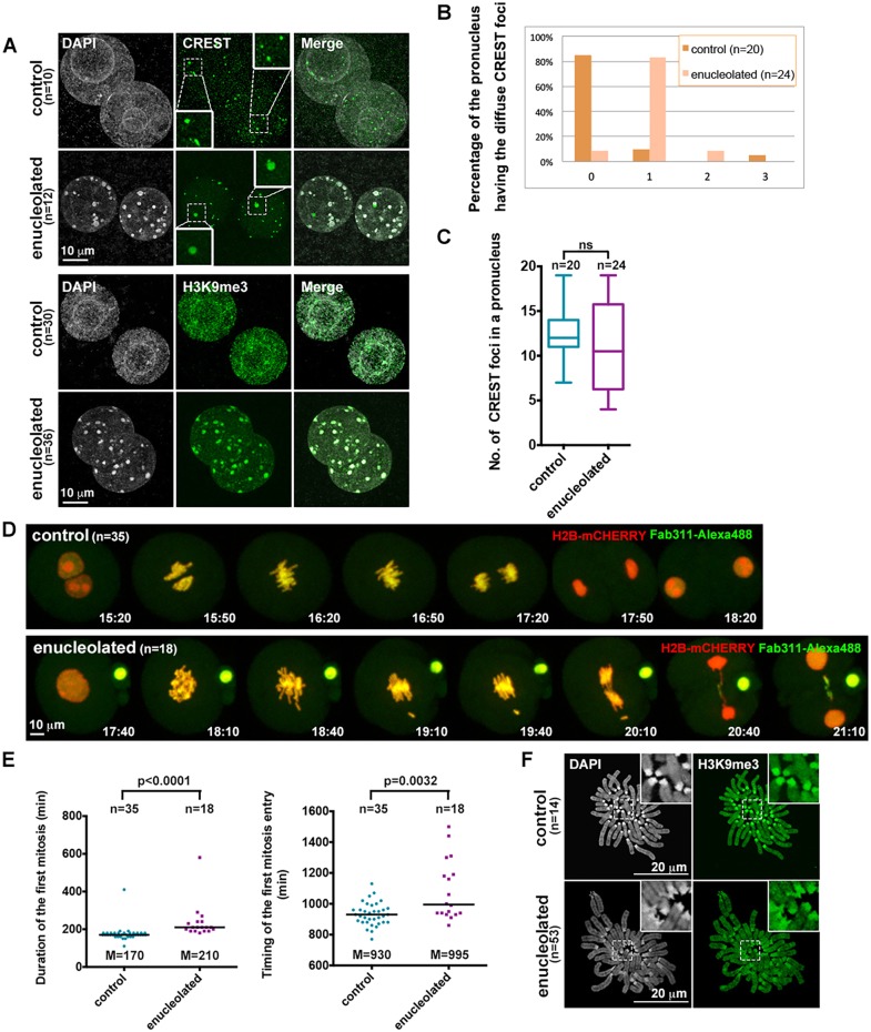
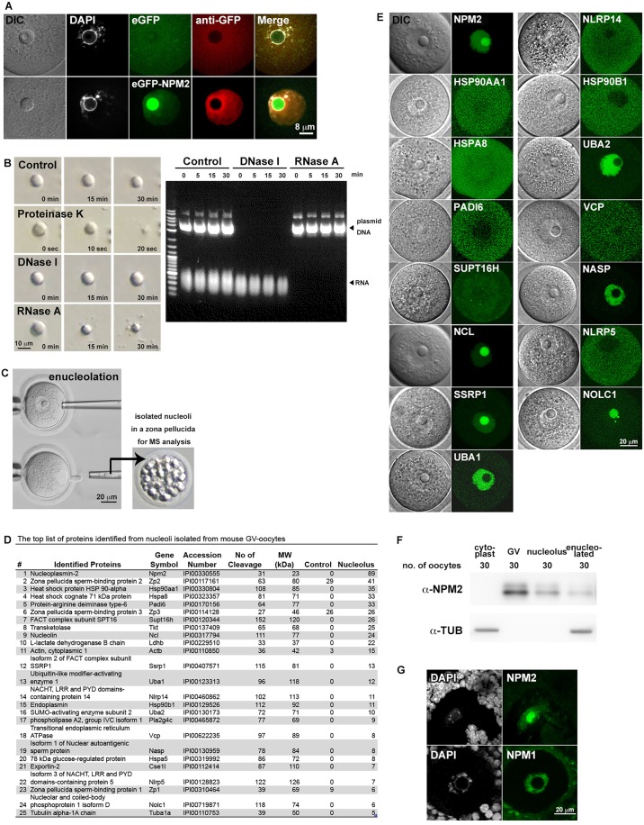
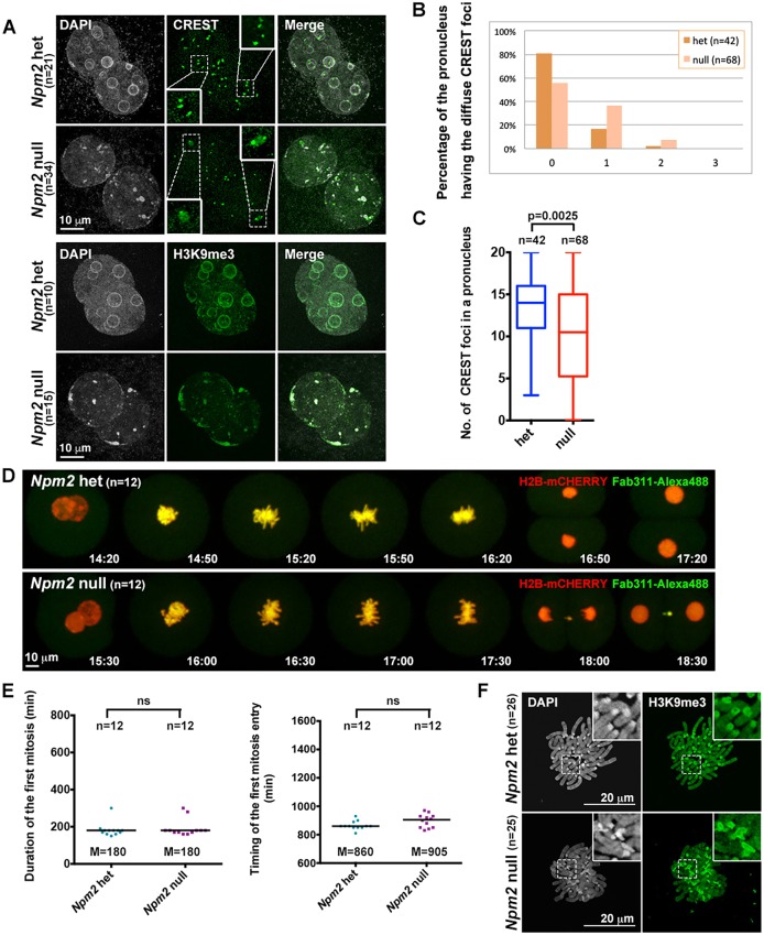
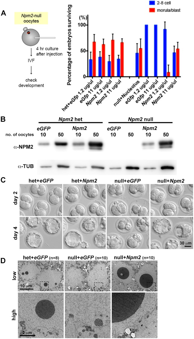
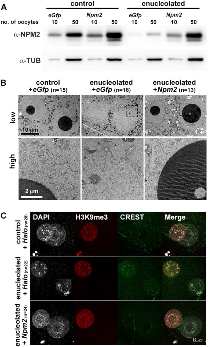
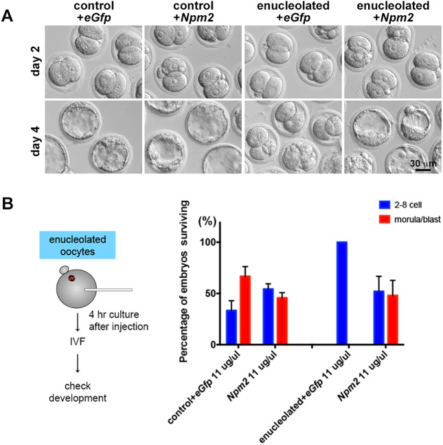
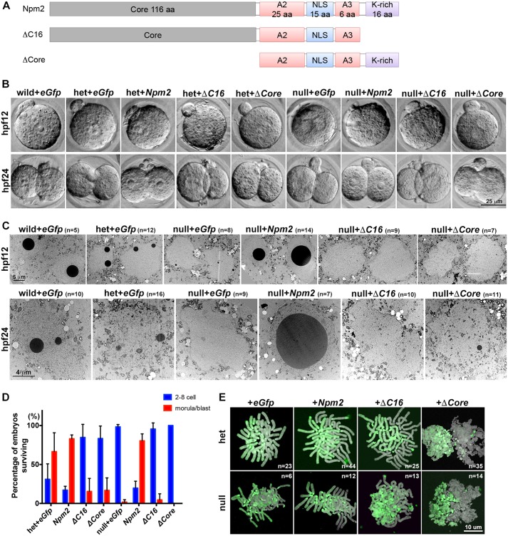
The mammalian oocyte nucleolus, the most prominent subcellular organelle in the oocyte, is vital in early development, yet its key functions and constituents remain unclear. We show here that the parthenotes/zygotes derived from enucleolated oocytes exhibited abnormal heterochromatin formation around parental pericentromeric DNAs, which led to a significant mitotic delay and frequent chromosome mis-segregation upon the first mitotic division. A proteomic analysis identified nucleoplasmin 2 (NPM2) as a dominant component of the oocyte nucleolus. Consistently, Npm2-deficient oocytes, which lack a normal nucleolar structure, showed chromosome segregation defects similar to those in enucleolated oocytes, suggesting that nucleolar loss, rather than micromanipulation-related damage to the genome, leads to a disorganization of higher-order chromatin structure in pronuclei and frequent chromosome mis-segregation during the first mitosis. Strikingly, expression of NPM2 alone sufficed to reconstitute the nucleolar structure in enucleolated embryos, and rescued their first mitotic division and full-term development. The nucleolus rescue through NPM2 required the pentamer formation and both the N- and C-terminal domains. Our findings demonstrate that the NPM2-based oocyte nucleolus is an essential platform for parental chromatin organization in early embryonic development.

Keywords: Heterochromatin; Nucleolus; Nucleoplasmin 2; Oocyte.


Conflict of interest statement

Competing interestsThe authors declare no competing or financial interests.

Figures

Fig. 1.
Fig. 1.
Loss of the oocyte nucleolus causes disorganization of the higher chromatin in pronuclei and chromosome mis-segregation during the first mitosis in parthenotes from enucleolated oocytes. (A) Immunostaining using parthenotes from enucleolated oocytes 9-12 h after activation with the antibodies a (green). Insets show the diffuse CREST signals (>2 µm2) in each pronucleus at 2-fold magnification of the regions shown in the dash-lined boxes. DNA was stained with DAPI (gray). Control, parthenotes from nucleolus-reinjected oocytes. (B) Percentage of pronuclei having diffuse CREST signals in parthenotes from control/enucleolated oocytes. The numbers on the x-axis of the graph indicate the number of diffuse signals in each pronucleus. (C) Number of CREST foci in each pronucleus of parthenotes from control/enucleolated oocytes. Boxes show the median, 25th and 75th percentiles, and bars show the 10th and 90th percentiles. The number of CREST foci was statistically analyzed by two-tailed Mann–Whitney test. ns, not significant. (D) Representative stills from live cell imaging of parthenotes from control/enucleolated oocytes. Chromosomes were labeled by H2B-mCHERRY (red) and chromosome condensation was visualized by Fab311-Alexa488 (green). Numbers indicate the time after artificial activation (hr:min). (E) The duration and entry timing of the first mitosis in parthenotes are plotted. Bars, median (M). Two-tailed Mann–Whitney test. (F) Representative images of chromosome spreads in parthenotes stained with H3K9me3 antibody (green) and DAPI (gray). Insets show 2.5-fold magnification of the regions shown in the dash-lined boxes. (A-F) n, the numbers of parthenotes measured in three independent experiments.
Fig. 2.
Fig. 2.
Analysis of the oocyte nucleolus components. (A) The compact structure of the oocyte nucleolus prevents antibody inflow into the structure. EGFP-NPM2 signals (green) in the oocyte nucleolus could not be stained by anti-GFP antibody (red). DNA was stained by DAPI (gray). DIC, differential interference contrast. (B) Components in the oocyte nucleolus were assessed by proteinase K, DNase I, and RNase A (left). The activity of the enzymes was checked at each time point indicated above the gel image. Digestion of DNA and RNA was checked by using 1% agarose gel electrophoresis (right). (C) The method of MS analysis using isolated nucleoli by enucleolation from GV-oocytes. The isolated nucleoli were kept in evacuated zona pellucidae and used for MS analysis. (D) List of identified nucleolus proteins. (E) Localization of MS candidate proteins tagged with eGFP (green) at the N-terminus. DIC, differential interference contrast. (F) NPM2 localization in the oocyte nucleolus was confirmed by western blotting. The oocyte number in each sample is indicated above the blots and α-tubulin (TUB) was used as a loading control (n=3). (G) NPM1 (green) localized at the nucleoplasm and NPM2 localized at the nucleolus in cryosections. DNA was stained by DAPI (gray).
Fig. 3.
Fig. 3.
Parthenotes from Npm2-null oocytes show defects similar to those from enucleolated oocytes. (A) Parthenotes from Npm2-null oocytes stained with the antibodies as indicated (green) and DAPI (gray). Insets show the diffuse CREST signals at 2-fold magnification. (B) Percentage of pronuclei having diffuse CREST signals in parthenotes from Npm2-het/-null oocytes. (C) Number of CREST foci in each pronucleus of parthenotes from Npm2-het/-null oocytes. Two-tailed Mann–Whitney test. ns, not significant. (D) Representative stills from live cell imaging of parthenotes from Npm2-het/-null oocytes as in Fig. 1C. (E) The duration and entry timing of the first mitosis in parthenotes are plotted. Bars, median (M). Two-tailed Mann–Whitney test. (F) Representative images of chromosome spreads in parthenotes stained with H3K9me3 antibody (green) and DAPI (gray). Insets show 2.5-fold magnification of the regions shown in the dash-lined boxes. (A-F) n, the numbers of parthenotes measured in three independent experiments.
Fig. 4.
Fig. 4.
NPM2 expression in oocytes at second metaphase rescues the defects of Npm2-null embryos in early embryonic development. (A) Schematic of the rescue experiments in Npm2-null oocytes. IVF, in vitro fertilization. The graph shows the percentage of mRNA-injected embryos that reached the blastocyst stage. Error bars, +s.d. (n>3 each). (B) NPM2 protein expression levels in Npm2-het/-null oocytes injected with the indicated mRNAs at the concentration of 11 µg/µl. The oocyte number in each sample is indicated above the blots; α-tubulin (TUB) was used as a loading control (n=3). (C) Representative images of embryos from Npm2-het/-null oocytes rescued by the indicated mRNAs at 2 and 4 days after IVF. (D) Transmission electron microscopy (TEM) images of pronuclei in zygotes from Npm2-het/-null oocytes rescued by Npm2-mRNA injection. Bottom panels show 5-fold magnifications of the regions in the dash-lined boxes. n, number of zygotes measured in three independent experiments.
Fig. 5.
Fig. 5.
Expression of NPM2 alone is sufficient to reconstitute the nucleolus structure of pronuclei in zygotes. (A) NPM2 protein expression levels in control/enucleolated oocytes injected with the indicated mRNA. The oocyte number in each sample is indicated above the blots and α-tubulin (TUB) was used as a loading control (n=3). Control, zygotes from nucleolus-reinjected oocytes. (B) Transmission electron microscopy (TEM) images of pronuclei in zygotes from control/enucleolated oocytes rescued by Npm2-mRNA injection. Bottom panels show 5-fold magnifications of the regions in the dash-lined boxes. (C) Immunostaining using zygotes from NPM2-expressing enucleolated oocytes 9-12 h after fertilization with antibodies as indicated. For injection control at MII oocytes, Halo mRNA at 10 µg/µl was injected into control and enucleolated oocytes as Npm2 mRNA. (B,C) n, number of zygotes measured in three independent experiments.
Fig. 6.
Fig. 6.
NPM2 expression rescues the defect caused by loss of the oocyte nucleolus in early embryonic development. (A) Representative images of embryos from control/enucleolated oocytes rescued by the indicated mRNAs at 2 and 4 days after IVF. (B) Schematic of the rescue experiments in enucleolated oocytes. IVF, in vitro fertilization. The graph shows the percentage of mRNA-injected embryos that reached the blastocyst stage. Error bars, +s.d. (n>3 each).
Fig. 7.
Fig. 7.
Both the N-terminal and the C-terminal domains are essential for the structural integrity and function of NPM2. (A) Schematic of NPM2-truncation mutants. NPM2 has a core domain, two acidic domains (A2 and A3), a nuclear localization signal (NLS) and a basic cluster, the K-rich motif. (B) Nucleolus formation in embryos at 12 h or 24 h post-fertilization (hpf) from Npm2-wild/-het/-null oocytes injected with the indicated mRNAs. (C) Representative TEM images in embryos from Npm2-wild/-het/-null oocytes injected with the indicated mRNAs. (D) Percentage of embryos from Npm2-het/-null oocytes injected with Npm2-truncated mutant mRNAs that reached the blastocyst stage. Bars, SD (n>3 each). (E) Representative images of chromosome spreads in zygotes from Npm2-het/-null oocytes injected with Npm2-truncated mutant mRNA. Chromosomes were stained with H3K9me3 antibody (green) and DAPI (gray). (C,E) n, the numbers of zygotes/embryos measured in three independent experiments.

Similar articles

Cited by

References

    1. Amin M. A., Matsunaga S., Uchiyama S. and Fukui K. (2008). Nucleophosmin is required for chromosome congression, proper mitotic spindle formation, and kinetochore-microtubule attachment in HeLa cells. FEBS Lett. 582, 3839-3844. 10.1016/j.febslet.2008.10.023 - DOI - PubMed
    1. Aoki F., Worrad D. M. and Schultz R. M. (1997). Regulation of transcriptional activity during the first and second cell cycles in the preimplantation mouse embryo. Dev. Biol. 181, 296-307. 10.1006/dbio.1996.8466 - DOI - PubMed
    1. Arney K. L., Bao S., Bannister A. J., Kouzarides T. and Surani M. A. (2002). Histone methylation defines epigenetic asymmetry in the mouse zygote. Int. J. Dev. Biol. 46, 317-320. - PubMed
    1. Banani S. F., Lee H. O., Hyman A. A. and Rosen M. K. (2017). Biomolecular condensates: organizers of cellular biochemistry. Nat. Rev. Mol. Cell Biol. 18, 285-298. 10.1038/nrm.2017.7 - DOI - PMC - PubMed
    1. Bergeron-Sandoval L.-P., Safaee N. and Michnick S. W. (2016). Mechanisms and consequences of macromolecular phase separation. Cell 165, 1067-1079. 10.1016/j.cell.2016.05.026 - DOI - PubMed