Review
. 2018 Apr 2;10(4):a029181.
doi: 10.1101/cshperspect.a029181.

Cell-Cell Junctions Organize Structural and Signaling Networks

Affiliations
Review

Cell-Cell Junctions Organize Structural and Signaling Networks

Miguel A Garcia et al. Cold Spring Harb Perspect Biol. .

Abstract

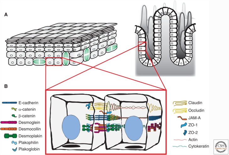
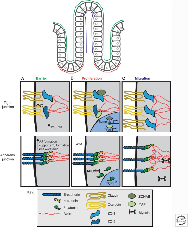
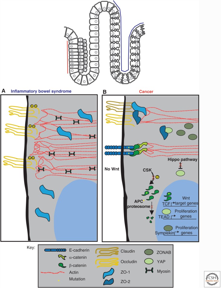
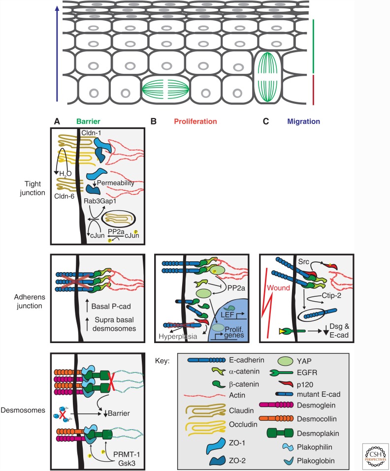
Cell-cell junctions link cells to each other in tissues, and regulate tissue homeostasis in critical cell processes that include tissue barrier function, cell proliferation, and migration. Defects in cell-cell junctions give rise to a wide range of tissue abnormalities that disrupt homeostasis and are common in genetic abnormalities and cancers. Here, we discuss the organization and function of cell-cell junctions primarily involved in adhesion (tight junction, adherens junction, and desmosomes) in two different epithelial tissues: a simple epithelium (intestine) and a stratified epithelium (epidermis). Studies in these tissues reveal similarities and differences in the organization and functions of different cell-cell junctions that meet the requirements for the specialized functions of each tissue. We discuss cell-cell junction responses to genetic and environmental perturbations that provide further insights into their roles in maintaining tissue homeostasis.


Figures

Figure 1.
Figure 1.
Tissue organization and cell junction composition in two different epithelia (intestine and epidermis). (A) The epidermis (left) is organized as stratified epithelium, and the intestine (right) is organized as a simple epithelium (one cell layer thick). (B) Composition and spatial organization of adhesive cell–cell junctions in epithelial cells. The tight junction (TJ) is localized to the most apical region of the cell and is composed of transmembrane proteins (claudin, occludin) and adaptor proteins (ZO-1 and ZO-2), which link to the underlying actin cytoskeleton. The adherens junction (AJ) is localized on the lateral membrane and is primarily composed of the transmembrane protein, E-cadherin, and adaptor proteins, β-catenin and α-catenin, which link to the underlying actin cytoskeleton. The desmosome (only one is shown, but many are located along the lateral membrane) is composed of transmembrane proteins (desmocollin and desmoglein) and adaptor proteins (plakoglobin, plakophillin, and desmoplakin), which links to cytokeratin intermediate filaments.
Figure 2.
Figure 2.
Cell–cell junction regulation of intestinal epithelial homeostasis. (A) Barrier function is primarily regulated by the tight junction (TJ) with indirect contributions by the adherens junction (AJ) through its role in TJ assembly. Phosphorylation of occludin by PKC- ζ results in occludin incorporation at the TJ. Phosphorylation of claudin results in its incorporation or removal from the TJ depending on the claudin type. (B) Proliferation is dually regulated by the TJ and AJ, which sequester proproliferation transcription factors, ZO-1-associated nucleic acid binding protein (ZONAB), β-catenin and Yes-associated-protein (YAP). (C) Cell migration is primarily mediated by alterations to components of the AJ, whereas changes in TJ protein homeostasis also occur. See text for details.
Figure 3.
Figure 3.
Disruption of homeostasis results in altered signaling at cell–cell junctions in intestinal epithelia. (A) In inflammatory bowel disease (IBD), reduction or loss of ZO-1 and claudin-1 from the tight junction (TJ) has been observed, resulting in a loss of barrier function. A hyperactive actomyosin contractile network has also been reported. (B) In cancer, deregulated signaling at the TJ and adherens junction (AJ) occurs, resulting in uncontrolled proliferation mediated by β-catenin, Yes-associated-protein (YAP), and ZO-1-associated nucleic acid binding protein (ZONAB). Mutations in adenomatous polyposis coli (APC) and in β-catenin result in β-catenin accumulation by bypassing degradation in the absence of Wnt. Mutations in E-cadherin also result in cancer. See text for details.
Figure 4.
Figure 4.
Cell junction regulation of homeostasis in epidermis. (A) Barrier function can be attributed to all junctional complexes. The tight junction (TJ) in the stratum granulosum prevent dehydration from the underlying tissues. Disruption of any claudin family member, particularly those that confer classic TJ strand organization, increases TJ and tissue permeability. At the adherens junctions (AJ), E-cadherin depletion results in a compensatory response by up-regulation of P-cadherin and desmosomes. Desmosomes are regulated by phosphorylation of desmosomal components, which is required for proper desmosome formation and cytokeratin association. Disruption in either the desmosome complex or cytokeratins leads to tissue lesions and blistering phenotypes. (B) Proliferation has been studied well with respect to AJ in the epidermis. The primary role E-cadherin in proliferation is to sequester transcription factors such as β-catenin from the nucleus. (C) Cell migration is primarily studied in the context of wound healing. E-cadherin is down-regulated at wound edges, proceeded by desmosome down-regulation perhaps to increase migration or release sequestered signaling proteins. See text for details.
Figure 5.
Figure 5.
Disruption of cell junctions results in altered homeostasis in the epidermis. Desmoglein targeted for cleavage or disruption causes subcutaneous defects in the epidermis. Staphylococcus aureus releases exfoliative toxins that cleave Dsg1 at the EC2 domain. IgGs that recognize the amino-terminal domain of Dsg3 trigger phosphorylation of desmoplakin and plakophilin, which reduced cytokeratin association and decreased plaque integrity at desmosomes. Similar to exfoliative toxins, the fiber knob domain of adenovirus binds the extracellular region of Dsg3 resulting in activation of ADAM17, which cleaves the extracellular domain of Dsg3. See text for details.

References

    1. Abraira VE, Ginty DD. 2013. The sensory neurons of touch. Neuron 79: 618–639. - PMC - PubMed
    1. Acehan D, Petzold C, Gumper I, Sabatini DD, Muller EJ, Cowin P, Stokes DL. 2008. Plakoglobin is required for effective intermediate filament anchorage to desmosomes. J Invest Dermatol 128: 2665–2675. - PubMed
    1. Al-Nafussi AI, Wright NA. 1982. Cell kinetics in the mouse small intestine during immediate postnatal life. Virchows Arch B Cell Pathol Incl Mol Pathol 40: 51–62. - PubMed
    1. Al-Sadi R, Khatib K, Guo S, Ye D, Youssef M, Ma T. 2011. Occludin regulates macromolecule flux across the intestinal epithelial tight junction barrier. Am J Physiol Gastrointest Liver Physiol 300: G1054–1064. - PMC - PubMed
    1. Albrecht LV, Zhang L, Shabanowitz J, Purevjav E, Towbin JA, Hunt DF, Green KJ. 2015. GSK3- and PRMT-1-dependent modifications of desmoplakin control desmoplakin-cytoskeleton dynamics. J Cell Biol 208: 597–612. - PMC - PubMed

Publication types