Case Reports
. 2017 Jul 3;214(7):1937-1947.
doi: 10.1084/jem.20160724. Epub 2017 Jun 9.

Human RELA haploinsufficiency results in autosomal-dominant chronic mucocutaneous ulceration

Affiliations
Case Reports

Human RELA haploinsufficiency results in autosomal-dominant chronic mucocutaneous ulceration

Yousef R Badran et al. J Exp Med. .

Abstract

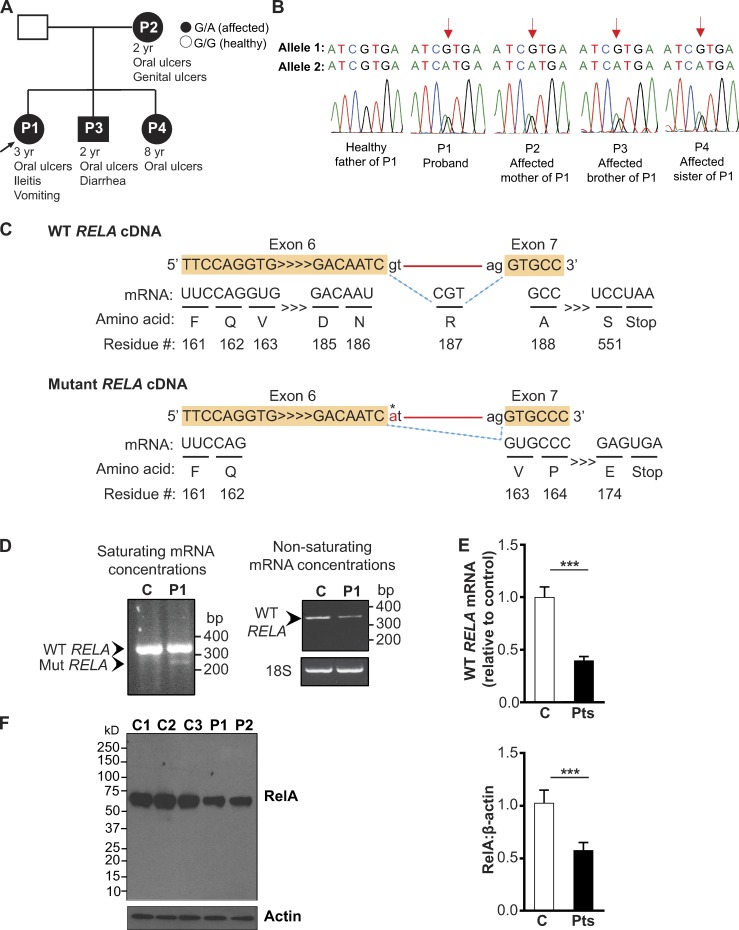
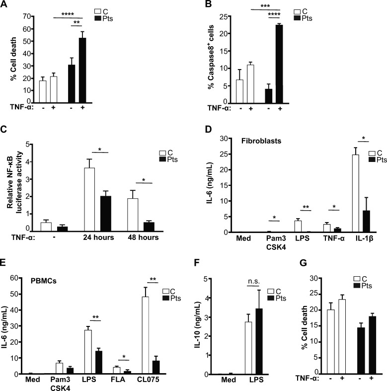
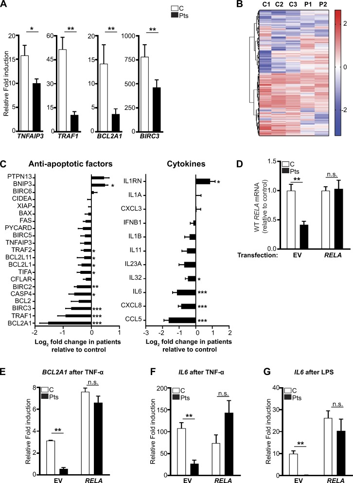
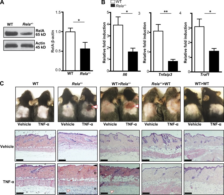
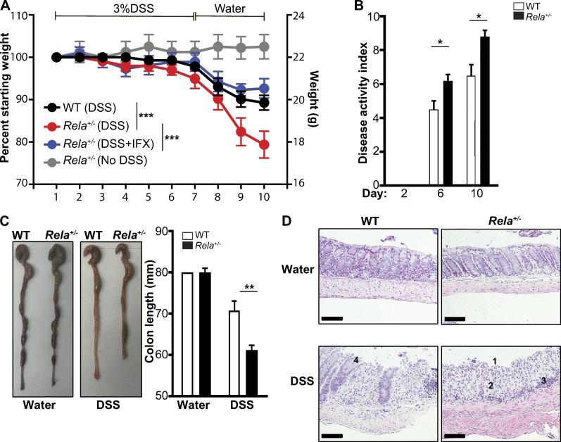
The treatment of chronic mucocutaneous ulceration is challenging, and only some patients respond selectively to inhibitors of tumor necrosis factor-α (TNF). TNF activates opposing pathways leading to caspase-8-mediated apoptosis as well as nuclear factor κB (NF-κB)-dependent cell survival. We investigated the etiology of autosomal-dominant, mucocutaneous ulceration in a family whose proband was dependent on anti-TNF therapy for sustained remission. A heterozygous mutation in RELA, encoding the NF-κB subunit RelA, segregated with the disease phenotype and resulted in RelA haploinsufficiency. The patients' fibroblasts exhibited increased apoptosis in response to TNF, impaired NF-κB activation, and defective expression of NF-κB-dependent antiapoptotic genes. Rela+/- mice have similarly impaired NF-κB activation, develop cutaneous ulceration from TNF exposure, and exhibit severe dextran sodium sulfate-induced colitis, ameliorated by TNF inhibition. These findings demonstrate an essential contribution of biallelic RELA expression in protecting stromal cells from TNF-mediated cell death, thus delineating the mechanisms driving the effectiveness of TNF inhibition in this disease.


Figures

Figure 1.
Figure 1.
Identification of a mutation in RELA that results in RelA haploinsufficiency. (A) Pedigree with RELA genotypes. (B) Sanger sequencing of the RELA mutation (arrow). (C) Schematic of WT and mutant RELA cDNA splicing (dotted blue lines) with the patients’ mutation (asterisk), interval nucleotides and aa (arrowheads), introns (red line). Alternative splicing of a cryptic splice site within exon 6 to the canonical acceptor splice site before exon 7 deletes 73 nucleotides at the 3′ of exon 6 and introduces a premature stop codon at residue 174. (D, left) RT-PCR, performed with saturating mRNA concentrations, identifies the alternatively spliced transcript (Mut RELA) in patient 1 (P1), but not in the control (Right) WT RELA transcript detected with RT-PCR under nonsaturating mRNA concentrations. Similar results were obtained in P2. Data are from one representative experiment of three independently performed. (E) WT RELA mRNA in P1 and P2 and three controls determined by qPCR using a primer complementary to the nucleotides in exon 6, specific to the WT RELA transcript. Gene expression was normalized to GAPDH. (F) Immunoblot of fibroblast lysates from controls (C1–C3) and patients (P1 and P2) using an N-terminal specific antibody against RelA, with densitometric quantification of RelA relative to β-actin. Data from E and F are pooled from three independent experiments. Columns and bars represent experiment means ± SEM. ***, P < 0.001, Student’s t test.
Figure 2.
Figure 2.
RelA haploinsufficiency is associated with impaired NF-κB signaling and increased sensitivity to TNF. (A) Cell death, determined by annexin-V and/or fixable viability-dye staining, after fibroblasts from three controls (C) and patients 1 and 2 (Pts) were treated with TNF for 24 h. (B) Percentage of cleaved caspase8+ cells among the annexin-V+ population after fibroblasts from three controls and patients 1 and 2 were treated with TNF for 24 h. (C) Relative NF-κB luciferase activity in fibroblasts from three controls and patients 1 and 2 after treatment with TNF. (D) IL-6 secretion from fibroblasts and (E) PBMCs from three controls and patients 1 and 2 in response to stimulation with Pam3CSK4 (TLR1/2 agonist), LPS (TLR4 agonist), flagellin (FLA, TLR5 agonist), CL075 (TLR7/8 agonist), TNF, or IL-1β. (F) IL-10 secretion after LPS stimulation of PBMCs from patients 1 and 2 and five controls. (G) Cell death in TNF-treated PBMCs from three controls and Patients 1 and 2. All data are pooled from 3 independent experiments. Columns and bars represent means ± SEM, n.s., not significant; *, P < 0.05; **, P < 0.01; ***, P < 0.001; ****, P < 0.0001; Student’s t test.
Figure 3.
Figure 3.
Decreased NF-κB regulated gene transcription in patient fibroblasts. (A) qPCR gene expression using TNF-treated fibroblasts from three controls (C) and two patients (Pts). (B) Heat map with unsupervised clustering showing the top 5% of genes with the most difference in TNF-treated fibroblasts from three controls and two patients, as determined by RNA-seq in one experiment. (C) Log2-fold change in the expression of indicated genes using RNA-seq data from two patients relative to three controls. (D) qPCR analysis of WT RELA expression in GFP+ fibroblasts from two controls and two patients after nucleofection with a vector encoding GFP or GFP-RELA. (E–G) RT-qPCR analysis of the expression of BCLA21 and IL6 in GFP+ fibroblasts from two controls and two patients after nucleofection with a vector encoding GFP or GFP-RELA and stimulated as indicated. Gene expression data in A and D–G were normalized to β-glucuronidase, expressed as the fold increase relative to unstimulated cells and were pooled from at least two independent experiments. Columns and bars represent means ± SEM. *, P < 0.05; **, P < 0.01; ***, P < 0.001; n.s., not significant.
Figure 4.
Figure 4.
RelA haploinsufficient mice develop cutaneous ulceration after TNF exposure. (A) Immunoblotting for RelA in WT and Rela+/− mice (n = 8 mice per genotype, pooled from three independent experiments). (B) qPCR analysis of NF-κB–regulated genes in WT and Rela+/− splenocytes after 24 h of TNF treatment. Gene expression was normalized to β-glucuronidase and expressed as the relative fold increase compared with unstimulated cells. Data are pooled from three independent experiments (n = 6 per genotype). (C, top) Photographs of shaved skin 24 h after s.c. injection of saline (vehicle) or TNF in the indicated genotypes. White arrows note ulceration. (Bottom) Hematoxylin and eosin stain of TNF-treated skin from WT and Rela+/− mice. The black arrow indicates loss of the epidermis. The white arrow indicates the inflammatory infiltrate. D, dermis; H, hypodermis. Bars, 500 µm. Data are from one representative experiment of three independently performed experiments. Means ± SEM are shown. *, P < 0.05; **, P < 0.01; Student’s t test.
Figure 5.
Figure 5.
RelA haploinsufficient mice develop significantly more severe DSS-induced colitis, which is ameliorated by TNF inhibition. (A) Weight loss in DSS-exposed WT, Rela+/−, and Rela+/− mice treated with infliximab. n = 5 mice per condition pooled from two independent experiments. ***, P < 0.001; two-way ANOVA. (B) DAI in DSS-treated WT and Rela+/− mice scores weight loss, stool consistency, and bleeding. n = 5 mice per condition pooled from two independent experiments. *, P < 0.05; Student’s t test. (C) Colon length of DSS-treated WT and Rela+/− mice at day 10. **, P < 0.01; Student’s t test. (D) Hematoxylin and eosin stain of colonic sections from DSS-treated WT and Rela+/− mice on day 10. Numbers show the following: 1, severe ulceration; 2, loss of crypt architecture; 3, inflammatory infiltrate; and 4, mild crypt dropout with sparse inflammation. Bars, 500 µm. Data in C and D are from one representative experiment of two independently performed. Bars represent means ± SEM.

References

    1. Alcamo E., Mizgerd J.P., Horwitz B.H., Bronson R., Beg A.A., Scott M., Doerschuk C.M., Hynes R.O., and Baltimore D.. 2001. Targeted mutation of TNF receptor I rescues the RelA-deficient mouse and reveals a critical role for NF-κB in leukocyte recruitment. J. Immunol. 167:1592–1600. 10.4049/jimmunol.167.3.1592 - DOI - PubMed
    1. Alex P., Zachos N.C., Nguyen T., Gonzales L., Chen T.-E., Conklin L.S., Centola M., and Li X.. 2009. Distinct cytokine patterns identified from multiplex profiles of murine DSS and TNBS-induced colitis. Inflamm. Bowel Dis. 15:341–352. 10.1002/ibd.20753 - DOI - PMC - PubMed
    1. Atreya I., Atreya R., and Neurath M.F.. 2008. NF-kappaB in inflammatory bowel disease. J. Intern. Med. 263:591–596. 10.1111/j.1365-2796.2008.01953.x - DOI - PubMed
    1. Beg A.A., Sha W.C., Bronson R.T., Ghosh S., and Baltimore D.. 1995. Embryonic lethality and liver degeneration in mice lacking the RelA component of NF-kappa B. Nature. 376:167–170. 10.1038/376167a0 - DOI - PubMed
    1. Boisson B., Laplantine E., Prando C., Giliani S., Israelsson E., Xu Z., Abhyankar A., Israël L., Trevejo-Nunez G., Bogunovic D., et al. 2012. Immunodeficiency, autoinflammation and amylopectinosis in humans with inherited HOIL-1 and LUBAC deficiency. Nat. Immunol. 13:1178–1186. 10.1038/ni.2457 - DOI - PMC - PubMed

Publication types

MeSH terms

Associated data