. 2017 Jun 9;7(1):3170.
doi: 10.1038/s41598-017-03460-y.

Amphiregulin contained in NSCLC-exosomes induces osteoclast differentiation through the activation of EGFR pathway

Affiliations

Amphiregulin contained in NSCLC-exosomes induces osteoclast differentiation through the activation of EGFR pathway

Simona Taverna et al. Sci Rep. .

Abstract

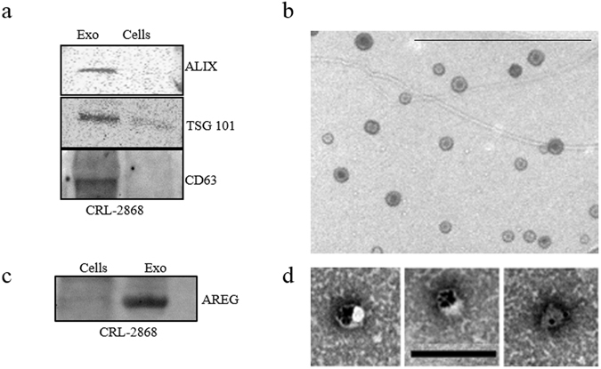
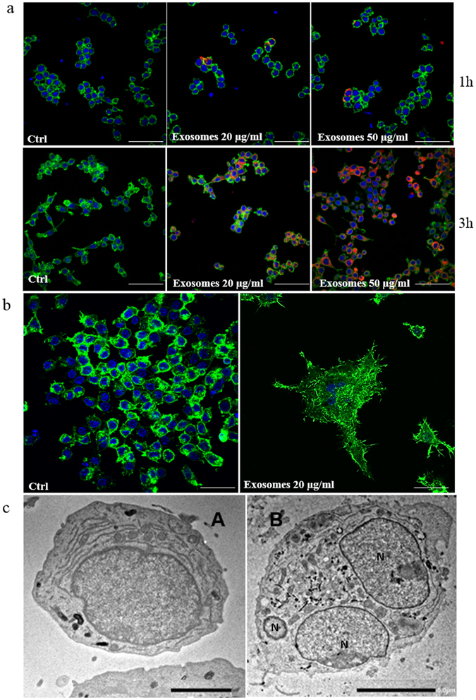
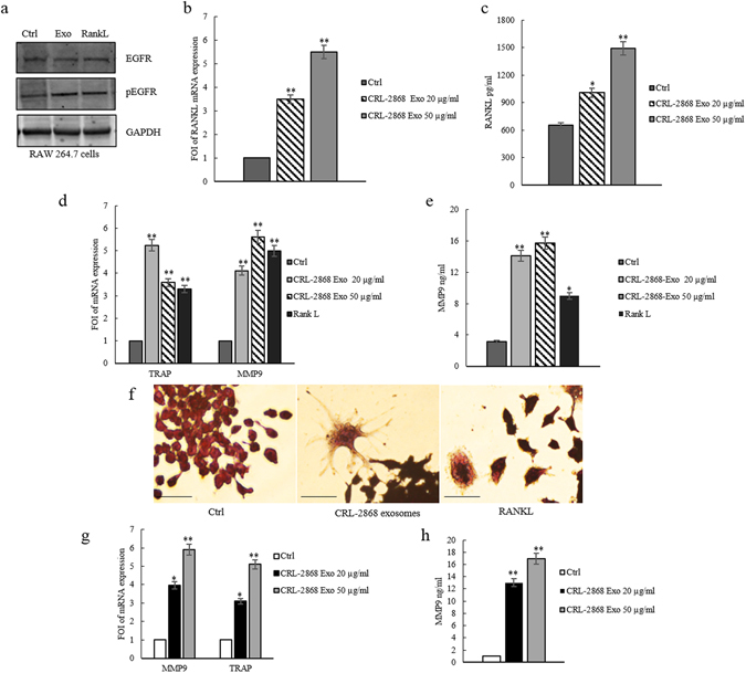
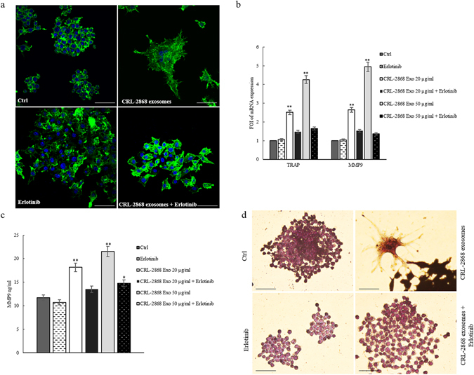
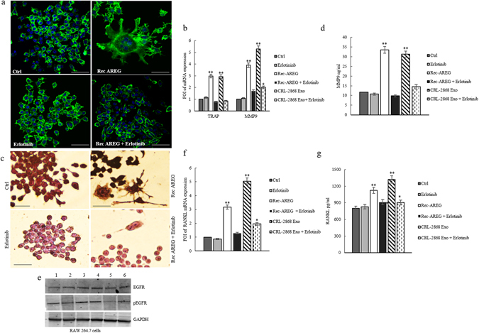
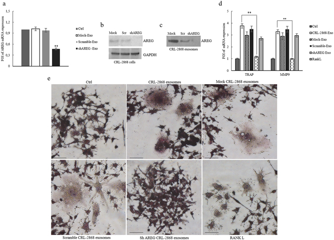
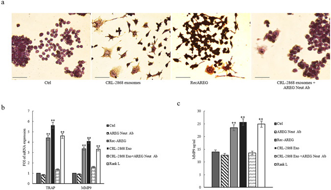
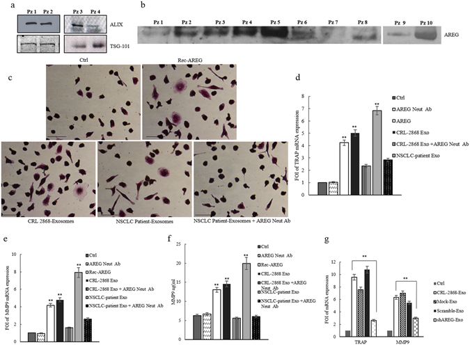
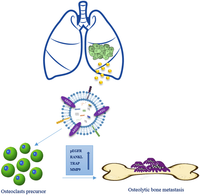
Non-small cell lung cancer (NSCLC) remains the leading cause of cancer-related deaths worldwide. The majority of patients are diagnosed in advanced disease stage. Bone metastasis is the most frequent complication in NSCLC resulting in osteolytic lesions. The perfect balance between bone-resorbing osteoclasts and bone-forming osteoblasts activity is lost in bone metastasis, inducing osteoclastogenesis. In NSCLC, the epidermal growth factor receptor (EGFR) pathway is constitutively activated. EGFR binds Amphiregulin (AREG) that is overexpressed in several cancers such as colon, breast and lung. Its levels in plasma of NSCLC patients correlate with poor prognosis and AREG was recently found as a signaling molecule in exosomes derived from cancer cell lines. Exosomes have a key role in the cell-cell communication and they were recently indicated as important actors in metastatic niche preparation. In the present work, we hypothesize a role of AREG carried by exosomes derived from NSCLC in bone metastasis induction. We observed that NSCLC-exosomes, containing AREG, induce EGFR pathway activation in pre-osteoclasts that in turn causes an increased expression of RANKL. RANKL is able to induce the expression of proteolytic enzymes, well-known markers of osteoclastogenesis, triggering a vicious cycle in osteolytic bone metastasis.


Conflict of interest statement

The authors declare that they have no competing interests.

Figures

Figure 1
Figure 1
CRL-2868 Exosomes characterization (a) Detection by western blotting of ALIX, TSG 101 and CD63 in 30 µg of exosomes purified conditioned medium of CRL-2868 cells compared to 30 µg of parental cells whole lysate. Original uncropped WBs were reported in Figure S5A. (b) Representative negative EM image of exosomes released by CRL-2868 cells. Scale bar 500 nm. (c) Detection by western blotting of AREG in 30 µg of CRL-2868 exosomes compared to 30 µg of parental cells whole lysate. Original uncropped WBs were reported in Figure S5B. (d) Gold labelling immune-electron microscopy and negative staining indicating the presence of AREG antigen on exosomes. Scale bar 200 nm.
Figure 2
Figure 2
CRL-2868 exosomes are internalized by RAW 264.7 cells and induce preosteoclasts morphological differentiation. (a) Confocal microscopy analysis of RAW 264.7 cells treated, for 1 and 3 hours, with 20 μg/ml (Exosomes 20 μg/ml) and 50 μg/ml (Exosomes 50 μg/ml) of CRL-2868 exosomes, compared to untreated RAW 264.7 cells (Ctrl). RAW 264.7 were stained with ActinGreen (green), nuclear counterstaining was performed using Hoescht (blue); exosomes were labelled with PKH26 (red). Scale bar 50 µm. (b) Confocal microscopy analysis of RAW 264.7 cells treated, for 6 days with 20 μg/ml (Exosomes 20 μg/ml) of CRL-2868 exosomes, compared to untreated RAW 264.7 cells (Ctrl). Scale bar 10 µm. (c) Comparative morphological features of pre-osteoclasts grown in the absence (A) and presence (B) of CRL-2868 exosomes and analyzed by TEM. Scale bars: A = 2 µm; B = 5 µm.
Figure 3
Figure 3
CRL-2868 exosomes induce the activation of EGFR pathway and osteoclast markers gene expression. (a) Western blotting analysis of pEGFR and EGFR in whole lysate of RAW 264.7 cells treated, for 6 days, with 20 μg/ml CRL-2868 exosomes (Exo) and RANKL (positive control) compared to untreated cells (Ctrl). Original uncropped WBs were reported in Figure S5C. GAPDH was used as loading control. (b) Evaluation by quantitative Real Time PCR of mRNA RANKL expression in RAW 264.7 cells treated, for 6 days, with 20 and 50 μg/ml CRL-2868 exosomes. (c) msRANKL protein levels assessed by ELISA, in RAW 264.7 cells treated, for 6 days, with 20 and 50 μg/ml CRL-2868 exosomes. (d) Evaluation by quantitative Real Time PCR of mRNA expression of TRAP and MMP9 in RAW 264.7 cells treated, for 6 days, with 20–50 μg/ml CRL-2868 exosomes and RANKL (positive control). (e) MMP9 protein level assessed by ELISA, in RAW 264.7 cells treated, for 6 days, with 20–50 μg/ml CRL-2868 exosomes and RANKL. Values are the mean ± SD of 3 three independent experiments *p ≤ 0.05, **p ≤ 0.01. (f) TRAP staining of RAW 264.7 cells incubated with CRL-2868 exosomes and 25 ng/ml of RANKL (positive control), for 6 days, compared with untreated cells. Scale bar 10 µm. (g) Evaluation by quantitative Real Time PCR of mRNA expression of TRAP and MMP9 in human primary pre-osteoclasts treated, for 4 days, with 20–50 μg/ml of CRL-2868 exosomes. (h) MMP9 protein levels assessed by ELISA, in human primary pre-osteoclasts treated, for 4 days, with 20–50 μg/ml CRL-2868 exosomes. Values are the mean ± SD in three independent experiments *p ≤ 0.05, **p ≤ 0.01.
Figure 4
Figure 4
Erlotinib reverts the effects of NSCLC-exosomes in osteoclast differentiation. (a) Confocal microscopy analysis of RAW 264.7 cells treated, for 6 days with: CRL-2868 exosomes, Erlotinib and CRL-2868 exosomes plus Erlotinib (CRL-2868 exosomes + Erlotinib) compared with RAW 264.7 control (Ctrl). Scale bar 10 µm. (b) Evaluation by quantitative Real Time PCR of mRNA expression of TRAP and MMP9 in RAW 264.7 cells treated, for 6 days, with: 20–50 μg/ml of CRL-2868 exosomes, Erlotinib and CRL-2868 exosomes plus Erlotinib. (c) MMP9 protein levels assessed by ELISA, in RAW 264.7 cells treated, for 6 days, with: 20–50 μg/ml of CRL-2868 exosomes, Erlotinib and CRL-2868 exosomes plus Erlotinib. Values are the mean ± SD of three independent experiments *p ≤ 0.05, **p ≤ 0.01. (d) TRAP staining of RAW 264.7 cells incubated with: CRL-2868 exosomes, Erlotinib, and CRL-2868 exosomes plus Erlotinib, for six days, compared with untreated RAW 264.7 cells. Scale bar 10 µm.
Figure 5
Figure 5
Rec-AREG induces osteoclast differentiation. (a) Confocal microscopy analysis of RAW 264.7 cells treated, for 6 days with: CRL-2868 exosomes, Rec-AREG and Rec-AREG plus Erlotinib (Rec-AREG + Erlotinib) compared with untreated RAW 264.7 cells (Ctrl). Scale bar 10 µm. (b) Evaluation by quantitative Real Time PCR of mRNA expression of TRAP and MMP9 in RAW 264.7 cells treated, for 6 days, with: 20–50 μg/ml of CRL-2868 exosomes, Rec-AREG and Rec-AREG plus Erlotinib (Rec-AREG + Erlotinib). (c) TRAP staining of RAW 264.7 cells incubated with: CRL-2868 exosomes, Rec-AREG, Erlotinib, CRL-2868 exosomes plus Erlotinib (CRL-2868 Exo + Erlotinib), Rec-AREG plus Erlotinib (Rec-AREG + Erlotinib), for 6 days, compared with untreated RAW 264.7 cells. (d) MMP9 protein level assessed by ELISA, in RAW 264.7 cells treated, for 6 days, with: CRL-2868 exosome, Rec-AREG, CRL-2868 exosomes plus Erlotinib (CRL-2868 Exo + Erlotinib), Rec-AREG plus Erlotinib (Rec-AREG + Erlotinib). Values are the mean ± SD of three independent experiments *p ≤ 0.05, **p ≤ 0.01. (e) Western blotting analysis of pEGFR and EGFR in whole lysate of RAW 264.7 cells treated, for 6 days, with: Erlotinib (2), Rec-AREG (3), CRL-2868 exosomes (4), Rec-AREG plus Erlotinib (5), CRL-2868 exosomes plus Erlotinib (6) compared to untreated cells (1). GAPDH was used as loading control Original uncropped WBs were reported in Figure S5D. (f) Evaluation by quantitative Real Time PCR of mRNA expression of mRNA RANKL expression in RAW 264.7 cells treated, for 6 days, with: CRL-2868 exosomes, Rec-AREG, CRL-2868 exosomes plus Erlotinib (CRL-2868 Exo + Erlotinib), Rec-AREG plus Erlotinib (Rec-AREG + Erlotinib). (g) RANKL protein levels assessed by ELISA, in RAW 264.7 cells treated, for 6 days, with: CRL-2868 exosomes, Rec-AREG, CRL-2868 exosomes plus Erlotinib (CRL-2868 Exo + Erlotinib), Rec-AREG plus Erlotinib (Rec-AREG + Erlotinib). Values are the mean ± SD of 3 three independent experiments *p ≤ 0.05, **p ≤ 0.01.
Figure 6
Figure 6
Knockdown of AREG in CRL-2868 cells. (a) Evaluation by real time PCR analysis of mRNA expression of AREG in CRL-2868 cells transfected with AREG shRNA plasmid (AREG-knockdown CRL-2868 cells), empty vector (Mock-CRL-2868 cells) and control AREG shRNA plasmid (Scramble-CRL-2868 cells). (b) Western blotting of AREG in whole lysate of Mock, Scramble, and AREG-knockdown CRL-2868 cells. GAPDH was used as loading control. Original uncropped WBs were reported in Figure S5E. (c) Western blotting of AREG in exosomes released by Mock, Scramble, and AREG-knockdown CRL-2868 cells. Original uncropped WBs were reported in Figure S5E. (d) Evaluation by real time PCR analysis of mRNA expression of TRAP and MMP9 in RAW 264.7 cells treated, for 6 days, with exosomes released by Control, Mock, Scramble, and AREG-knockdown CRL-2868 cells and RANKL. Values are the mean ± SD of three independent experiments *p ≤ 0.05, **p ≤ 0.01. (e) RAW 264.7 cells were incubated with exosomes released by Control, Mock, Scramble, and AREG-knockdown CRL-2868 cells and RANKL, for 6 days, stained for TRAP and compared with untreated cells (Ctrl).
Figure 7
Figure 7
Neutralizing AREG antibodies revert osteoclast differentiation induced by NSCLC-exosomes. (a) TRAP staining of RAW 264.7 cells incubated with: CRL-2868 exosomes, Rec-AREG, CRL-2868 exosomes plus AREG neutralizing antibodies, for 6 days, stained for TRAP and compared with untreated cells (Ctrl). Scale bar 10 µm. (b) Evaluation by real Time PCR analysis of mRNA expression of TRAP and MMP9 in RAW 264.7 cells treated, for 6 days, with: CRL-2868 exosomes, Rec-AREG, AREG neutralizing antibodies, RANKL, CRL-2868 exosomes plus AREG neutralizing antibodies. (c) MMP9 protein levels assessed by ELISA, in RAW 264.7 cells treated, for 6 days, with: CRL-2868 exosomes, Rec-AREG, AREG neutralizing antibodies, RANKL, CRL-2868 exosomes plus AREG neutralizing antibodies. Values are the mean ± SD of three independent experiments *p ≤ 0.05, **p ≤ 0.01.
Figure 8
Figure 8
Exosomes from NSCLC plasma patients modulate osteoclastogenesis in human primary osteoclasts. (a) Representative western blotting of ALIX and TSG 101 in 30 µg of exosomes isolated from plasma (1, 5 ml) of four NSCLC patients (Pz 1:stage I, Pz 2:stage IB, Pz 3:stage IIIA, Pz 4:stage IIIA). Original uncropped WBs were reported in Figure S5F. (b) Representative western blotting of AREG in 30 µg of exosomes isolated from plasma of ten NSCLC patients at different disease stages (Pz 1:stage I, Pz 2:stage IB, Pz 3:stage III, Pz 4:stage IIIA, Pz 5:stage IV, Pz 6:stage IV, Pz 7:stage IV, Pz 8:stage IV, Pz 9:stage IV, Pz 10:stage IV). Original uncropped WBs were reported in Figure S5G. (c) TRAP staining of human pOCs cultured in differentiation medium and incubated with: REC-AREG, CRL-2868 exosomes, NSCLC-patient exosomes, NSCLC-patient exosomes plus AREG neutralizing antibodies. Scale bar 10 µm. Evaluation by Real Time PCR of TRAP (d) and MMP9 (e) mRNA expression, in primary human pre-osteoclasts, treated for 4 days, with: CRL-2868 exosomes, AREG neutralizing antibodies, NSCLC patient exosomes, CRL-2868 exosomes plus AREG neutralizing antibodies, NSCLC patients exosomes plus AREG neutralizing antibodies. (f) MMP9 protein levels assessed by ELISA, in primary human pre-osteoclasts treated for 4 days with: CRL-2868 exosomes, AREG neutralizing antibodies, NSCLC patient exosomes, CRL-2868 exosomes plus AREG neutralizing antibodies, NSCLC patients exosomes plus AREG neutralizing antibodies. (g) Evaluation by real time PCR analysis of mRNA expression of TRAP and MMP9 in primary human pre-osteoclasts treated for 4 days with: exosomes released by Control, Mock, Scramble, and AREG-knockdown CRL-2868 cells. Values are the mean ± SD of three independent experiments *p ≤ 0.05, **p ≤ 0.01.
Figure 9
Figure 9
Working hypothesis of the effects of NSCLC exosomes in osteoclast differentiation. CRL-2868 cells released exosomes enriched in AREG that activating EGFR pathway in a pre-osteoclast model induced morphological differentiation and RANKL expression at mRNA and protein levels that in turn is able to increase the expression of TRAP and MMP9 well-known markers of osteoclastogenesis.

Similar articles

Cited by

References

    1. Sugiura H, Yamada K, Sugiura T, Hida T, Mitsudomi T. Predictors of survival in patients with bone metastasis of lung cancer. Clin Orthop Relat Res. 2008;466:729–736. doi: 10.1007/s11999-007-0051-0. - DOI - PMC - PubMed
    1. Riess JW, Wakelee HA. Metastatic non-small cell lung cancer management: novel targets and recent clinical advances. Clin Adv Hematol Oncol. 2012;10:226–234. - PubMed
    1. Perisano C, et al. Soft tissue metastases in lung cancer: a review of the literature. Eur Rev Med Pharmacol Sci. 2012;16:1908–1914. - PubMed
    1. Tsuya A, Kurata T, Tamura K, Fukuoka M. Skeletal metastases in non-small cell lung cancer: a retrospective study. Lung Cancer. 2007;57:229–232. doi: 10.1016/j.lungcan.2007.03.013. - DOI - PubMed
    1. Roato, I. et al. Spontaneous osteoclastogenesis is a predictive factor for bone metastases from non-small cell lung cancer. Lung Cancer61, doi:10.1016/j.lungcan.2007.10.016 (2008). - PubMed

Publication types

MeSH terms