

Real-time imaging of intestinal bacterial β-glucuronidase activity by hydrolysis of a fluorescent probe
- PMID: 28600512
- PMCID: PMC5466677
- DOI: 10.1038/s41598-017-03252-4
Real-time imaging of intestinal bacterial β-glucuronidase activity by hydrolysis of a fluorescent probe
Abstract
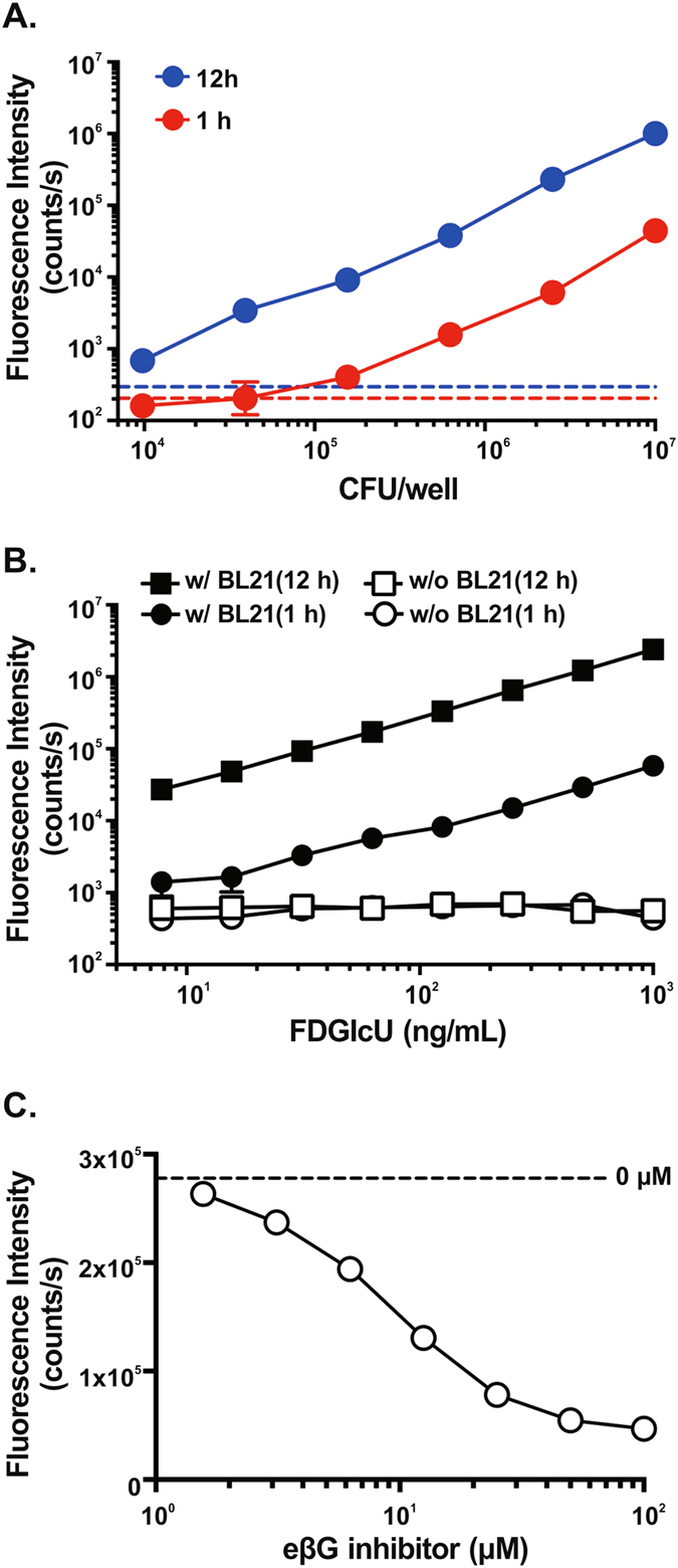
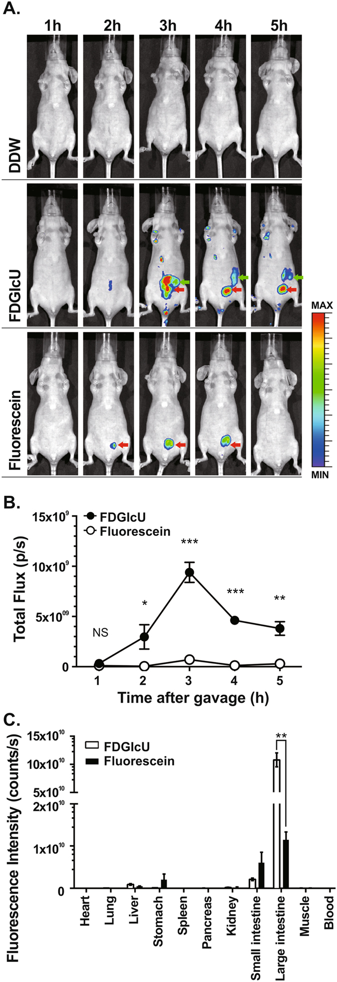
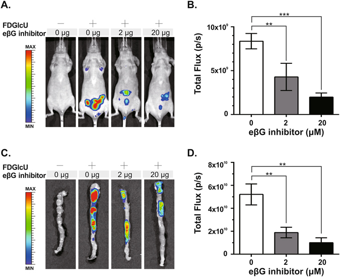
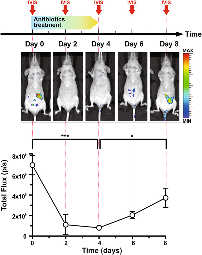
Intestinal bacterial β-glucuronidase (βG) hydrolyzes glucuronidated metabolites to their toxic form in intestines, resulting in intestinal damage. The development of a method to inhibit βG is thus important but has been limited by the difficulty of directly assessing enzyme activity in live animals. Here, we utilized a fluorescent probe, fluorescein di-β-D-glucuronide (FDGlcU), to non-invasively image the intestinal bacterial βG activity in nude mice. In vitro cell-based assays showed that the detection limit is 104 colony-forming units/well of βG-expressing bacteria, and that 7.81 ng/mL of FDGlcU is enough to generate significant fluorescent signal. In whole-body optical images of nude mice, the maximum fluorescence signal for βG activity in intestines was detected 3 hours after gavage with FDGlcU. Following pretreatment with a bacterial βG inhibitor, the fluorescence signal was significantly reduced in abdomens and excised intestines images. For a 4-day antibiotic treatment to deplete intestinal bacteria, the FDGlcU-based images showed that the βG activity was decreased by 8.5-fold on day 4 and then gradually increased after treatment stopped. The results suggested that FDGlcU-based imaging revealed the in vitro and in vivo activity of intestinal bacterial βG, which would facilitate pharmacodynamic studies of specific bacterial βG inhibitors in animal studies.
Conflict of interest statement
The authors declare that they have no competing interests.
Figures
Similar articles
-
Gene expression imaging by enzymatic catalysis of a fluorescent probe via membrane-anchored beta-glucuronidase.Gene Ther. 2007 Apr;14(7):565-74. doi: 10.1038/sj.gt.3302896. Epub 2007 Jan 18. Gene Ther. 2007. PMID: 17235292
-
N'-Fluorescein-N''-[4-O-(β-d-glucopyranuronic acid)-3-difluoromethylphenyl]-S-methylthiourea (FITC-TrapG) and N'-(p-aminophenyl)thioether of IR-820-N''-[4-O-(β-d-glucopyranuronic acid)-3-difluoromethylphenyl]-S-methylthiourea (NIR-TrapG).2012 Apr 10 [updated 2012 May 10]. In: Molecular Imaging and Contrast Agent Database (MICAD) [Internet]. Bethesda (MD): National Center for Biotechnology Information (US); 2004–2013. 2012 Apr 10 [updated 2012 May 10]. In: Molecular Imaging and Contrast Agent Database (MICAD) [Internet]. Bethesda (MD): National Center for Biotechnology Information (US); 2004–2013. PMID: 22593947 Free Books & Documents. Review.
-
Impediments to enhancement of CPT-11 anticancer activity by E. coli directed beta-glucuronidase therapy.PLoS One. 2015 Feb 17;10(2):e0118028. doi: 10.1371/journal.pone.0118028. eCollection 2015. PLoS One. 2015. PMID: 25688562 Free PMC article.
-
PET imaging of β-glucuronidase activity by an activity-based 124I-trapping probe for the personalized glucuronide prodrug targeted therapy.Mol Cancer Ther. 2014 Dec;13(12):2852-63. doi: 10.1158/1535-7163.MCT-14-0212. Epub 2014 Oct 2. Mol Cancer Ther. 2014. PMID: 25277385
-
Intestinal bacterial β-glucuronidase as a possible predictive biomarker of irinotecan-induced diarrhea severity.Pharmacol Ther. 2019 Jul;199:1-15. doi: 10.1016/j.pharmthera.2019.03.002. Epub 2019 Mar 1. Pharmacol Ther. 2019. PMID: 30831128 Review.
Cited by
-
Gastrointestinal tract and neuroendocrine system responses of young turkeys to the early administration of antibiotics or feeding a diet containing a coccidiostat.Poult Sci. 2022 Nov;101(11):102098. doi: 10.1016/j.psj.2022.102098. Epub 2022 Aug 2. Poult Sci. 2022. PMID: 36087440 Free PMC article.
-
EGFR monoclonal antibody panitumumab inhibits chronic proliferative cholangitis by downregulating EGFR.Int J Mol Med. 2019 Jul;44(1):79-88. doi: 10.3892/ijmm.2019.4190. Epub 2019 May 9. Int J Mol Med. 2019. PMID: 31115490 Free PMC article.
-
Vancomycin relieves tacrolimus-induced hyperglycemia by eliminating gut bacterial beta-glucuronidase enzyme activity.Gut Microbes. 2024 Jan-Dec;16(1):2310277. doi: 10.1080/19490976.2024.2310277. Epub 2024 Feb 8. Gut Microbes. 2024. PMID: 38332701 Free PMC article.
-
Recent progress in the imaging detection of enzyme activities in vivo.RSC Adv. 2019 Aug 13;9(44):25285-25302. doi: 10.1039/c9ra04508b. eCollection 2019 Aug 13. RSC Adv. 2019. PMID: 35530057 Free PMC article. Review.
-
Effect of Chromium Nanoparticles and Switching from a High-Fat to a Low-Fat Diet on the Cecal Microenvironment in Obese Rats.Nutrients. 2023 Jul 12;15(14):3118. doi: 10.3390/nu15143118. Nutrients. 2023. PMID: 37513536 Free PMC article.
References
-
- Kehrer DF, et al. Modulation of irinotecan-induced diarrhea by cotreatment with neomycin in cancer patients. Clin. Cancer Res. 2001;7:1136–1141. - PubMed
Publication types
MeSH terms
Substances
LinkOut - more resources
Full Text Sources
Other Literature Sources
