

Real-time imaging of intestinal bacterial β-glucuronidase activity by hydrolysis of a fluorescent probe
- PMID: 28600512
- PMCID: PMC5466677
- DOI: 10.1038/s41598-017-03252-4
Real-time imaging of intestinal bacterial β-glucuronidase activity by hydrolysis of a fluorescent probe
Abstract
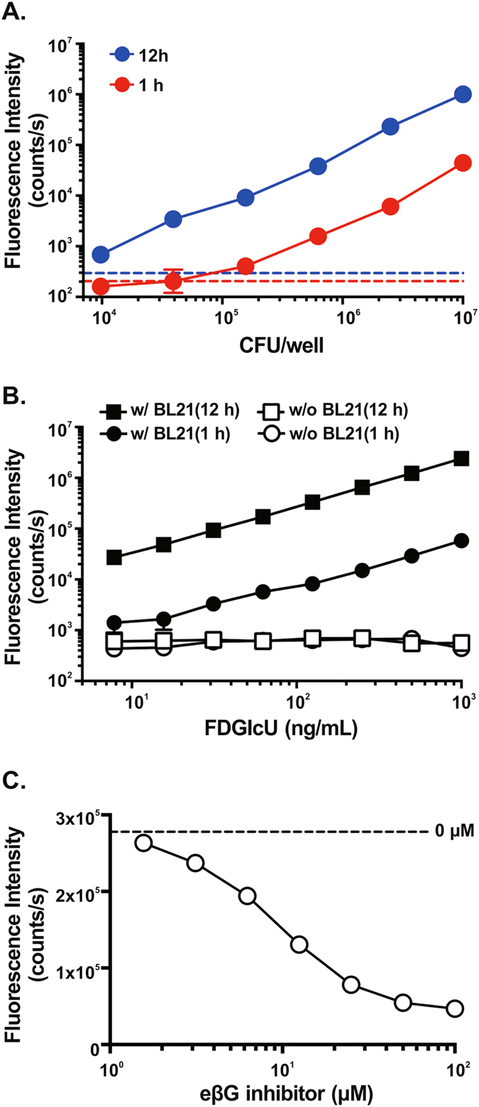
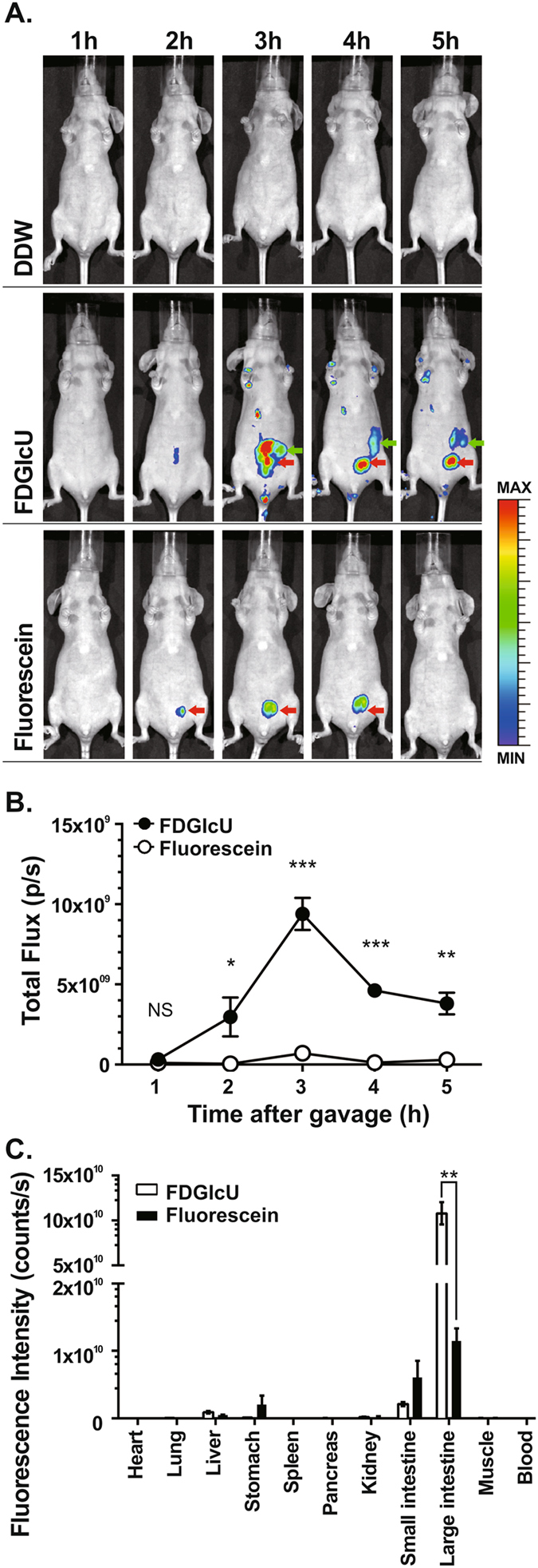
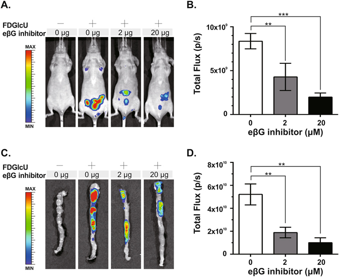
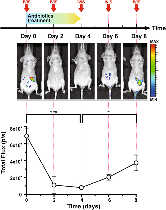
Intestinal bacterial β-glucuronidase (βG) hydrolyzes glucuronidated metabolites to their toxic form in intestines, resulting in intestinal damage. The development of a method to inhibit βG is thus important but has been limited by the difficulty of directly assessing enzyme activity in live animals. Here, we utilized a fluorescent probe, fluorescein di-β-D-glucuronide (FDGlcU), to non-invasively image the intestinal bacterial βG activity in nude mice. In vitro cell-based assays showed that the detection limit is 104 colony-forming units/well of βG-expressing bacteria, and that 7.81 ng/mL of FDGlcU is enough to generate significant fluorescent signal. In whole-body optical images of nude mice, the maximum fluorescence signal for βG activity in intestines was detected 3 hours after gavage with FDGlcU. Following pretreatment with a bacterial βG inhibitor, the fluorescence signal was significantly reduced in abdomens and excised intestines images. For a 4-day antibiotic treatment to deplete intestinal bacteria, the FDGlcU-based images showed that the βG activity was decreased by 8.5-fold on day 4 and then gradually increased after treatment stopped. The results suggested that FDGlcU-based imaging revealed the in vitro and in vivo activity of intestinal bacterial βG, which would facilitate pharmacodynamic studies of specific bacterial βG inhibitors in animal studies.
Conflict of interest statement
The authors declare that they have no competing interests.
Figures
References
-
- Kehrer DF, et al. Modulation of irinotecan-induced diarrhea by cotreatment with neomycin in cancer patients. Clin. Cancer Res. 2001;7:1136–1141. - PubMed
Publication types
MeSH terms
Substances
LinkOut - more resources
Full Text Sources
Other Literature Sources
