. 2017 Jun 9;7(1):3143.
doi: 10.1038/s41598-017-03244-4.

REG3β modifies cell tumor function by impairing extracellular vesicle uptake

Affiliations

REG3β modifies cell tumor function by impairing extracellular vesicle uptake

Laia Bonjoch et al. Sci Rep. .

Abstract

Extracellular vesicles (EVs), including exosomes and microvesicles, are nano-sized membrane vesicles containing proteins and nucleic acids, which act as intercellular messengers. They play an important role in a variety of physiological processes, as well as in pathological situations such as inflammation or cancer. Here, we show that in the case of pancreatic ductal adenocarcinoma (PDAC), the healthy pancreatic tissue surrounding the tumor releases REG3β, a lectin that binds to the glycoproteins present in the surface of EVs, thus interfering with their uptake and internalization by target cells. In vitro, the disruption of the signaling mediated by EVs due to the presence of REG3β, prevents the EV-induced phenotypic switch in macrophages, inhibits the increased cell migration of cancer cells and reverses a number of metabolomic changes promoted by EVs. In vivo, the uptake of REG3β+ EVs by tumor cells is significantly impaired. Furthermore, it results in an increase of circulating REG3β+ EVs in blood of pancreatic cancer patients. Our findings highlight the effect of a lectin released by the healthy pancreatic tissue surrounding the tumor in modulating the EV-mediated interactions between different cell types in PDAC.


Figures

Figure 1
Figure 1
REG3β inhibits the uptake of EVs both in vitro and in vivo. (a) Transmission electron microscopy images of 120,000 × g pelleted THP1-EVs and MPC-EVs. 2x magnification in the lower right corner to appreciate the double membrane. Scale bars: 200 nm. (b) Representative Western blot of EVs samples and cell lysates to confirm the presence of classical exosome markers (CD81, ALIX, TSG101) and the absence of endoplasmic reticulum contamination (Calnexin). (c,d) Fluorescence microscopy of THP-1 macrophages (c) and MIA PaCa-2 cells (MPC) (d) incubated, respectively, with 3 µg/ml of PKH26-labeled MPC EVs or THP1-EVs and increasing concentrations of REG3β. Nuclei counterstained with DAPI. On the right, quantification of the amount of EVs internalization via fluorimetric reading (n = 4). Data are expressed as mean ± SEM. *P < 0.05, **P < 0.01, ***P < 0.001, compared to 0 ng/ml REG3β. ANOVA with Tukey’s post-test was used to calculate P-values. Scale bars: 50 µm. (e) PKH26-labeled EVs injected into tumor xenografts were uptaken by tumor cells (−REG3β) but remained in the intercellular space when pretreated with REG3β (+REG3β). Nuclei counterstained with DAPI. 4x magnification in the top right corner. Scale bars: 50 µm.
Figure 2
Figure 2
REG3β interacts with EVs through its lectin domain. (a) PKH26-labeled THP1-EVs binding to plates coated with anti-REG3β antibody and saturated with 500 ng/ml of REG3β. The indicated concentrations of EVs were used. Non-specific binding was assessed in the absence of REG3β. Data are depicted as relative expression to saturation (5 ng/µl of EV) (n = 4). (b,c) PKH26-labeled EVs (1 ng/µl of EV protein) binding to plates (b) or magnetic beads (c) coated with anti-REG3β antibody and incubated with the indicated concentrations of REG3β. Data are depicted as relative expression to maximum binding (500 ng/ml of REG3β). (d) Anti-REG3β immunogold labeling of THP1-EVs incubated with REG3β (500 ng/ml). Non-specific binding was tested in the absence of REG3β. Scale bar: 200 nm. (e) Competitive assay of PKH26-labeled THP1-EVs binding to REG3β as in (B), but in the presence of 1 mg/ml mannose, 1 mg/ml mannan or 5 mM of NAG. (n = 4). ***P < 0.001 compared to non-sugar binding (F test). In all panels, data are expressed as mean ± SEM. R 2 represents goodness of fit to the one-site binding hyperbola model.
Figure 3
Figure 3
EVs blocked by REG3β lose their capability to modulate some cell functions. (a) qPCR analysis of different pro-inflammatory (IL-1β, IL-8, CCL2, CXCL2) and anti-inflammatory/regulatory (MRC-1, TGFβ) markers on THP-1 macrophages treated with 500 ng/ml of REG3β, and 3 µg/ml of MPC-EVs (EV) or REG3β-blocked MPC-EVs (EV-REG3β) for 24 h (n = 6). Data are depicted as relative expression to the GAPDH housekeeping gene. (b) Scratch-wound healing assay of MIA PaCa-2 cells incubated with 500 ng/ml of REG3β, and 3 µg/ml of THP1-EVs (EV) or REG3β-blocked EVs (EV-REG3β) for 24 h (n = 6). Down, quantification of cell migration. Scale bars: 200 µm. In all panels, data are expressed as mean ± SEM. ANOVA with Tukey’s post-test was used to calculate P-values. *P < 0.05, **P < 0.01, ***P < 0.001 compared to non-treated (C), + P < 0.05, ++ P < 0.01, +++ P < 0.001 compared to EV.
Figure 4
Figure 4
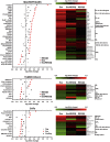
Impaired metabolic switch in the three main cell types of tumor microenvironment due to EV blockage by REG3β. Metabolomic analysis of those metabolites which the REG3β-blocking of signal transduction through EVs prevented increases or decreases in their dysregulation. Heatmaps of metabolites grouped according to their chemical class in order to illustrate the most frequent altered families by this blocking mechanism. AA (aminoacids), AC (acylcarnitines), Carbs (carbohydrates), CBA (carboxylic acids), GL (glycerolipids), GPL (glycerophospholipids), NEFA (non-esterified fatty acids), ns (nucleosides), nt (nucleotides), Pu (purines), Pyr (pyrimidines), SL (sphingolipids).
Figure 5
Figure 5
REG3β+ EVs are released to bloodstream in PDAC patients. (a) Immunolocalization by immunohistochemistry of REG3β in human PDAC. REG3β is absent in tumor cells but strongly expressed by the healthy acinar cells surrounding the tumor. (b) Quantification of EV levels in plasma through a Bradford assay. Circulating EVs were isolated from samples of healthy donors (C) (n = 15) and PDAC patients (n = 15). Data are expressed as mean ± SEM. Student’s t-test used to calculate P-values. *P < 0.05. (c) Human PKH26-labeled EVs (1 ng/µl of EV protein) binding to plates coated with anti-REG3β antibody. Data are depicted as PDAC EVs binding levels relative to healthy donors samples. Significance determined using a two-tailed Mann–Whitney U test. ***P < 0.001. (d,e) Representative immunogold labeling of EVs from healthy donors (D) or PDAC patients (E) with anti-REG3β. Scale bars: 200 nm.

References

    1. Valadi H, et al. Exosome-mediated transfer of mRNAs and microRNAs is a novel mechanism of genetic exchange between cells. Nature cell biology. 2007;9:654–9. doi: 10.1038/ncb1596. - DOI - PubMed
    1. Tickner JA, Urquhart AJ, Stephenson S-A, Richard DJ, O’Byrne KJ. Functions and therapeutic roles of exosomes in cancer. Frontiers in oncology. 2014;4:127. doi: 10.3389/fonc.2014.00127. - DOI - PMC - PubMed
    1. Melo SA, et al. Glypican-1 identifies cancer exosomes and detects early pancreatic cancer. Nature. 2015;523:177–182. doi: 10.1038/nature14581. - DOI - PMC - PubMed
    1. Costa-Silva B, et al. Pancreatic cancer exosomes initiate pre-metastatic niche formation in the liver. Nature cell biology. 2015;17:816–826. doi: 10.1038/ncb3169. - DOI - PMC - PubMed
    1. Whiteside TL. Immune modulation of T-cell and NK (natural killer) cell activities by TEXs (tumour-derived exosomes) Biochemical Society transactions. 2013;41:245–51. doi: 10.1042/BST20120265. - DOI - PMC - PubMed

Publication types

Substances