Multi-layered silk film coculture system for human corneal epithelial and stromal stem cells
- PMID: 28600807
- PMCID: PMC5723569
- DOI: 10.1002/term.2499
Multi-layered silk film coculture system for human corneal epithelial and stromal stem cells
Abstract
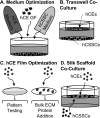
With insufficient options to meet the clinical demand for cornea transplants, one emerging area of emphasis is on cornea tissue engineering. In the present study, the goal was to combine the corneal stroma and epithelium into one coculture system, to monitor both human corneal stromal stem cell (hCSSC) and human corneal epithelial cell (hCE) growth and differentiation into keratocytes and differentiated epithelium in these three-dimensional tissue systems in vitro. Coculture conditions were first optimized, including the medium, air-liquid interface culture, and surface topography and chemistry of biomaterial scaffold films based on silk protein. The silk was used as scaffolding for both stromal and epithelial tissue layers because it is cell compatible, can be surface patterned, and is optically clear. Next, the effects of proliferating and differentiating hCEs and hCSSCs were studied in this in vitro system, including the effects on cell proliferation, matrix formation by immunochemistry, and gene expression by quantitative reverse transcription-polymerase chain reaction. The incorporation of both cell types into the coculture system demonstrated more complete differentiation and growth for both cell types compared to the corneal stromal cells and corneal epithelial cells alone. Silk films for corneal epithelial culture were optimized to combine a 4.0-μm-scale surface pattern with bulk-loaded collagen type IV. Differentiation of each cell type was in evidence based on increased expression of corneal stroma and epithelial proteins and transcript levels after 6 weeks in coculture on the optimized silk scaffolds.
Keywords: coculture; cornea; silk; tissue engineering.
Copyright © 2017 John Wiley & Sons, Ltd.
Figures


Similar articles
-
3D Functional Corneal Stromal Tissue Equivalent Based on Corneal Stromal Stem Cells and Multi-Layered Silk Film Architecture.PLoS One. 2017 Jan 18;12(1):e0169504. doi: 10.1371/journal.pone.0169504. eCollection 2017. PLoS One. 2017. PMID: 28099503 Free PMC article.
-
Substance P and patterned silk biomaterial stimulate periodontal ligament stem cells to form corneal stroma in a bioengineered three-dimensional model.Stem Cell Res Ther. 2017 Nov 13;8(1):260. doi: 10.1186/s13287-017-0715-y. Stem Cell Res Ther. 2017. PMID: 29132420 Free PMC article.
-
The Effect of Micro- and Nanoscale Surface Topographies on Silk on Human Corneal Limbal Epithelial Cell Differentiation.Sci Rep. 2019 Feb 6;9(1):1507. doi: 10.1038/s41598-018-37804-z. Sci Rep. 2019. PMID: 30728382 Free PMC article.
-
Characterization, isolation, expansion and clinical therapy of human corneal epithelial stem/progenitor cells.J Stem Cells. 2014;9(2):79-91. J Stem Cells. 2014. PMID: 25158157 Review.
-
[Corneal cell therapy-an overview].Ophthalmologe. 2017 Aug;114(8):705-715. doi: 10.1007/s00347-017-0454-6. Ophthalmologe. 2017. PMID: 28204869 Review. German.
Cited by
-
Silk Film Stiffness Modulates Corneal Epithelial Cell Mechanosignaling.Macromol Chem Phys. 2021 Apr;222(7):2170013. doi: 10.1002/macp.202170013. Epub 2021 Apr 5. Macromol Chem Phys. 2021. PMID: 34149247 Free PMC article.
-
Generation of Patterned Cocultures in 2D and 3D: State of the Art.ACS Omega. 2023 Sep 13;8(38):34249-34261. doi: 10.1021/acsomega.3c02713. eCollection 2023 Sep 26. ACS Omega. 2023. PMID: 37780002 Free PMC article. Review.
-
The Human Tissue-Engineered Cornea (hTEC): Recent Progress.Int J Mol Sci. 2021 Jan 28;22(3):1291. doi: 10.3390/ijms22031291. Int J Mol Sci. 2021. PMID: 33525484 Free PMC article. Review.
-
From hard tissues to beyond: Progress and challenges of strontium-containing biomaterials in regenerative medicine applications.Bioact Mater. 2025 Mar 6;49:85-120. doi: 10.1016/j.bioactmat.2025.02.039. eCollection 2025 Jul. Bioact Mater. 2025. PMID: 40124596 Free PMC article. Review.
-
Silk-Based Biomaterials for Designing Bioinspired Microarchitecture for Various Biomedical Applications.Biomimetics (Basel). 2023 Jan 28;8(1):55. doi: 10.3390/biomimetics8010055. Biomimetics (Basel). 2023. PMID: 36810386 Free PMC article. Review.
References
-
- America EBAo. 2014 Eye Banking Statistical Report. 2015 from http://www.restoresight.org/wp-content/uploads/2015/03/2014_Statistical_....
-
- Arai T, Freddi G, Innocenti R, et al. Biodegradation of Bombyx mori silk fibroin fibers and films. Journal of Applied Polymer Science. 2004;91:2383–2390.
-
- Biazar E, Baradaran-Rafii A, Heidari-keshel S, et al. Oriented nanofibrous silk as a natural scaffold for ocular epithelial regeneration. Journal of Biomaterials Science-Polymer Edition. 2015;26:1139–1151. - PubMed
Publication types
MeSH terms
Substances
Grants and funding
LinkOut - more resources
Full Text Sources
Other Literature Sources
Medical

