. 2017 Oct:13:255-265.
doi: 10.1016/j.redox.2017.05.015. Epub 2017 Jun 1.

Altered bioenergetics and enhanced resistance to oxidative stress in human retinal pigment epithelial cells from donors with age-related macular degeneration

Affiliations

Altered bioenergetics and enhanced resistance to oxidative stress in human retinal pigment epithelial cells from donors with age-related macular degeneration

Deborah A Ferrington et al. Redox Biol. 2017 Oct.

Abstract

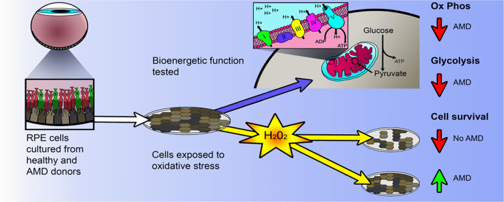
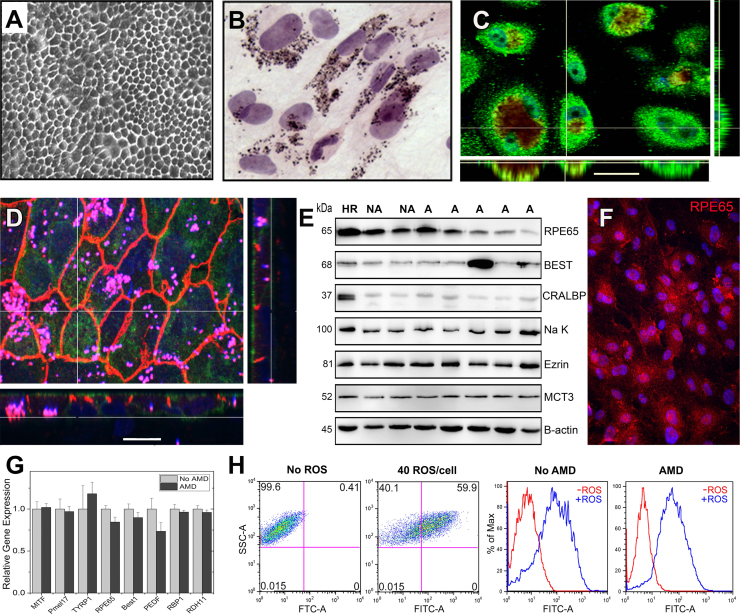
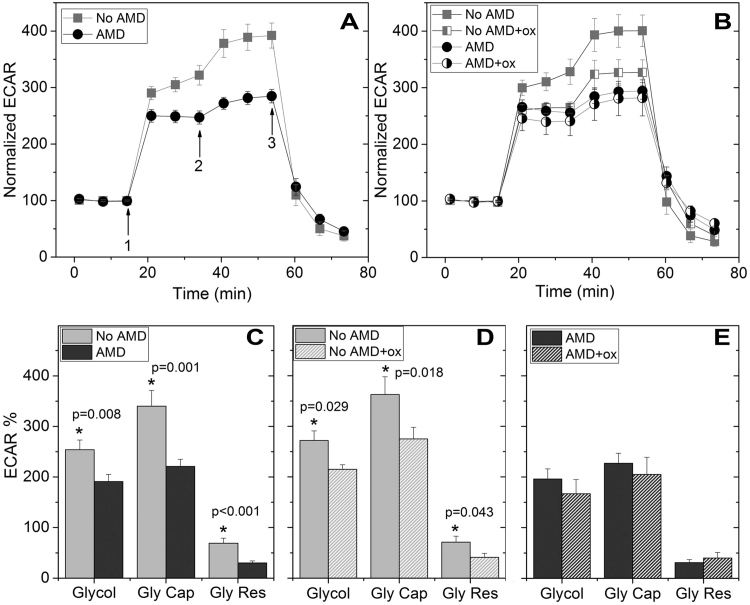
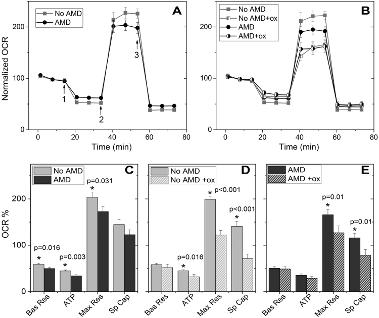
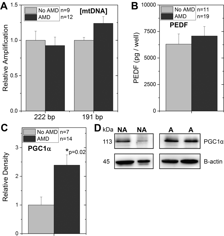
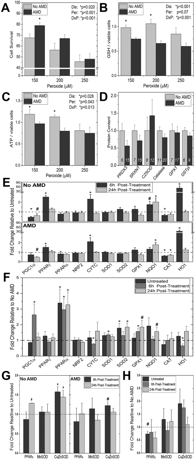
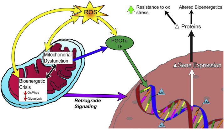
Age-related macular degeneration (AMD) is the leading cause of blindness among older adults. It has been suggested that mitochondrial defects in the retinal pigment epithelium (RPE) underlies AMD pathology. To test this idea, we developed primary cultures of RPE to ask whether RPE from donors with AMD differ in their metabolic profile compared with healthy age-matched donors. Analysis of gene expression, protein content, and RPE function showed that these cultured cells replicated many of the cardinal features of RPE in vivo. Using the Seahorse Extracellular Flux Analyzer to measure bioenergetics, we observed RPE from donors with AMD exhibited reduced mitochondrial and glycolytic function compared with healthy donors. RPE from AMD donors were also more resistant to oxidative inactivation of these two energy-producing pathways and were less susceptible to oxidation-induced cell death compared with cells from healthy donors. Investigation of the potential mechanism responsible for differences in bioenergetics and resistance to oxidative stress showed RPE from AMD donors had increased PGC1α protein as well as differential expression of multiple genes in response to an oxidative challenge. Based on our data, we propose that cultured RPE from donors phenotyped for the presence or absence of AMD provides an excellent model system for studying "AMD in a dish". Our results are consistent with the ideas that (i) a bioenergetics crisis in the RPE contributes to AMD pathology, and (ii) the diseased environment in vivo causes changes in the cellular profile that are retained in vitro.

Keywords: 6 max) Age-related macular degeneration; Antioxidants; Glycolytic function; Mitochondrial function; Oxidative stress; Retinal pigment epithelium.


Figures

fx1
Graphical abstract
Fig. 1
Fig. 1
Characterization of primary human RPE cultures derived from adult donors. (A) Phase microscopy image shows confluent RPE form a monolayer with a cobblestone appearance. (B) H&E stained RPE cells visualize the pigment granules inside the cytoplasm (C,D) Confocal microscopy images of RPE cultured on transwell inserts for one month are shown. En face views of the RPE monolayer shown as maximum intensity projections through the z-axis. Also shown are cross-sections (locations shown by the white line) through the z-plane of multiple optical slices. (C) Bestrophin (green) is expressed on the basal surface. Pigment granules seen due to auto fluorescence (red) Nuclei are stained using DAPI (blue). (Scale bar 30 µm) (D) The Na-K ATPase (green) is expressed on the apical surface. ZO-1 staining (orange) marks cell borders; punctate pigment granules appear pink. Nuclei are stained using DAPI (blue). (Scale bar 15 µm) (E) RPE cultures from non-diseased (No AMD) and AMD donors contain many prototypic RPE proteins as demonstrated on Western immunoblots. Molecular mass for each protein is shown on the left. HR is RPE homogenate from a human donor. β-actin is the loading control. CRALBP, cellular retinaldehyde-binding protein; MCT3, monocarboxylate transporter 3. (F) Immunohistochemistry and fluorescent microcopy was used to detect the expression of the prototypic RPE protein, the 65 kDa Retinal Pigment Epithelium-Specific Protein RPE65. (G) RT-qPCR analysis was performed on RPE cultures from non-diseased (No AMD) (n=4) and AMD (n=7) donors. Graph shows AMD delta CT relative to the mean delta CT of the non-diseased group. Data are the mean (±SEM) normalized values. MITF, microphthalmia-associated transcription factor; Pmel17, pre-melanosome protein 1; TYRP1, tyrosinase related protein 1; Best1, Bestrophin; PEDF, pigment epithelial derived factor; RBP1,Retinol binding protein 1; RDH11, Retinal dehydrogenase. (H) FACs analysis measuring the phagocytosis of FITC-labeled rod OS by RPE from a healthy (No AMD) and an AMD donor. Dot plots (left) and histograms for cells without and with the addition of OS are shown. Data are mean (± SEM). (* denotes p<0.05).
Fig. 2
Fig. 2
RPE from donors with AMD show reduced glycolytic function and resistance to oxidative inactivation. (A,B) Trace shows extracellular acidification rate (ECAR) normalized to baseline for cells from non-diseased and AMD donors with no treatment (A, No AMD n=9; AMD n=9) and following 24 h incubation with 500 µM hydrogen peroxide (B, No AMD n=7; AMD n=6). Arrows indicate injection of glucose (1), oligomycin (2), and 2-deoxyglucose (2-DG,3). (C,D,E) Parameters of glycolytic function calculated from data shown in A or B. Probability values for significant differences, as determined by t-test comparing No AMD with AMD (C) or paired t-test comparing glycolytic function with and without oxidation for individual samples (D,E), is provided on the graphs. See Supplement Fig. 1A for the method of calculation. All data are mean (± SEM). (* denotes p<0.05).
Fig. 3
Fig. 3
RPE from donors with AMD show reduced mitochondrial function. (A,B) Trace from an XF96 Extracellular Flux Analyzer shows the oxygen consumption rate (OCR) normalized to baseline for cells from non-diseased and AMD donors with no treatment (A, No AMD n=14; AMD n=19) and following 24 h incubation with 500 µM hydrogen peroxide (B, No AMD n=10; AMD n=15). Arrows indicate injection of oligomycin (1), FCCP (2) and antimycin and rotenone (3) to perturb mitochondrial function. (C,D,E) Parameters of mitochondrial function were calculated from data shown in A or B. Probability values for significant differences, as determined by t-test comparing No AMD with AMD (C) or paired t-test comparing values with and without oxidation for individual samples (D,E), is provided on the graphs. Bas Res=basal respiration; Max Res=maximal respiration; Sp Cap=spare capacity. See Supplement Fig. 1B for the method of calculation. All data are mean (± SEM). (* denotes p<0.05).
Fig. 4
Fig. 4
Investigating the loss in mitochondrial function for RPE from AMD donors. (A) Mitochondrial content was estimated from the real-time PCR amplification of the mitochondrial genome (Cyt b (222 bp) and16S rRNA (197 bp)) relative to amplification of the β-globin nuclear gene (a measure of total RPE). Ratios (mtDNA/ β-globin) were normalized to the mean ratio for non-diseased (No AMD) donors. (B) Graph shows results of ELISA measuring pigment epithelium derived factor (PEDF) secreted by RPE for donors without AMD (n=11) or with AMD (n=19). (C,D) Western blots were used to evaluate the content of PGC1α in cultures from donors with or without AMD. Results of densitometry (C) and representative western blot (D) are shown. * p=0.02, RPE from AMD donors were significantly higher than cells from donors without AMD. All data are mean (± SEM). (* denotes p<0.05).
Fig. 5
Fig. 5
Investigating the resistance to oxidation stress in RPE from AMD donors. (A,B,C) RPE cultures were incubated with 0, 150 µm, 200 µm, or 250 µm hydrogen peroxide for 24 h. (A) Cell survival (No AMD n=9; AMD n=16) was measured using the CyQuant Direct Cell Proliferation Assay. (B) ATP content (No AMD n=8; AMD n=14) was determined using the ATPlite luminescence ATP detection assay. (C) GSH content (No AMD n=9; AMD n=16) was determined using the GSH-Glo Glutathione assay. Data are hydrogen peroxide treated relative to untreated controls. Results from the two-way ANOVA (main effects were disease and hydrogen peroxide) are shown on each graph. (D) Data are from densitometry of Western immunoblots using antibodies specific for multiple antioxidants. Results are normalized to the mean density for donors with No AMD. The number of donors for each analysis is shown within the bars. (E) mRNA was isolated from untreated controls and cells exposed to 300 µm hydrogen peroxide for either 6 h or 24 h. Results are the fold change in expression relative to untreated controls (dashed line). (F) The same Ct values used in (E) were used to calculate fold change in expression of AMD relative to the average for No AMD samples (dashed line). (G) Proteins were isolated using RIPA buffer from untreated controls and cells exposed to 300 µm hydrogen peroxide at 6 h or 24hrs. Results are the fold change of densitometry relative to untreated controls (dashed line). (H) The same densitometry values were used to calculate the fold change relative to No AMD donors. See Materials and Methods for fold change calculations. All data are mean (± SEM). ( * denotes p<0.05 and # denotes 0.10 > p > 0.05).
Fig. 6
Fig. 6
Model for AMD related changes in RPE. AMD is associated with mitochondrial dysfunction and a bioenergetic crisis, documented by decreased OxPhos and glycolysis. Mitochondrial dysfunction leads to increased ROS production. PGC1a and redox sensitive transcription factors (TF) respond to environmental changes caused by mitochondrial dysfunction. In combination with mitochondrial retrograde signaling, these pathways alter gene expression and protein content of the cell. The epigenetic landscape, denoted by CpG methylation (Me) adds another layer of complexity to gene expression, and is altered due to an oxidative challenge. These changes lead to an increased resistance to oxidative stress and an altered bioenergetics profile.

Similar articles

Cited by

References

    1. Alliance for Eye and Vision Research, 20th Anniversary Special Report. October. 〈http://www.eyeresearch.org/pdf/ValueBrochure/Value_of_Vision_Research_B..., 2013.
    1. Bhattacharya S., Chaum E., Johnson D.A., Johnson L.R. Age-related susceptibility to apoptosis in human retinal pigment epithelial cells is triggered by disruption of p53-Mdm2 association. Invest Ophthalmol. Vis. Sci. 2012;53:8350–8366. - PMC - PubMed
    1. Blaauwgeers H.G.T., Holtkamp G.M., Rutten H. Polarized vascular endothelial growth factor secretion by human retinal pigment epithelium and localization of vascular endothelial growth factor receptors on the inner choriocapillaris. Am. J. Pathol. 1999;155:421–428. - PMC - PubMed
    1. Blenkinsop T.A., Salero E., Stern J.H., Temple S. The culture and maintenance of functional retinal pigment epithelial monolayers from adult human eyes. Methods Mol. Biol. 2016;945:45–55. - PubMed
    1. Carver K.A., Yang D. N-Acetylcysteine amide protects against oxidative stress- induced microparticle release from human retinal pigment epithelial cells. Invest Ophthalmol. Vis. Sci. 2016;57:360–371. - PMC - PubMed

Publication types

MeSH terms

Substances