. 2017 Jun 10;17(1):409.
doi: 10.1186/s12885-017-3399-x.

Granulin-epithelin precursor interacts with 78-kDa glucose-regulated protein in hepatocellular carcinoma

Affiliations

Granulin-epithelin precursor interacts with 78-kDa glucose-regulated protein in hepatocellular carcinoma

Chi Wai Yip et al. BMC Cancer. .

Abstract

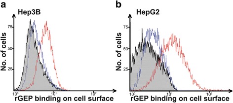
Background: Granulin-epithelin precursor (GEP) is a secretory growth factor, which has been demonstrated to control cancer growth, invasion, drug resistance and immune escape. Our previous studies and others also demonstrated its potential in targeted therapy. Comprehensive characterization of GEP partner on cancer cells are warranted. We have previously shown that GEP interacted with heparan sulfate on the surface of liver cancer cells and the interaction is crucial for GEP-mediated signaling transduction. This study aims to characterize GEP protein partner at the cell membrane with the co-immunoprecipitation and mass spectrometry approach.

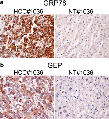
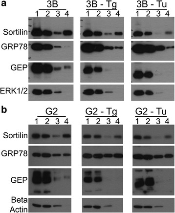
Methods: The membrane fraction from liver cancer model Hep3B was used for capturing binding partner with the specific monoclonal antibody against GEP. The precipitated proteins were analyzed by mass spectrometry. After identifying the GEP binding partner, this specific interaction was validated in additional liver cancer cell line HepG2 by co-immunoprecipitation using GRP78 and GEP antibodies, respectively, as the bait. GRP78 transcript levels in hepatocellular carcinoma (HCC) clinical samples (n = 77 pairs) were examined by real-time quantitative RT-PCR. GEP and GRP78 protein expressions were investigated by immunohistochemistry on paraffin sections.

Results: We identified the GEP-binding protein as 78-kDa glucose-regulated protein (GRP78, also named heat shock 70-kDa protein 5, HSPA5). This interaction was validated in independent HCC cell lines. Increased GRP78 mRNA levels were demonstrated in liver cancer tissues compared with the paralleled liver tissues (t-test, P = 0.002). GRP78 and GEP transcript levels were significantly correlated (Spearman's correlation, P = 0.001), and the proteins were also detectable in the cytoplasm of liver cancer cells by immunohistochemical staining.

Conclusions: GRP78 and GEP are interacting protein partners in liver cancer cells and may play a role in GEP-mediated cancer progression in HCC.

Keywords: 78-kDa glucose-regulated protein; Granulin-epithelin precursor; Hepatocellular carcinoma; Mass spectrometry; Protein interaction.


Figures

Fig. 1
Fig. 1
Binding of rGEP on the surface of HCC cells includes a fraction of non-HS binding. After EDTA detachment, HCC cells (a) Hep3B and (b) HepG2 were incubated with rGEP for cell surface binding. The HS-rGEP interaction was displaced by heparin. Residual binding (blue line) of rGEP and control binding (red line) of rGEP were detected by anti-His antibody and quantified by flow cytometry. Grey area represents the background fluorescent signal of cells without rGEP incubation. Representative histograms from three independent replicates are shown
Fig. 2
Fig. 2
Co-immunoprecipitation using GRP78 antibody and GEP antibody respectively. Two sets of co-IP were performed using GRP78 antibody and GEP antibody, respectively, as baits. Each set of experiment contains lysate with specific antibody (I), antibody alone (II) and lysate alone (III). The proteins absorbed in the protein G beads were extracted by boiling in SDS sampling buffer and were loaded in each lane. Immunoblotting (IB) targeting GEP and GRP78 were performed. Representative blots from three independent experiments are shown
Fig. 3
Fig. 3
GRP78 and GEP protein expression in clinical samples by immunohistochemical staining. HCC tissue and non-tumor (NT) liver tissue from patients were formalin-fixed and paraffin-embedded. Sections were deparaffinised and hydrated. Specific antibodies to (a) GRP78 and (b) GEP were added and detected by HRP-conjugated second antibody. The sections were counterstained with hematoxylin and imaged at 400× magnification. Representative images from three individual clinical sample pairs are shown
Fig. 4
Fig. 4
Transcript levels of GRP78 and GEP in HCC samples. a GRP78 expression was up-regulated in HCC with reference to their corresponding non-tumor (NT) (t-test, P = 0.002). (b-d) GRP78 and GEP transcript levels were significantly correlated when compared their HCC mRNA, NT mRNA and the HCC versus NT fold ratio. e Kaplan-Meier analysis on recurrence-free survival. Patients were segregated based on their GRP78 mRNA levels (log rank test, P = 0.022). The cut-off value of GRP78 expression level was determined by the Youden index
Fig. 5
Fig. 5
Biotinylation of cell surface proteins reflects the localization of GRP78 and GEP under thapsigargin/tunicamycin treatments in (a) Hep3B and (b) HepG2. Sortilin serves as positive control for cell surface localization; while ERK1/2 and β-actin are negative controls. 1, before loading to avidin column; 2, flow through from the column; 3, wash from the column; 4, elution of the biotinylated cell surface proteins. Starting materials is 1.5 mg/ml. Loading volume of 1 & 2 are 10 μl. Loading volume of 3 & 4 are 20 μl. Tg, 300 nM thapsigargin; Tu, 1.5 μg/ml tunicamycin. Representative blots from three independent experiments are shown

References

    1. Zhu J, Nathan C, Jin W, Sim D, Ashcroft GS, Wahl SM, et al. Conversion of proepithelin to epithelins: roles of SLPI and elastase in host defense and wound repair. Cell. 2002;111:867–878. doi: 10.1016/S0092-8674(02)01141-8. - DOI - PubMed
    1. Baker M, Mackenzie IR, Pickering-Brown SM, Gass J, Rademakers R, Lindholm C, et al. Mutations in progranulin cause tau-negative frontotemporal dementia linked to chromosome 17. Nature. 2006;442:916–919. doi: 10.1038/nature05016. - DOI - PubMed
    1. Cruts M, Gijselinck I, van der Zee J, Engelborghs S, Wils H, Pirici D, et al. Null mutations in progranulin cause ubiquitin-positive frontotemporal dementia linked to chromosome 17q21. Nature. 2006;442:920–924. doi: 10.1038/nature05017. - DOI - PubMed
    1. Daniel R, Daniels E, He Z, Bateman A. Progranulin (acrogranin/PC cell-derived growth factor/granulin-epithelin precursor) is expressed in the placenta, epidermis, microvasculature, and brain during murine development. Dev Dyn. 2003;227:593–599. doi: 10.1002/dvdy.10341. - DOI - PubMed
    1. He Z, Ong CH, Halper J, Bateman A. Progranulin is a mediator of the wound response. Nat Med. 2003;9:225–229. doi: 10.1038/nm816. - DOI - PubMed

Substances