Review
. 2017:41:1-50.
doi: 10.1016/bs.enz.2017.03.005. Epub 2017 Apr 22.

The Importance of Being Modified: The Role of RNA Modifications in Translational Fidelity

Affiliations
Review

The Importance of Being Modified: The Role of RNA Modifications in Translational Fidelity

Paul F Agris et al. Enzymes. 2017.

Abstract

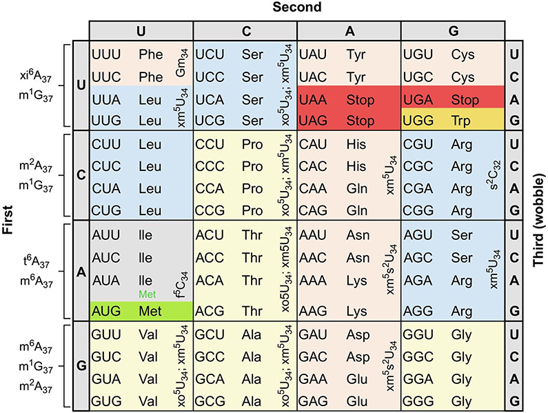
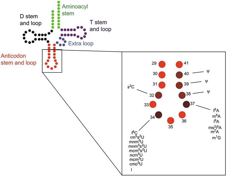
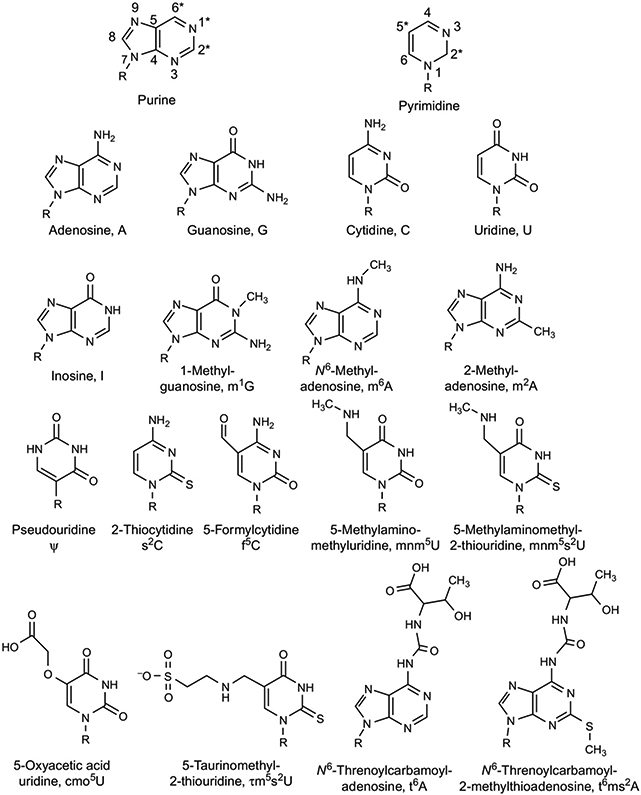
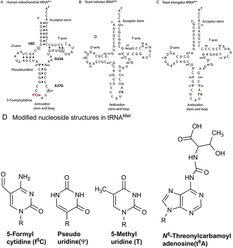
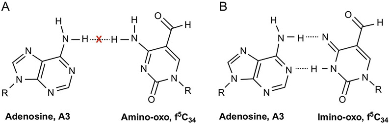
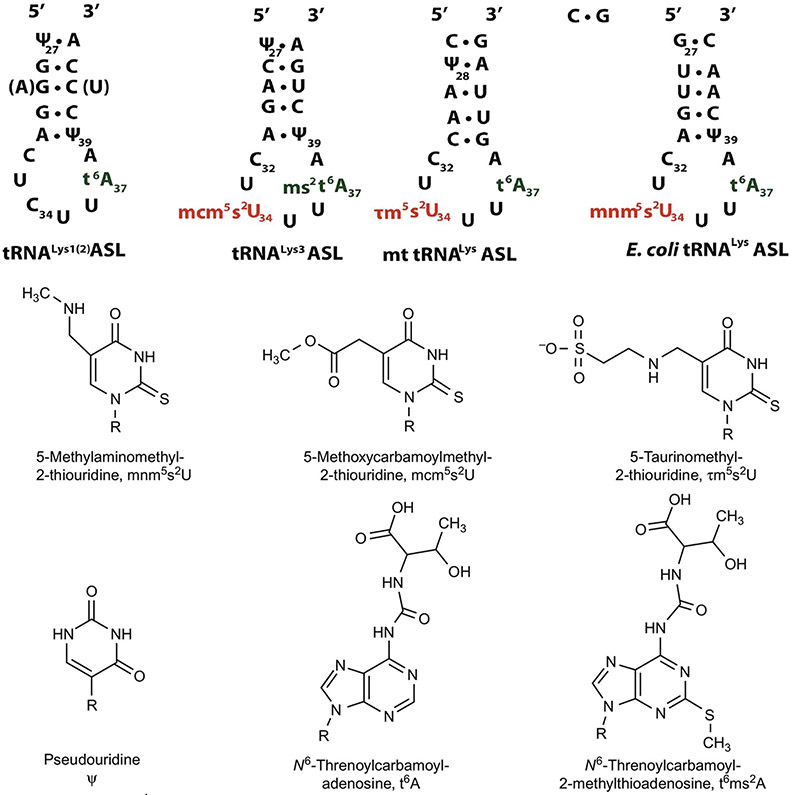
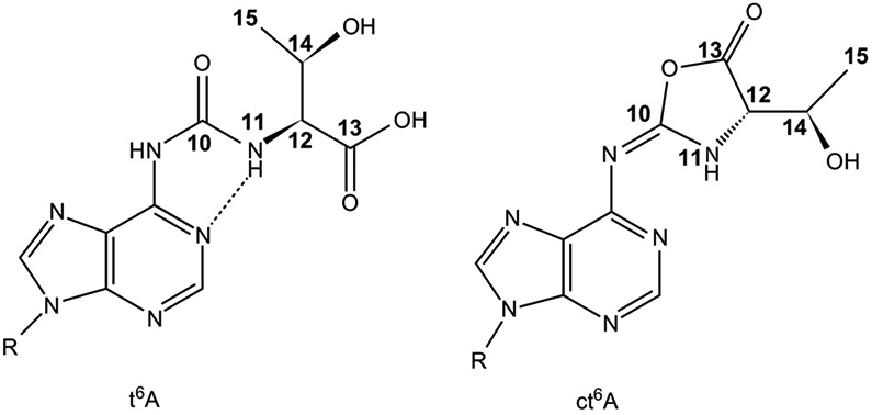
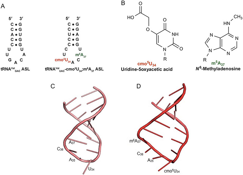
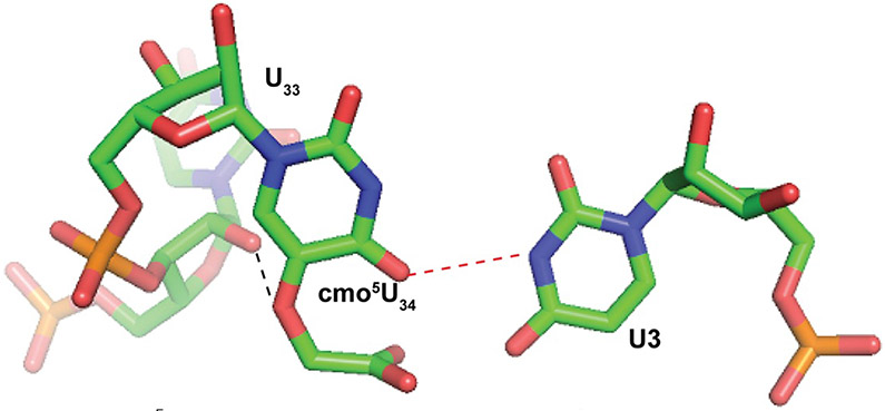
The posttranscriptional modifications of tRNA's anticodon stem and loop (ASL) domain represent a third level, a third code, to the accuracy and efficiency of translating mRNA codons into the correct amino acid sequence of proteins. Modifications of tRNA's ASL domain are enzymatically synthesized and site specifically located at the anticodon wobble position-34 and 3'-adjacent to the anticodon at position-37. Degeneracy of the 64 Universal Genetic Codes and the limitation in the number of tRNA species require some tRNAs to decode more than one codon. The specific modification chemistries and their impact on the tRNA's ASL structure and dynamics enable one tRNA to decode cognate and "wobble codons" or to expand recognition to synonymous codons, all the while maintaining the translational reading frame. Some modified nucleosides' chemistries prestructure tRNA to read the two codons of a specific amino acid that shares a twofold degenerate codon box, and other chemistries allow a different tRNA to respond to all four codons of a fourfold degenerate codon box. Thus, tRNA ASL modifications are critical and mutations in genes for the modification enzymes and tRNA, the consequences of which is a lack of modification, lead to mistranslation and human disease. By optimizing tRNA anticodon chemistries, structure, and dynamics in all organisms, modifications ensure translational fidelity of mRNA transcripts.

Keywords: Codon degeneracy and decoding; Human disease and tRNA modifications; Modification chemistry and structure; Modifications 3′-adjacent to the anticodon; Modifications prestructure tRNA; Wobble position tRNA modifications.


Figures

FIG. 1
FIG. 1
The Universal Genetic Code. Twofold degenerate codons are highlighted in tan; threefold (Ile) in gray, fourfold in yellow, and sixfold in blue. The single codons of Met and Trp are highlighted in green and orange, respectively, and the three stop codons are highlighted in red. The figure is annotated with the abbreviations for those modified nucleosides found in the anticodon domain of tRNAs responding to the codons and discussed in this review. The chemical structures and full names of the modifications are found in Fig. 3.
FIG. 2
FIG. 2
tRNA and its anticodon stem and loop (ASL) domain. Left: General cloverleaf structure of a 76-nucleotide tRNA with the aminoacyl-accepting stem in green, dihydrouridine (D) stem and loop in black, ASL in red, extra loop in blue, and ribothymidine (T) stem and loop in plum. Right: An enlargement of the ASL domain, with the abbreviations of the important modifications discussed in this review listed. A darker color hue in plum represents more important modification sites.
FIG. 3
FIG. 3
Chemical structures of the major and modified nucleosides of tRNA. Top row: General structures of purine and pyrimidine nucleosides with numbering of the atoms. R represents ribose and the asterisk (*) marks the most important and/or frequent sites of modification. Second row: Conventional RNA nucleosides and their letter abbreviations. Third through fifth rows: The modified nucleosides discussed in this review with their shorthand notions [14,15].
FIG. 4
FIG. 4
Nucleoside sequence and cloverleaf structures of tRNAMet. (A) Human mitochondrial tRNAMet displaying the condensed variable loop and the shortened D- and T-arms. Nucleotides affected by disease-causing point mutations are boxed. (B) Saccharomyces cerevisiae initiator tRNAMet. Ar(p) at position-64 refers to the 2′-O-ribosyl phosphate modification, which is an identity element of initiator tRNAMet in yeast [60]. (C) S. cerevisiae elongator tRNAMet; 5-methyluridine at position-54, indicated by T, is an important determinant of elongator tRNAMet. (D) Chemical structures of the modifications f5C34, Ψ at various noted positions, and t6A37 found in the three methionyl tRNA [61].
FIG. 5
FIG. 5
The tautomeric forms of 5-formylcytidine, f5C34. Tautomerism of f5C34 allows for the stereochemistry of the Watson–Crick base pairing between tRNA's f5C34 and A3 of the AUA codon to approximate that of a canonical U●A pair. (A) Steric repulsion between the common amino-oxo form of f5C34 N4H and A3N6H marked by an X. (B) Favorable interactions between the imino-oxo form of f5C34 and A3[70]. Dotted lines indicate hydrogen bonding between nucleoside residues [70].
FIG. 6
FIG. 6
tRNALys ASL sequences and the chemical structures of their modified. Top: Sequences and secondary structures of the anticodon stem and loop (ASL) domains of human tRNALys1,2CUU, tRNALys3, human mitochondrial tRNALysUUU, and E. coli tRNALysUUU. Nucleosides in parenthesis indicate differences between tRNALys1 and tRNALys2 sequences. Bottom: Chemical structures of the modified nucleosides in the ASLs above and their shorthand notations [92,93].
FIG. 7
FIG. 7
N6-Threonylcarbamoyladenosine and its cyclic derivative, t6A and ct6A. Left: Chemical structure of t6A with the atoms of the modification numbered. The hydrogen bonding that produces a pseudocyclic conformation of the modified nucleoside is shown as a dashed line. Right: The cyclic form of t6A, ct6A, with the corresponding atoms numbered [88,133].
FIG. 8
FIG. 8
The primary sequences and nucleoside modifications of the E. coli tRNAVal isoacceptors and the structures of ASLValUAC and ASLValUAC-cmo5U34;m6A37 compared. (A) The anticodon domain primary sequences of the three tRNAVal isoacceptors from E. coli and their modifications: uridine-5-oxyacetic acid at position-34, red and N6-methyladenosine at position-37, green. The two tRNAVal species containing the GAC anticodon have identical ASLs with variations in sequence in other regions of the tRNA molecule. (B) The chemical structures, names, and abbreviations of the modifications that appear in the E. coli tRNAValUAC-cmo5U34;m6A37. (C) Structure of unmodified ASLValUAC, pink. (D) Fully modified ASLValUAC containing the modifications cmo5U34 and m6A37, ASLValUAC-cmo5U34;m6A37, red, showing the base stacking of cmo5U34 and an open loop structure. The structures of the unmodified and modified ASLValUAC were determined by NMR [45] (Protein Data Bank 2JR4 for unmodified ASLVal3UAC and 2JRG for ASLVal3UAC-cmo5U34;m6A37).
FIG. 9
FIG. 9
cmo5U34○U3 wobble pair. Base pairing between cmo5U34 of the ASLValUAC and U3 of the GUU codon [46] (Protein Data Bank 2UUB). A hydrogen bond occurs between the 2′-OH of the invariant U33 (background) of the ASLValUAC and O5 of cmo5U34 (black dashed line), structuring cmo5U34 for base pairing with U3. The hydrogen bond that allows for the base pair between O2 of cmo5U34 and N2 of U3 (red dashed line).
FIG. 10
FIG. 10
The primary sequences and nucleoside modifications of the E. coli tRNAArg isoacceptors. (A) The anticodon domain primary sequences of the five tRNAArg isoacceptors from E. coli and their modifications: 2-thiocytidine at position-32 (blue); inosine and 5-methylaminomethyluridine at position-34 (red); 2-methyladenosine, N6-threonylcarbamoyladenosine, and 1-methylguanosine at position-37 (green); and pseudouridine at position-40 (orange). (B) The chemical structures, names, and abbreviations of the modifications that appear in the five E. coli tRNAArg isoacceptors.

Similar articles

Cited by

References

    1. Liu Z, Gutierrez-Vargas C, Wei J, Grassucci RA, Sun M, Espina N, Madison-Antenucci S, Tong L, Frank J Determination of the ribosome structure to a resolution of 2.5 A by single-particle cryo-EM. Protein Sci. 2017;26(1):82–92. - PMC - PubMed
    1. Zimmerman E, Yonath A Biological implications of the ribosome's stunning stereochemistry. ChemBioChem. 2009;10(1):63–72. - PubMed
    1. Greber BJ, Ban N Structure and function of the mitochondrial ribosome. Annu. Rev. Biochem 2016;85:103–132. - PubMed
    1. Brocchieri L, Karlin S Protein length in eukaryotic and prokaryotic proteomes. Nucleic Acids Res. 2005;33(10):3390–3400. - PMC - PubMed
    1. Kurland C, Hughes D, Ehrenberg M Limitations of translational accuracy. In: Neidhardt FC, Curtis JL III Ingraham R, Lin ECC, Low JKB, Magasarák B, Reznikoff WS, Riley M, Schaachler M, Umbarger HE, eds. Escherichia coli and Salmonella: Cellular and Molecular Biology. Washington, DC: ASM Press; 1996:979–1004.