Review
. 2017:41:89-115.
doi: 10.1016/bs.enz.2017.03.003. Epub 2017 Apr 12.

TrmD: A Methyl Transferase for tRNA Methylation With m1G37

Affiliations
Review

TrmD: A Methyl Transferase for tRNA Methylation With m1G37

Ya-Ming Hou et al. Enzymes. 2017.

Abstract

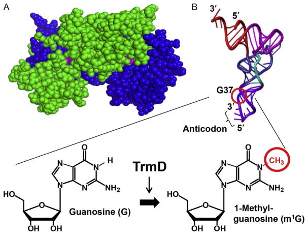
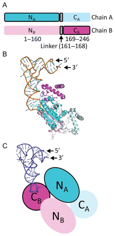
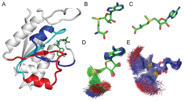
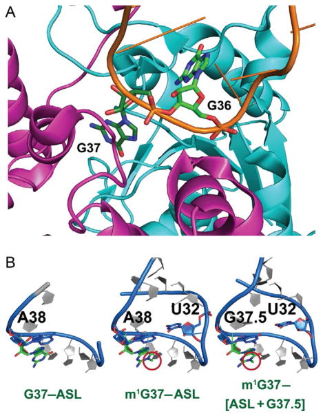
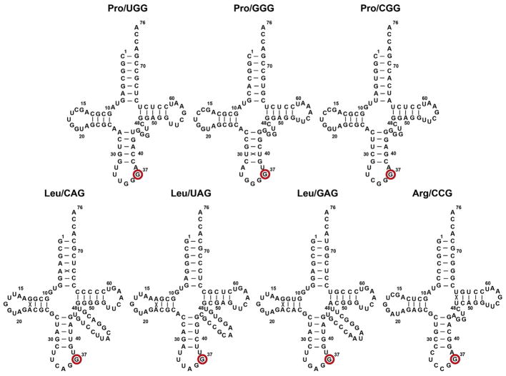
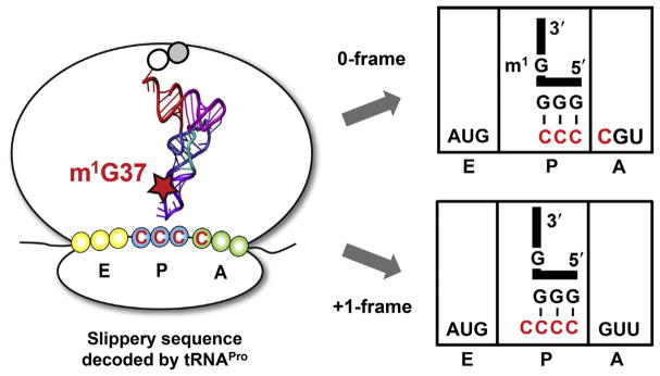
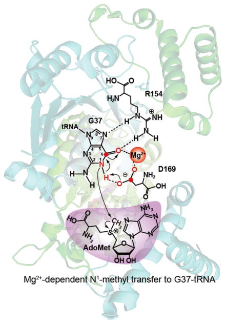
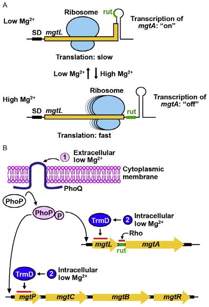
TrmD is an S-adenosyl methionine (AdoMet)-dependent methyl transferase that synthesizes the methylated m1G37 in tRNA. TrmD is specific to and essential for bacterial growth, and it is fundamentally distinct from its eukaryotic and archaeal counterpart Trm5. TrmD is unusual by using a topological protein knot to bind AdoMet. Despite its restricted mobility, the TrmD knot has complex dynamics necessary to transmit the signal of AdoMet binding to promote tRNA binding and methyl transfer. Mutations in the TrmD knot block this intramolecular signaling and decrease the synthesis of m1G37-tRNA, prompting ribosomes to +1-frameshifts and premature termination of protein synthesis. TrmD is unique among AdoMet-dependent methyl transferases in that it requires Mg2+ in the catalytic mechanism. This Mg2+ dependence is important for regulating Mg2+ transport to Salmonella for survival of the pathogen in the host cell. The strict conservation of TrmD among bacterial species suggests that a better characterization of its enzymology and biology will have a broad impact on our understanding of bacterial pathogenesis.

Keywords: Frameshifts; Mg(2+) transport; Protein trefoil knot; S-adenosyl methionine; m(1)G37-tRNA; tRNA(Pro).


Figures

Fig. 1
Fig. 1
TrmD structure and activity. (A) The obligated dimer structure of TrmD (PDB: IUAK), showing chain A in green and chain B in blue and the two AdoMet molecules in pink at each dimer interface. (B) TrmD catalyzes methylation to the N1 position of G37 in tRNA, using AdoMet as the methyl donor and producing m1G37 on the 3′-side of the tRNA anticodon. The position of G37 and synthesis of m1G37 on the L-shaped tRNA tertiary structure is circled.
Fig. 2
Fig. 2
Domain structure of TrmD. (A) Each chain of the TrmD dimer has three distinct domains: an N-terminal domain (residues 1–160) for binding AdoMet, a flexible linker (residues 161–168) for binding G37 of tRNA, and a C-terminal domain (residues 169–246). In the dimer of TrmD, one active site is formed by the N-terminal domain of chain A (NA, dark blue with a black outline) and the C-terminal of chain B (CB, dark pink with a black outline). The other active site, which is not operational, is formed by the N-terminal domain of chain B (NB, light pink without an outline) with the C-terminal domain of chain A (CA, light blue without an outline). The sequence and length of each domain is highly conserved among TrmD enzymes across diverse organisms of the bacterial domain. (B) Each TrmD dimer (chain A in blue and chain B in pink) binds one tRNA molecule in the crystal structure of the ternary complex (PDB: 4YVI). The G37 base of the tRNA is inserted to the active site formed by the NA domain and the CB domain. (C) A cartoon diagram of the TrmD dimer, showing that one active site is assembled between the NA (in dark blue) and the CB (in dark pink), while the anticodon region of tRNA (in purple) is bound to the flexible linker of chain B. The darker color with a black outline indicates the active unit of the dimer structure.
Fig. 3
Fig. 3
The trefoil knot structure of TrmD. (A) The trefoil knot is formed starting with the central β3 strand, which turns into β4 through a loop. The β4 makes a turn into β5, which makes a circular insertion through the loop to come out with another loop that binds the adenine base of AdoMet (PDB: 4YVI). The three β stands β3, β4, and β5, together with β1 and β2 (not labeled), form the central β sheet in the N-terminal domain of each monomeric chain in TrmD. (B) The bent conformation of AdoMet in the catalytically active monomer. (C) The open and extended conformation of AdoMet in the catalytically inactive monomer, which is similar to the structure observed in Trm5. (D) The bent conformation of AdoMet has rigid constraints and maintains the bent shape. (E) The open conformation of AdoMet has high flexibility and can extend from the bent shape to the open shape.
Fig. 4
Fig. 4
G37 in TrmD and in the tRNA anticodon stem–loop structure. (A) G37 binding to TrmD organizes the flexible linker of chain B (magenta) and projects G36 to the trefoil knot of chain A (cyan) to stabilize the entire tRNA molecule bound to the enzyme (PDB: 4YVI). (B) The unmethylated G37 in the anticodon stem–loop (ASL) structure of tRNAPro/CGG (G37–ASL) prevents the interaction between U32 and A38 and renders the 5′-side of the ASL disordered and invisible (left, PDB: 4P70). The methylated m1G37 remodels the ASL structure (m1G37–ASL) to allow U32–A38 base pairing as in a canonical structure (middle, PDB: 4LT8). The insertion of G37.5 to the ASL, resulting in the structure of m1G37-[ASL + G37.5], disrupts U32–A38 pairing to form the aberrant U32–G37.5 pairing in a disordered structure (right, PDB: 4L47) similar to that in the G37–ASL structure.
Fig. 5
Fig. 5
Sequence and cloverleaf structure of E. coli tRNA species that are substrates for TrmD, including the Leu/UAG isoacceptor that is most likely a substrate as well. The G37 base to be methylated to m1G37 is marked with a red circle. The numbering is based on the structure of yeast tRNAPhe [88].
Fig. 6
Fig. 6
Suppression of ribosomal +1-frameshifting by m1G37-tRNA. On a slippery mRNA sequence AUG-CCC-C, the methylated m1G37-tRNAPro maintains the correct reading frame (0-frame), whereas the unmethylated G37-tRNAPro has a high propensity to shift to the +1-frame, which most frequently occurs during the tRNA sitting at the P-site next to an empty A-site. The tRNA is shown in the L-shape with the anticodon and G37 or m1G37 highlighted.
Fig. 7
Fig. 7
The proposed Mg2+ in the catalytic mechanism of TrmD, involving an eight-membered ring that consists of the N1, C6, and O6 of the guanine ring, the hydrogen bond to the imino group of N1, the two coordination bonds of Mg2+, and the three atoms of the carboxylate of the general base D169. Atomic numbering is indicated for guanine.
Fig. 8
Fig. 8
A proposed mechanism of Mg2+ sensing of Salmonella. (A) Regulation of mgtA transcription in response to Mg2+ level is mediated by the translation speed of the 5′-leader mRNA mgtL sequence, which codes for a short peptide. At low Mg2+, ribosomal translation of mgtL is slow, sequestering the rut sequence in a stem–loop structure and excluding the Rho-dependent transcription termination to permit transcription of mgtA in the “on” state. At high Mg2+, ribosomal translation of mgtL is fast, exposing the rut sequence and resulting in the Rho-dependent transcription termination of mgtA. (B) The two-component system PhoPQ provides a sensor for extracellular Mg2+. At low extracellular Mg2+, the activated PhoQ phosphorylates PhoP to turn on transcription of the Mg2+ transporter gene mgtA as well as the virulence operon mgtCBR, where mgtB is another Mg2+ transporter gene. Transcription of mgtA is then regulated by translation of the 5′-leader sequence mgtL, which encodes a small peptide. Rapid translation of mgtL exposes the Rho-utilization (rut) sequence and terminates transcription ahead of mgtA. In contrast, slow and incomplete translation of mgtL sequesters the rut sequence and allows transcription into the structural gene. The Mg2+-dependent methyl transferase activity of TrmD can act as an intracellular sensor that regulates the speed of translation of mgtL.

References

    1. Keffer-Wilkes LC, Veerareddygari GR, Kothe U. RNA modification enzyme TruB is a tRNA chaperone. Proc Natl Acad Sci USA. 2016;113:14306–14311. - PMC - PubMed
    1. Winsor GL, Lam DK, Fleming L, Lo R, Whiteside MD, Yu NY, Hancock RE, Brinkman FS. Pseudomonas Genome Database: improved comparative analysis and population genomics capability for Pseudomonas genomes. Nucleic Acids Res. 2011;39:D596–D600. - PMC - PubMed
    1. de Berardinis V, Vallenet D, Castelli V, Besnard M, Pinet A, Cruaud C, Samair S, Lechaplais C, Gyapay G, Richez C, Durot M, Kreimeyer A, et al. A complete collection of single-gene deletion mutants of Acinetobacter baylyi ADP1. Mol Syst Biol. 2008;4:174. - PMC - PubMed
    1. Forsyth RA, Haselbeck RJ, Ohlsen KL, Yamamoto RT, Xu H, Trawick JD, Wall D, Wang L, Brown-Driver V, Froelich JM, KGC, King P, et al. A genome-wide strategy for the identification of essential genes in Staphylococcus aureus. Mol Microbiol. 2002;43:1387–1400. - PubMed
    1. Baba T, Ara T, Hasegawa M, Takai Y, Okumura Y, Baba M, Datsenko KA, Tomita M, Wanner BL, Mori H. Construction of Escherichia coli K-12 in-frame, single-gene knockout mutants: the Keio collection. Mol Syst Biol. 2006;2:2006.0008. - PMC - PubMed

MeSH terms

LinkOut - more resources