. 2017 Jun 10;8(5):461-473.
doi: 10.1007/s12975-017-0539-1. Online ahead of print.

Cerebellar Exposure to Cell-Free Hemoglobin Following Preterm Intraventricular Hemorrhage: Causal in Cerebellar Damage?

Affiliations

Cerebellar Exposure to Cell-Free Hemoglobin Following Preterm Intraventricular Hemorrhage: Causal in Cerebellar Damage?

Alex Adusei Agyemang et al. Transl Stroke Res. .

Abstract

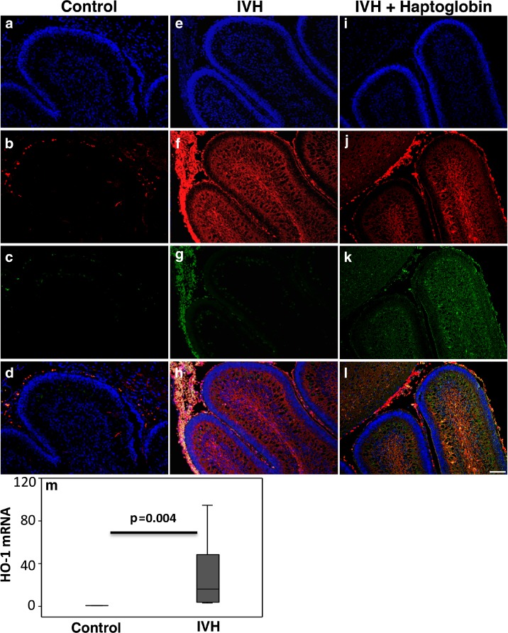
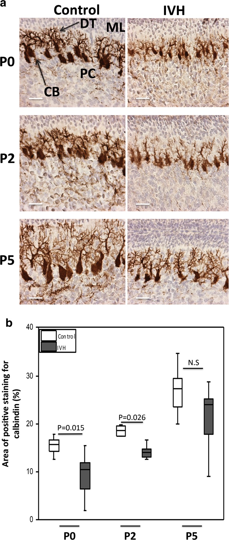
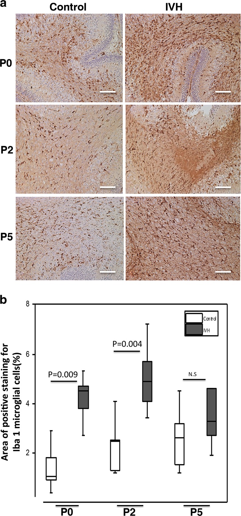
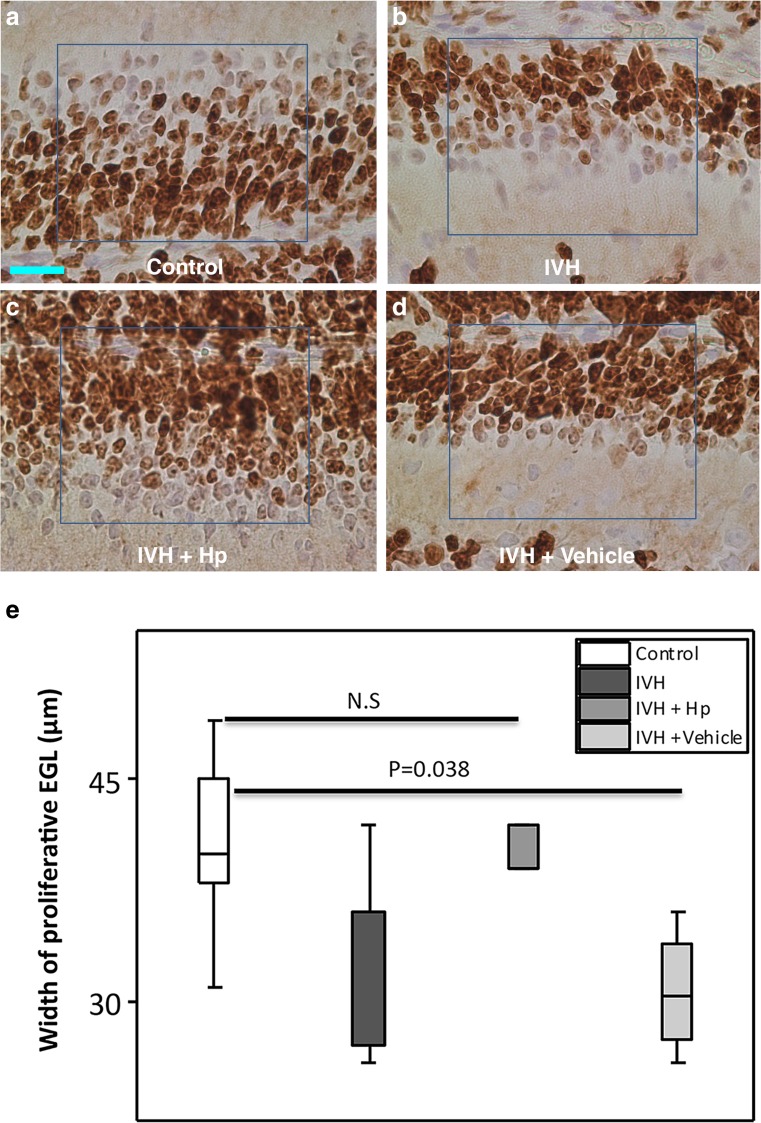
Decreased cerebellar volume is associated with intraventricular hemorrhage (IVH) in very preterm infants and may be a principal component in neurodevelopmental impairment. Cerebellar deposition of blood products from the subarachnoid space has been suggested as a causal mechanism in cerebellar underdevelopment following IVH. Using the preterm rabbit pup IVH model, we evaluated the effects of IVH induced at E29 (3 days prior to term) on cerebellar development at term-equivalent postnatal day 0 (P0), term-equivalent postnatal day 2 (P2), and term-equivalent postnatal day 5 (P5). Furthermore, the presence of cell-free hemoglobin (Hb) in cerebellar tissue was characterized, and cell-free Hb was evaluated as a causal factor in the development of cerebellar damage following preterm IVH. IVH was associated with a decreased proliferative (Ki67-positive) portion of the external granular layer (EGL), delayed Purkinje cell maturation, and activated microglia in the cerebellar white matter. In pups with IVH, immunolabeling of the cerebellum at P0 demonstrated a widespread presence of cell-free Hb, primarily distributed in the white matter and the molecular layer. Intraventricular injection of the Hb scavenger haptoglobin (Hp) resulted in a corresponding distribution of immunolabeled Hp in the cerebellum and a partial reversal of the damaging effects observed following IVH. The results suggest that cell-free Hb is causally involved in cerebellar damage following IVH and that blocking cell-free Hb may have protective effects.

Keywords: Cerebellum; External granular layer; Haptoglobin; Hemoglobin; Intraventricular hemorrhage.


Conflict of interest statement

Conflict of Interest

All authors declare that they have no conflict of interest.

Ethical Approval

All applicable national and institutional guidelines for the care and use of animals were followed.

Figures

Fig. 1
Fig. 1
Study outline. A diagram summarizing the experimental procedure. The experiment consisted of the following steps: preterm delivery of rabbit pups by caesarean section, induction of IVH by intraperitoneal glycerol administration, verification of IVH or sham control by the use of high-frequency ultrasound, randomization into study groups, intraventricular administration of Hp or vehicle solution, termination of pups, and collection of cerebellar tissue. For details about each step, see “Materials and Methods”
Fig. 2
Fig. 2
Immunofluorescence labeling of Hb and the administered human Hp. Representative images are from rabbit pups at P0. Images illustrate the detected immunofluorescence labeling, performed by double immunofluorescence labeling of Hb (red) and Hp (green) together with a DAPI nuclear staining (blue), in animals with no IVH (Control), in animals with IVH (IVH), and in animals with IVH that received human Hp injections (IVH + Haptoglobin). Antibody specificity tests showed that the antibodies against Hb and human Hp bound to their true targets (see Fig. 1 in the Data supplement). ad Control animal: Images b and c show the lack of Hb and Hp labeling and the autofluorescence mainly from whole erythrocytes (RBCs) restricted to the subarachnoid space and some blood vessels (d). eh IVH: In pups with IVH, the Hb labeling (red) was extensive, widely distributed in the molecular layer and white matter and to some degree in the EGL. Whole erythrocytes in the subarachnoid space surrounding the cerebellar lobuli were also intensely labeled and gave rise to green autofluorescence (g), observed as yellowish in the merged image (h). Hb labeling intermingled with dense nuclear regions (intense DAPI staining) appears as pink (bottom images). il IVH + Haptoglobin: j and k show immunofluorescence labeling of Hb (red) and human Hp (green) following intraventricular injection of Hp at E29. j shows the widespread distribution of cell-free Hb (red), corresponding to that in IVH animals (f), and the domination coexistence of Hp in K (green), primarily in the molecular layer, white matter, and the EGL as shown in the merged image (l). Hp labeling was scarce in the subarachnoid space (k and l), in which Hb labeling of RBCs was extensive (j and l). Thus, the cell-free Hb and Hp are clearly distinguishable from the cell body–associated Hb labeling and autofluorescence. Scale bar = 50 μm. m HO-1 mRNA expression in the cerebellum was investigated at P0 following IVH. Following IVH, heme-degrading protein HO-1 mRNA was upregulated (IVH, dark gray bar, n = 7) as compared to the controls (n = 5). mRNA expression for HO-1 was normalized against GAPDH and is given as fold change. The fold change values were calculated by normalizing against samples from control pups. Results are presented as box plots displaying medians and 25th and 75th percentiles. Differences between no IVH and IVH at P0 were analyzed using the Mann–Whitney U test
Fig. 3
Fig. 3
Reduced width of proliferative EGL following preterm IVH. a Images of EGL of the developing cerebellum from which quantitative measurements were made of the proliferative width in the respective groups at the time points studied. The image shows the Ki67-positive outer portion of the EGL where proliferation of granule cell precursors occurs and the deeper portion, which hosts the differentiation of granule cell precursors to mature granule cells. Scale bar = 50 μm. b GCP proliferation in the outer portion of the EGL of the developing cerebellum was investigated following IVH by Ki67 staining. Measurement of the width of proliferative EGL was done in cerebellar tissue sections of both sham controls (control, white bars; n at P0 = 6, n at P2 = 6, n at P5 = 5) and IVH pups (dark gray bars; n at P0 = 5, n at P2 = 6, n at P5 = 6) at P0, P2, and P5. Results are presented as box plots displaying medians and 25th and 75th percentiles. Statistical differences between groups for respective time points were analyzed using the Mann–Whitney U test
Fig. 4
Fig. 4
Impaired Purkinje cell maturation following preterm IVH. a Immunostaining of calbindin, a calcium-binding protein, was used as a marker of Purkinje cell development in the molecular layer of the developing cerebellum. Calbindin stains are seen as brown to dark brown. Decreased calbindin immunoreactivity was observed in IVH pups (brown) compared to controls (intense dark brown). Observation of neuronal morphology revealed smaller neuronal cell bodies and underdeveloped Purkinje dendrites in IVH pups compared to controls at postnatal time points of P0, P2, and P5. ML molecular layer, PC Purkinje cell, DT dendrites, CB cell bodies; scale bar = 50 μm. b Grading of Purkinje cell development by measurement of percentage area of positive calbindin staining was done in cerebellar tissue sections of both control (white bars; n at P0 = 6, n at P2 = 6, n at P5 = 5) and IVH pups (dark gray bars; n at P0 = 6, n at P2 = 6, n at P5 = 6) at P0, P2, and P5, as described in “Materials and Methods.” Results are presented as box plots displaying medians and 25th and 75th percentiles. Statistical differences between groups for respective time points were analyzed using the Mann–Whitney U test
Fig. 5
Fig. 5
Microglial activation in the cerebellar white matter following preterm IVH. a Immunolabeling to confirm upregulation of Iba1 (seen as brown to dark brown) expression, a marker of microglial activation was used as a qualitative marker of reactive microglia cellular response in the white matter of the developing cerebellum. Increased Iba1 immunoreactivity was observed in IVH pups compared to controls at P0, P2, and P5. Observation of microglial morphology revealed an amoeboid shape with long processes in the IVH pups. Scale bar = 50 μm. b Measurement of percentage area of positive Iba1 staining was done in cerebellar tissue sections of both control (white bars; n at P0 = 6, n at P2 = 6, n at P5 = 5) and IVH pups (dark gray bars; n at P0 = 5, n at P2 = 6, n at P5 = 6) at P0, P2, and P5, as described in “Materials and Methods.” Results are presented as box plots displaying medians and 25th and 75th percentiles. Statistical differences between groups for respective time points were analyzed using the Mann–Whitney U test
Fig. 6
Fig. 6
Intraventricular Hp administration protects against impaired Purkinje cell development following preterm IVH. ad Following intraventricular Hp administration at P0, a higher intensity of calbindin immunoreactivity, relatively larger Purkinje cell bodies, and developed dendrites were observed in the Hp-administered IVH pups as compared to pups with IVH only or vehicle-treated IVH pups. Scale bar = 50 μm. e Grading of Purkinje cell development by measurement of percentage area of positive calbindin staining was done in cerebellar tissue sections at P0 of control pups (white bars, n = 6), IVH pups (dark gray bars, n = 6), and following intraventricular injection of Hp in pups with IVH (IVH + Hp, gray bars, n = 6) or vehicle solution (IVH + Vehicle, light gray bars, n = 4). Results are presented as box plots displaying medians and 25th and 75th percentiles. Differences between IVH + Hp vs. control and IVH + Vehicle vs. control were analyzed using the Kruskal–Wallis test followed by pairwise comparison with significance values adjusted for multiple comparisons
Fig. 7
Fig. 7
Intraventricular Hp administration protects against reduction in width of proliferative EGL following preterm IVH. ad Following intraventricular Hp administration at P0, a higher intensity of Ki67 immunoreactivity was observed in the Hp-administered IVH pups as compared to pups with IVH only or vehicle-treated IVH pups. Scale bar = 20 μm. e Measurement of the width of Ki67-positive proliferative EGL was performed in cerebellar tissue sections at P0 of control pups (white bars, n = 6), IVH pups (dark gray bars, n = 5), and following intraventricular injection of Hp in pups with IVH (IVH + Hp, gray bars, n = 5) or vehicle solution (IVH + Vehicle, light gray bars, n = 4). Results are presented as box plots displaying medians and 25th and 75th percentiles. Differences between IVH + Hp vs. control and IVH + Vehicle vs. control were analyzed using the Kruskal–Wallis test followed by pairwise comparison with significance values adjusted for multiple comparisons

References

    1. Ballabh P. Intraventricular hemorrhage in premature infants: mechanism of disease. Pediatr Res. 2010;67:1–8. doi: 10.1203/PDR.0b013e3181c1b176. - DOI - PMC - PubMed
    1. Schmahmann JD, Smith EE, Eichler FS, Filley CM. Cerebral white matter: neuroanatomy, clinical neurology, and neurobehavioral correlates. Ann N Y Acad Sci. 2008;1142:266–309. doi: 10.1196/annals.1444.017. - DOI - PMC - PubMed
    1. Volpe JJ. Cerebellum of the premature infant: rapidly developing, vulnerable and clinically important. J Child Neurol. 2009;24:1085–1104. doi: 10.1177/0883073809338067. - DOI - PMC - PubMed
    1. Parker J, Mitchell A, Kalpakidou A, Walshe M, Jung HY, Nosarti C, et al. Cerebellar growth and behavioural & neuropsychological outcome in preterm adolescents. Brain. 2008;131:1344–1351. doi: 10.1093/brain/awn062. - DOI - PubMed
    1. Limperopoulos C, Chilinaryan G, Sullivan N, Guizard N, Robertson RL, Du Plessis AJ. Injury to the premature cerebellum: outcome is related to remote cortical development. Cereb Cortex. 2014;24:728–736. doi: 10.1093/cercor/bhs354. - DOI - PMC - PubMed

LinkOut - more resources