. 2017 Jul 5;25(7):1130-1138.e6.
doi: 10.1016/j.str.2017.05.006. Epub 2017 Jun 8.

Structure of the ACF7 EF-Hand-GAR Module and Delineation of Microtubule Binding Determinants

Affiliations

Structure of the ACF7 EF-Hand-GAR Module and Delineation of Microtubule Binding Determinants

Thomas R Lane et al. Structure. .

Abstract

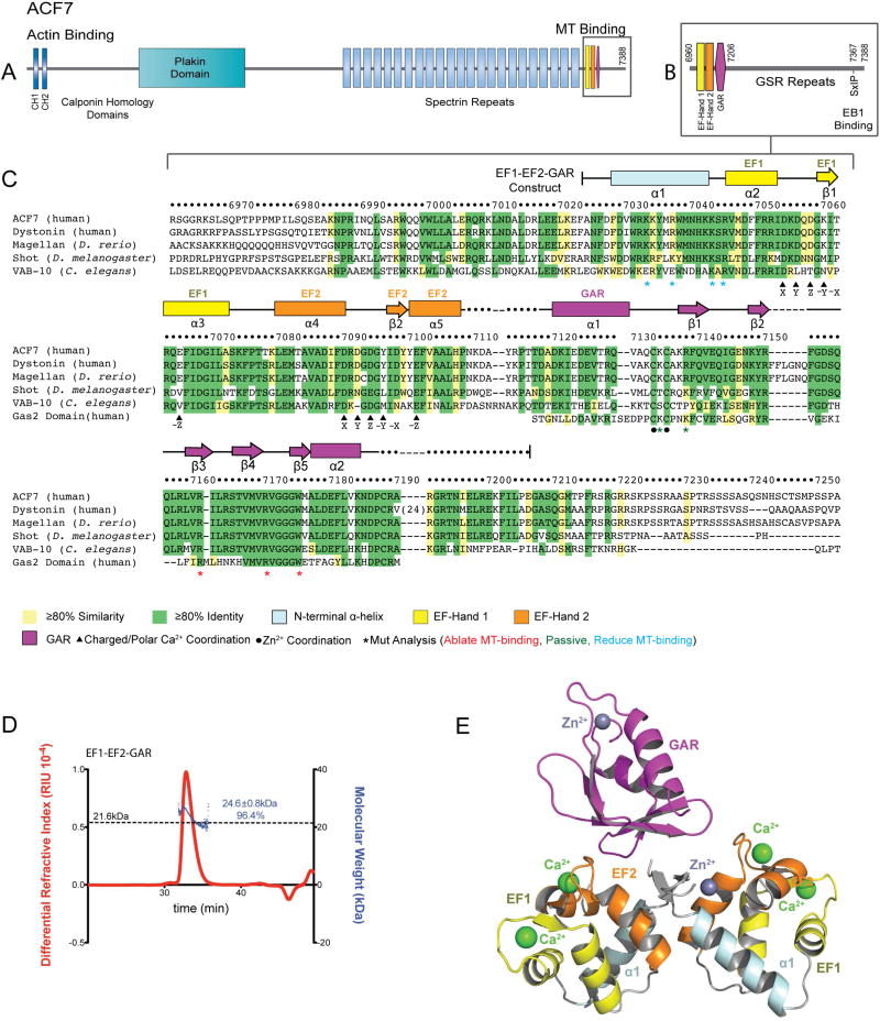
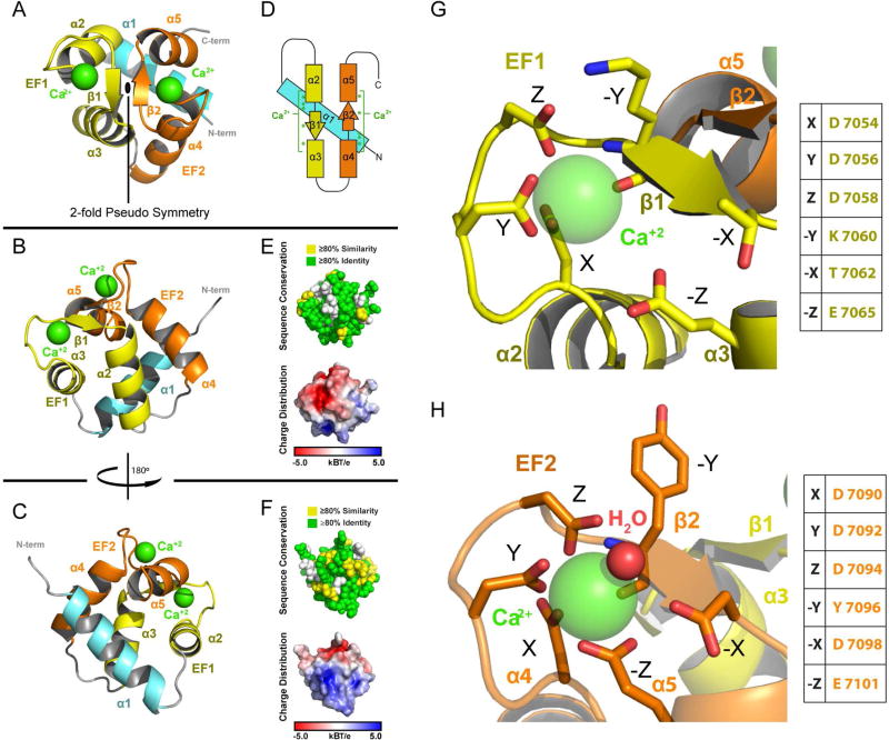
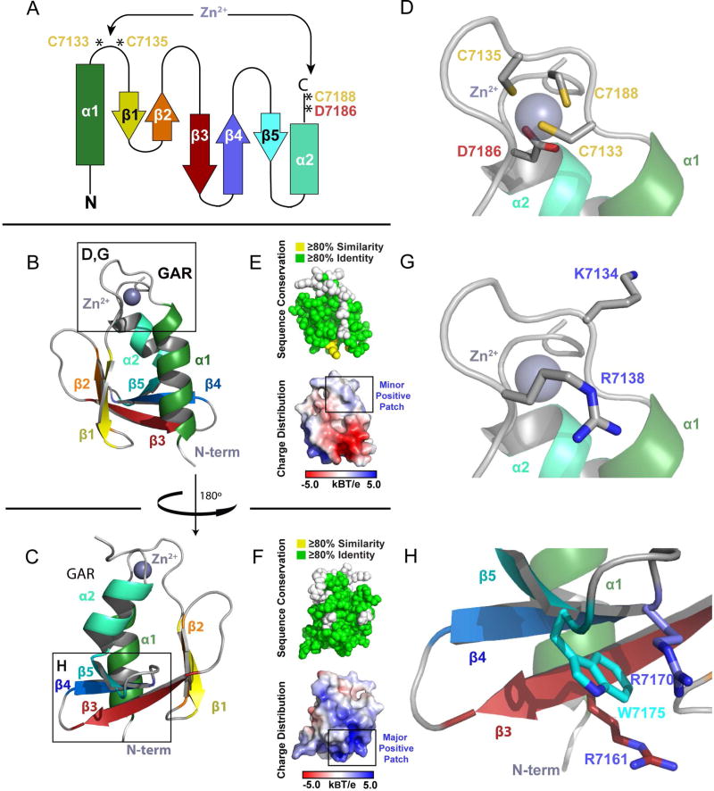
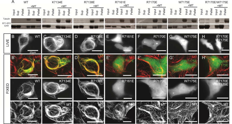
Spectraplakins are large molecules that cross-link F-actin and microtubules (MTs). Mutations in spectraplakins yield defective cell polarization, aberrant focal adhesion dynamics, and dystonia. We present the 2.8 Å crystal structure of the hACF7 EF1-EF2-GAR MT-binding module and delineate the GAR residues critical for MT binding. The EF1-EF2 and GAR domains are autonomous domains connected by a flexible linker. The EF1-EF2 domain is an EFβ-scaffold with two bound Ca2+ ions that straddle an N-terminal α helix. The GAR domain has a unique α/β sandwich fold that coordinates Zn2+. While the EF1-EF2 domain is not sufficient for MT binding, the GAR domain is and likely enhances EF1-EF2-MT engagement. Residues in a conserved basic patch, distal to the GAR domain's Zn2+-binding site, mediate MT binding.

Keywords: ACF7; EF Hand; GAR; Gas2; MACF1; actin; microtubule; spectraplakin.


Figures

Figure 1
Figure 1. Architecture of the ACF7 EF1-EF2-GAR Module
(A) hACF7 domain architecture and zoom view (B) of the MT-binding region. (C) Sequence alignment of the EF1-EF2-GAR module from human, D. rerio, D. melanogaster, and C. elegans spectraplakins, and human Gas2. hACF7 EF1-EF2-GAR 2° structure and residue number are depicted above, residues mutated are indicated below. (D) SEC-MALS analysis of the EF1-EF2-GAR module. The molecular weight of a monomer is indicated by the dashed line. The peak measured accounts for 96.4% of the total mass eluted. (E) Model of the EF1-EF2-GAR modules observed in the ASU. See also Figure S1.
Figure 2
Figure 2. EF1-EF2 Domain Structure
(A–C) Structure of the EF1-EF2 domain in different orientations. Following the N-terminal α-helix (cyan) are the EF1 (yellow) and EF2 (orange) helix-turn-helix motifs. Calcium atoms are shown in green. (D) Secondary structure topology of the EF1-EF2 domain showing the location of residues that coordinate calcium (green asterisks). (E,F) The EF1-EF2 domain illustrating cross-species conservation as delineated in Figure 1C (above, spherical representation) and displaying electrostatic surface potential (below, surface representation). Domain orientation in (E) and (F) correspond to the orientation in (B) and (C) respectively. (G, H) Zoom views of the EF1 and EF2 calcium binding sites. Canonical EF-Hand residues are indicated. See also Figure S2.
Figure 3
Figure 3. The GAR Domain is a Novel α/β Sandwich that Coordinates Zinc
(A) Secondary structure topology of the GAR domain α/β sandwich showing the location of the residues that coordinate zinc. (B) Ribbon diagram of the GAR domain as colored in A. (C) The GAR domain as shown in (B), rotated 180° about the y-axis. (D) Zoom view of the zinc binding site boxed in (B), showing the Cys2-Asp-Cys residues that coordinate zinc. (E,F) The GAR domain illustrating cross-species conservation as delineated in Figure 1C (above, spherical representation) and displaying electrostatic surface potential (below, surface representation). Domain orientation in (E) and (F) correspond to the orientation in (B) and (C) respectively. (G,H) Zoom view of two positively charged regions on the GAR domain, corresponding to the boxed regions in (B) and (C) respectively. See also Figure S2.
Figure 4
Figure 4. The GAR β3-β5 Basic Region Mediates MT Binding
(A) MT co-sedimentation assays of WT and mutant EF1-EF2-GAR constructs. (B–H) Live cell analysis of GFP-EF1-EF2-GAR WT and mutant constructs in HEK293 cells. (B’-H’) Analysis of GFP-EF1-EF2-GAR WT and mutant constructs for MT co-localization in fixed HEK293 cells. All scale bars: 20 µm. See also Figures S3, S4.

References

    1. Adams PD, Afonine PV, Bunkóczi G, Chen VB, Davis IW, Echols N, Headd JJ, Hung L-W, Kapral GJ, Grosse-Kunstleve RW, et al. PHENIX: a comprehensive Python-based system for macromolecular structure solution. Acta Crystallogr. Sect. D Biol. Crystallogr. 2010;66:213–221. - PMC - PubMed
    1. Applewhite DA, Grode KD, Duncan MC, Rogers SL. The actin-microtubule cross-linking activity of Drosophila Short stop is regulated by intramolecular inhibition. Mol. Biol. Cell. 2013;24:2885–2893. - PMC - PubMed
    1. Babu YS, Bugg CE, Cook WJ. Structure of calmodulin refined at 2.2 Å resolution. J. Mol. Biol. 1988;204:191–204. - PubMed
    1. Baker NA, Sept D, Joseph S, Holst MJ, McCammon JA. Electrostatics of nanosystems: application to microtubules and the ribosome. Proc. Natl. Acad. Sci. U. S. A. 2001;98:10037–10041. - PMC - PubMed
    1. Bencze KZ, Yoon T, Millán-Pacheco C, Bradley PB, Pastor N, Cowan JA, Stemmler TL. Human frataxin: iron and ferrochelatase binding surface. Chem. Commun. 2007:1798–1800. - PMC - PubMed

Publication types

LinkOut - more resources