. 2017 Sep:295:222-232.
doi: 10.1016/j.expneurol.2017.06.013. Epub 2017 Jun 9.

CNS disease diminishes the therapeutic functionality of bone marrow mesenchymal stem cells

Affiliations

CNS disease diminishes the therapeutic functionality of bone marrow mesenchymal stem cells

Alex Sargent et al. Exp Neurol. 2017 Sep.

Abstract

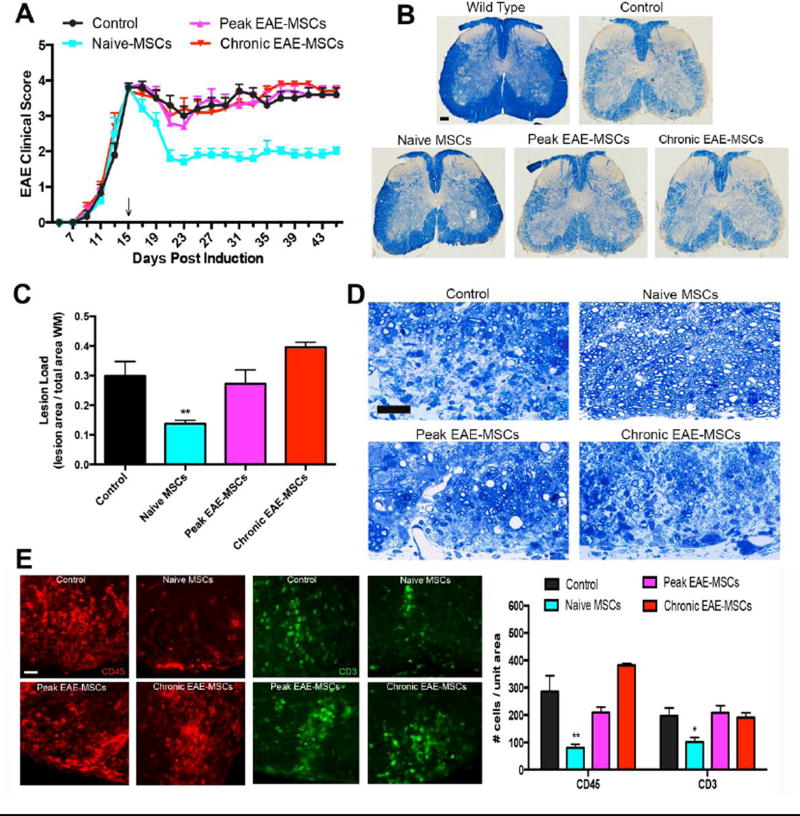
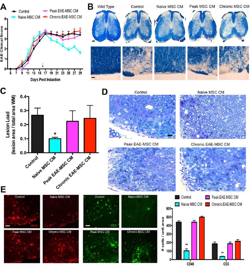
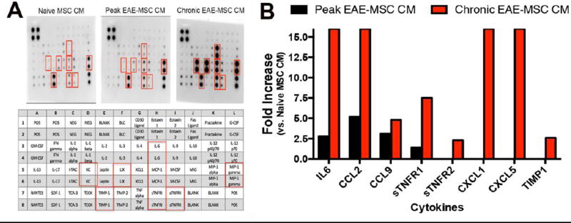
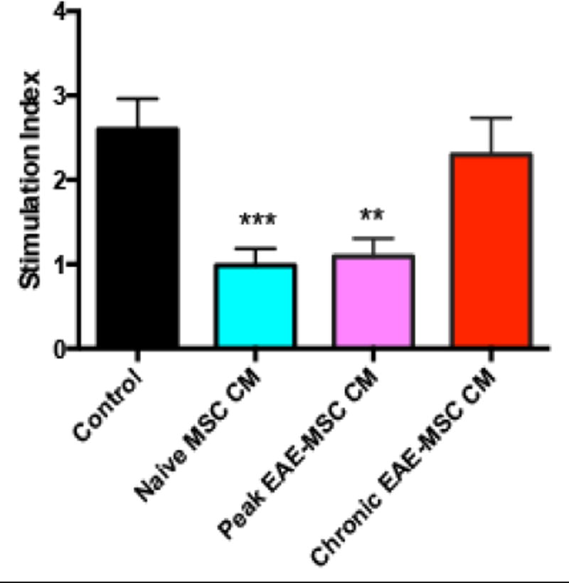
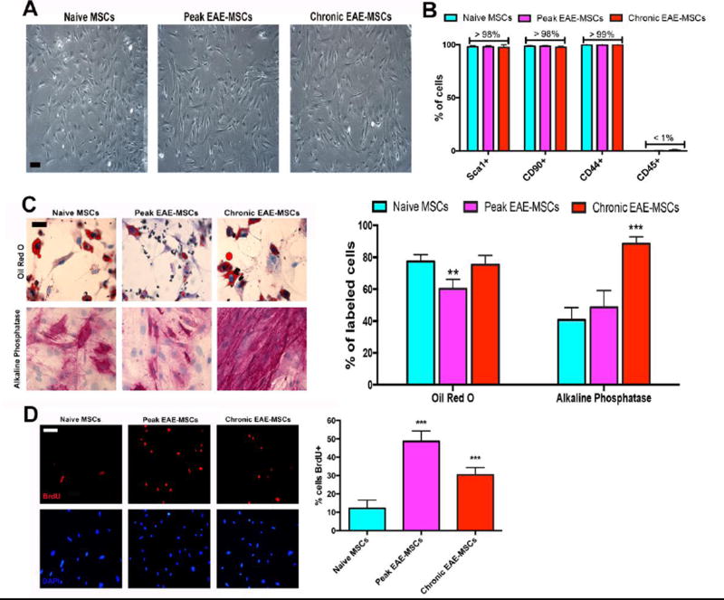
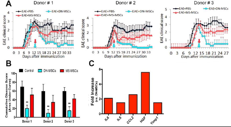
Mesenchymal stem cells (MSCs) have emerged as a potentially powerful cellular therapy for autoimmune diseases including multiple sclerosis (MS). Based on their success in treating animal models of MS like experimental autoimmune encephalomyelitis (EAE), MSCs have moved rapidly into clinical trials for MS. The majority of these trials use autologous MSCs derived from MS patients, although it remains unclear how CNS disease may affect these cells. Here, we report that bone marrow MSCs derived from EAE mice lack therapeutic efficacy compared to naïve MSCs in their ability to ameliorate EAE. Treatment with conditioned medium from EAE-MSCs also fails to modulate EAE, and EAE-MSCs secrete higher levels of many pro-inflammatory cytokines compared to naïve MSCs. Similarly, MSCs derived from MS patients have less therapeutic efficacy than naïve MSCs in treating EAE and secrete higher levels of some of the same pro-inflammatory cytokines. Thus diseases like EAE and MS diminish the therapeutic functionality of bone marrow MSCs, prompting reevaluation about the ongoing use of autologous MSCs as a treatment for MS.

Keywords: Experimental autoimmune encephalomyelitis; Mesenchymal stem cells (MSCs); Multiple sclerosis.


Figures

Figure 1
Figure 1. Transplanted EAE-MSCs fail to improve functional recovery or CNS pathology in EAE mice
(A) Infusion of naïve MSCs leads to improved functional recovery in MOG35–55 EAE-mice, while treatment with peak or chronic EAE-MSCs does not improve functional recovery versus saline-treated controls. 0.8 × 10^6 MSCs were intravenously injected into mice 15 days after EAE induction (arrow). Data shown represents mean + SEM, n=12 mice per group, from 3 independent experiments. (B) Representative images of solochrome cyanine stained spinal cord sections showing myelin loss in EAE mice 30 days after treatment with naïve MSCs versus peak or chronic EAE-MSCs. (C) EAE mice treated with naïve MSCs have less myelin loss and lower lesion load, while mice treated with EAE-MSCs have a higher lesion load that is comparable to controls. Data shown = mean + SEM, with lesion load quantified from 6 solochrome cyanine stained sections per mouse, 3 mice per group; **P<0.01, One-way ANOVA. (D) Representative images of Toluidine blue stained spinal cord sections from EAE mice 30 days after treatment confirms that mice treated with naïve MSCs have more myelinated axons and less inflammatory infiltrates than controls. By contrast, animals treated with peak or chronic EAE-MSCs show no appreciable difference from controls. (E) EAE-mice treated with naïve MSCs have significantly lower numbers of CD45 positive inflammatory cells (red) and CD3 positive T-cells (green) in their spinal cords 30 days after treatment, while mice treated with peak or chronic EAE-MSCs show no significant difference from controls. Data shown in graph = mean + SEM, quantified from 4 sections per animal, 3 animals per group. *P<0.05, **P<0.01, One-way ANOVA. Scale bars in (B) = 500um, (D) = 25 um, (E) = 20um.
Figure 2
Figure 2. Conditioned medium (CM) from EAE-MSCs fails to improve functional recovery or CNS pathology in EAE mice
(A) Intravenous infusion of conditioned medium (CM) from naïve MSCs leads to improved functional recovery in MOG35–55 EAE-mice, while infusion of CM from either peak or chronic EAE-MSCs does not improve functional recovery relative to controls (EAE mice that received unconditioned medium). 0.5 mg conditioned medium was given 16 days after EAE induction (arrow), data shown represents mean + SEM, n=11–12 mice per group, from 3 independent experiments. (B) Representative images of solochrome cyanine stained spinal cord sections showing myelin loss in EAE mice 14 days after they received CM from naïve MSCs versus peak or chronic EAE-MSCs. (C) EAE mice treated with naïve MSC CM have less myelin loss and lower lesion load, while mice treated with EAE-MSC CM have a higher lesion load comparable to controls. Data shown = mean + SEM, with lesion load quantified from 6 solochrome cyanine stained sections per mouse, 3 mice per group; *P<0.05, One-way ANOVA. (D) Toluidine blue staining of spinal cord sections in EAE mice 14 days after treatment shows more myelinated axons in mice that received naïve MSC CM, while mice receiving EAE-MSC CM have less myelinated axons and more inflammatory infiltrates similar to controls. (D) EAE-mice treated with naïve MSC-CM have significantly lower numbers of CD45 positive inflammatory cells (red) and CD3 positive T-cells (green) in their spinal cords 14 days after treatment, whereas mice treated with EAE-MSC CM show no significant difference from controls. Data shown in graph = mean + SEM, quantified from 5 sections per animal, 3 animals per group. **P<0.01, One-way ANOVA. Scale bars in (B) (Top)= 500um, (Bottom) = 25um (D) = 25 um, (E) = 20um.
Figure 3
Figure 3. EAE-MSCs secrete higher levels of pro-inflammatory cytokines
(A) Representative images of antibody arrays treated with conditioned medium (CM) from naïve MSCs, peak EAE-MSCs, or chronic EAE-MSCs. Cytokines found to be up-regulated in the arrays are indicated by red boxes and identified in the array diagram depicted below; CCL2 = MCP-1, CCL9 = MIP-1 gamma, CXCL1 = KC, and CXCL5 = LIX. (B) Fold changes of proteins increased in conditioned medium from peak or chronic EAE-MSCs relative to naïve MSCs; note conditioned medium from EAE-MSCs contains higher levels of pro-inflammatory cytokines, including IL6, CCL2, and CCL9. Fold changes were calculated by densitometric quantification of spot intensity values from 3 separate antibody arrays per group, n = 3 experiments. Fold changes were capped at 16, with no fold decreases of any cytokines tested observed in the arrays.
Figure 4
Figure 4. Effects of EAE versus naïve MSC CM on MOG-stimulated splenocytes
Conditioned medium from naïve MSCs or peak EAE-MSCs suppresses MOG induced restimulation of splenocytes in-vitro, whereas conditioned medium from chronic EAE-MSCs fails to significantly inhibit splenocyte proliferation. Proliferation was assessed 48 hours after MOG stimulation (20uM) via BrdU Elisa. Data shown = mean + SEM, n = 3 experiments, **P<0.01, ***P<0.005, One-way ANOVA.
Figure 5
Figure 5. Comparison of cultured EAE-MSCs with naïve MSCs
(A) Phase contrast images of passage 3 naïve MSCs versus peak and chronic EAE-MSCs showing cells have similar morphologies. (B) Both peak and chronic EAE-MSCs express common MSC markers and are CD45 negative like naïve MSCs. Data shown = mean + SEM, n = 3 experiments. (C) (Left) Representative images of naïve and EAE-MSCs differentiated into Oil-Red O positive adipocytes and Alkaline Phosphatase (ALP) positive osteoblasts. (Right) Slightly fewer peak EAE-MSCs differentiate into Oil-Red O stained adipocytes compared to naïve or chronic EAE MSCs, whereas differentiation of chronic EAE-MSCs into ALP-positive osteoblasts is significantly increased. Data shown = mean + SEM, n = 2 experiments. **P<0.01, ***P<0.005, One-way ANOVA. (D) (Left) Representative images of BrdU labeled naïve MSCs versus peak and chronic EAE-MSCs after a 16 hour BrdU pulse. (Right) The proportion of cells labeled with BrdU (16 hour pulse) is significantly higher for peak and chronic EAE-MSCs versus naïve MSCs. Data shown = mean + SEM, n = 3 experiments, ***P<0.005, One-way ANOVA. Scale bar in (A) = 50um, (C) and (D) = 20 um.
Figure 6
Figure 6. MS-MSCs lack therapeutic efficacy compared to naïve MSCs in modulating EAE
(A) MSCs derived from MS patients (MS-MSCs) are less effective at improving functional recovery when transplanted into EAE mice compared to naïve MSCs (DN-MSCs) derived from healthy donors. MOG35–55 induced EAE mice were infused with 1×10^6 MSCs at 14 days post EAE induction (arrows), with MS-MSCs derived from 3 separate donors diagnosed with relapsing-remitting MS and DN-MSCs derived from corresponding sex and age-matched healthy donors. Data shown = mean ± SEM, n = 11–13 mice per group. (B) EAE mice treated with DN-MSCs from each of the respective donors show a significant reduction in cumulative disease scores (area under the curve), whereas EAE mice treated with MS-MSCs show no significant difference versus saline treated controls. Data shown = mean + SEM, **P<0.01, One-way ANOVA. (C) Fold changes of proteins increased in conditioned medium from MS-MSCs relative to naïve MSCs; note conditioned medium from MS-MSCs contains higher levels of pro-inflammatory cytokines, including IL-6, IL-8, and CCL2. Fold changes were calculated by comparing spot intensity values from 2 separate antibody arrays per group, with each array treated with an independent sample. No fold decreases of any cytokine tested were observed in the arrays.

References

    1. Bai L, Lennon DP, Caplan AI, DeChant A, Hecker J, Kranso J, Zaremba A, Miller RH. Hepatocyte growth factor mediates mesenchymal stem cell–induced recovery in multiple sclerosis models. Nat. Neurosci. 2012;15:862–870. doi: 10.1038/nn.3109. - DOI - PMC - PubMed
    1. Bai L, Lennon DP, Eaton V, Maier K, Caplan AI, Miller SD, Miller RH. Human bone marrow-derived mesenchymal stem cells induce Th2-polarized immune response and promote endogenous repair in animal models of multiple sclerosis. Glia. 2009;57:1192–1203. doi: 10.1002/glia.20841. - DOI - PMC - PubMed
    1. Caplan AI, Dennis JE. Mesenchymal stem cells as trophic mediators. Journal of cellular biochemistry. 2006;98(5):1076–84. doi: 10.1002/jcb20886. - DOI - PubMed
    1. Chen X, Armstrong MA, Li G. Mesenchymal stem cells in immunoregulation. Immunol. Cell Biol. 2006;84:413–421. doi: 10.1111/j.1440-1711.2006.01458.x. - DOI - PubMed
    1. Cohen JA. Mesenchymal stem cell transplantation in multiple sclerosis. Journal of the neurological sciences. 2013;330(0):43–49. doi: 10.1016/j.jns.2012.12.00. - DOI - PMC - PubMed