Review
. 2017;93(6):361-377.
doi: 10.2183/pjab.93.022.

Ultra-Early Phase pathologies of Alzheimer's disease and other neurodegenerative diseases

Affiliations
Review

Ultra-Early Phase pathologies of Alzheimer's disease and other neurodegenerative diseases

Hitoshi Okazawa. Proc Jpn Acad Ser B Phys Biol Sci. 2017.

Abstract

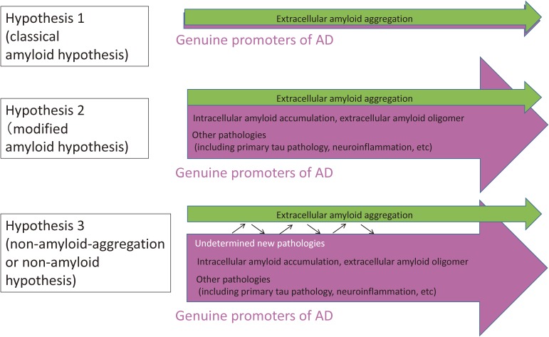
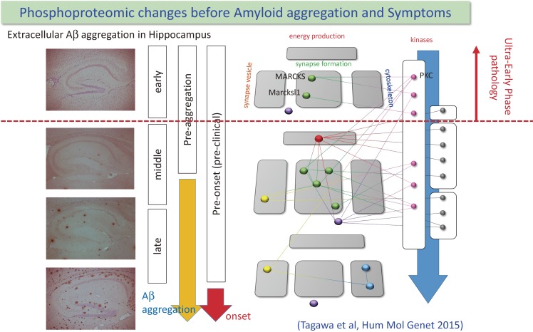
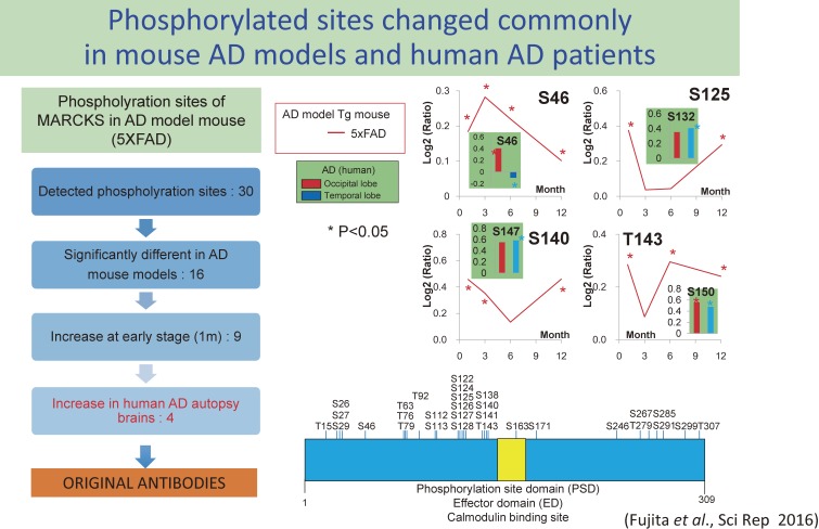
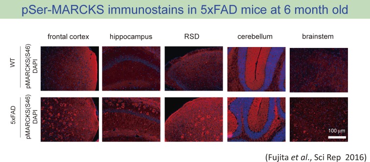
The concept of neurodegenerative diseases and the therapeutics targeting these intractable diseases are changing rapidly. Protein aggregation as the top of pathological cascade is now challenged, and many alternative ideas are proposed. Early molecular pathologies before microscopic detection of diseases protein aggregates, which I propose to call "Ultra-Early Phase pathologies or phase 0 pathologies", are the focus of research that might explain the failures of clinical trials with anti-Aβ antibodies against Alzheimer's disease. In this review article, I summarize the critical issues that should be successfully and consistently answered by a new concept of neurodegeneration. For reevaluating old concepts and reconstructing a new concept of neurodegeneration that will replace the old ones, non-biased comprehensive approaches including proteome combined with systems biology analyses will be a powerful tool. I introduce our recent efforts in this orientation that have reached to the stage of non-clinical proof of concept applicable to clinical trials.

Keywords: Alzheimer’s disease; Huntington’s disease; Ultra-Early Phase pathology; amyloid hypothesis; intracellular amyloid; neurodegeneration.


Figures

Figure 1.
Figure 1.
Prototypes of hypotheses for Alzheimer’s disease (AD) molecular pathology. Hypothesis 1 assumes that extracellular amyloid aggregation is the only one pathological process essential and sufficient for AD. Progression can be explained by extracellular amyloid aggregation and the other pathologies are not necessary for the progression of AD. Hypothesis 2 assumes that several other pathologies play essential and critical roles in the AD progression. The relationships of pathological domains to extracelluar amyloid aggregation are variable. Intracellular amyloid accumulation and amyloid oligomer are relatively close to extracellular amyloid aggregation. Meanwhile the relationship of tau or inflammation to amyloid aggregation is more indirect, and the exact mechanisms connecting them to amyloid aggregation are not understood completely. Hypothesis 3 is more aggressive. In this case, although amyloid aggregation occurs in the group of various Alzheimer’s diseases as diagnostic criteria, many and even undetermined pathologies exist other than extracellular amyloid aggregation, and their contributions to the AD progression are larger than extracellular amyloid aggregation or might be even the main or sufficient promoter of AD.
Figure 2.
Figure 2.
Chronological relationship among extracellular amyloid aggregation, cognitive/memory symptom and protein phosphorylation changes that were identified from comprehensive phosphoproteome analysis is shown. Surprisingly, Phosphorylation of two synapse-related phosphoproteins and one splicing related protein were changed before initiation of Amyloid-beta aggregation. Protein-protein interactions among identified proteins or between kinases and substrates are indicated with lines. Reproduced from Tagawa et al., Hum Mol Genet 2015 with modifications.
Figure 3.
Figure 3.
Strategies to narrow down the important phosphorylation sites of MARCKS in the AD pathology.29) We detected 30 phospho-sites in MARCKS by our mass spectroscopy analysis (right lower scheme). 16 phospho-sites were significantly changed in AD model mice, and 9 phospho-sites among them were changed at 1 month of age in 5xFAD mice. 4 in 9 phspho-sites were commonly changed in human postmortem brains (left scheme). Chronological changes of phosphorylation at the 4 phospho-sites are shown (right upper scheme).
Figure 4.
Figure 4.
Immunohistochemistry of 5xFAD mice with the antibody against phospho-Ser46-MARCKS showed amyloid plaque-like staining pattern.29) The pattern was not detected in age-matched background mice.
Figure 5.
Figure 5.
Comparison between electron microscopy image of amyloid plaque and immunohistochemistry image from co-staining of amyloid-beta and pSer46-MARCKS. pSr46-MARCKS stains correspond to the localization of degenerative neurites surrounding amyloid aggregates. The image are reproduced from Greenfield’s Neuropathology Seventh Edition (Volume II, page 210)66) and Fujita et al., Scientific Reports 2016.29)
Figure 6.
Figure 6.
Multiple functions of HMGB1 inside and outside of cells. HMGB1 accelerates autophagy and contributes to nuclear transcription and nuclear/mitochondrial DNA damage repair. Meanwhile HMGB1 interacts with membrane receptors of inflammatory cells like TLRs and RAGE and triggers inflammation. Thus HMGB1 protects cells from inside but damages cells from outside. The scheme was reproduced from Ito et al., EMBO Mol Med 2015.36)
Figure 7.
Figure 7.
(A) Nuclear bodies of PQBP1. PQBP1-EGFP (left) and PQBP1-RFP (right) are expressed in cell lines. Reproduced from Okazawa et al., Brain Res Bullet 2001.45) (B) PONDR analysis of the PQBP-1 that predicts C-terminal domain is disordered. Reproduced from Takahashi et al., Biochem Biophys Acta 2009.67) (C) The result from CD analysis of PQBP1 is typical for intrinsically disordered protein. Reproduced from Rees et al., Biophys J 2012.68) (D) Net charge versus hydropathy analysis predict PQBP1 is disordered. Plots are generated from database of folded (gray squares) and unfolded (open circles) proteins. PQBP-1 indicated with a black circle. Reproduced from Rees et al., Biophys J 2012.68)
Figure 8.
Figure 8.
Concept of the therapy with anti-HMGB1 antibody against AD.29) Before formation of amyloid aggregates in the brain, HMGB1 is released from neurons accumulating intracellular amyloid or from hyper-excitatory neurons. The released HMGB1 trigger MARCKS phosphorylation at Ser46 through TLR4 and induces the synapse and neurite instability that leads to cognitive impairment. Anti-HMGB1 antibody blocks the action of HMGB1 and inhibits the progressive neurite degeneration. The similar pathological process based on the HMGB1-TLR4-MARCKS axis continues aftre cell death and extracellular amyloid aggregation.

Similar articles

Cited by

References

    1. Halliday G., Flowers D., Baum L. (1994) Analysis of staining methods for different cortical plaques in Alzheimer’s disease. Acta Neuropathol. 87, 174–186. - PubMed
    1. Ponte P., Gonzalez-DeWhitt P., Schilling J., Miller J., Hsu D., Greenberg B., Davis K., Wallace W., Lieberburg I., Fuller F. (1988) A new A4 amyloid mRNA contains a domain homologous to serine proteinase inhibitors. Nature 331, 525–527. - PubMed
    1. Kitaguchi N., Takahashi Y., Tokushima Y., Shiojiri S., Ito H. (1988) Novel precursor of Alzheimer’s disease amyloid protein shows protease inhibitory activity. Nature 331, 530–532. - PubMed
    1. Salbaum J.M., Weidemann A., Lemaire H.G., Masters C.L., Beyreuther K. (1988) The promoter of Alzheimer’s disease amyloid A4 precursor gene. EMBO J. 7, 2807–2813. - PMC - PubMed
    1. Manning R.W., Reid C.M., Lampe R.A., Davis L.G. (1988) Identification in rodents and other species of an mRNA homologous to the human beta-amyloid precursor. Brain Res. 427, 293–297. - PubMed

MeSH terms