

Multifaceted Mechanisms of WY-14643 to Stabilize the Blood-Brain Barrier in a Model of Traumatic Brain Injury
- PMID: 28603485
- PMCID: PMC5445138
- DOI: 10.3389/fnmol.2017.00149
Multifaceted Mechanisms of WY-14643 to Stabilize the Blood-Brain Barrier in a Model of Traumatic Brain Injury
Abstract
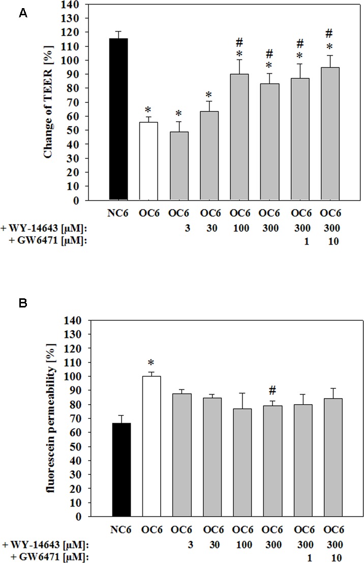
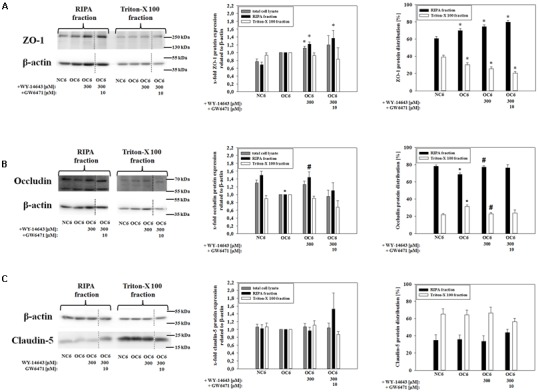
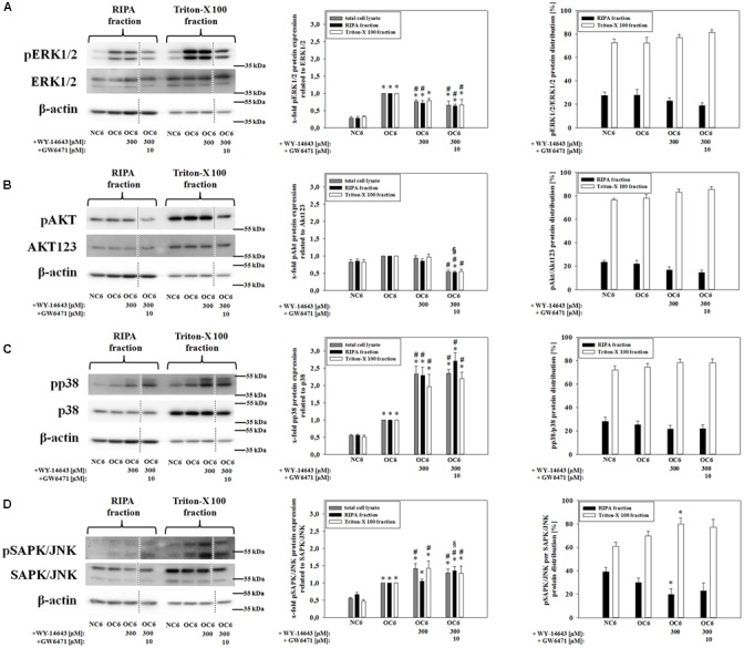
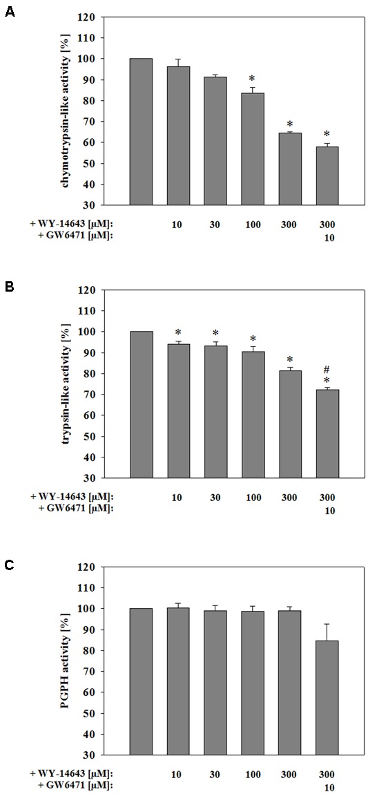
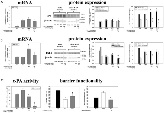
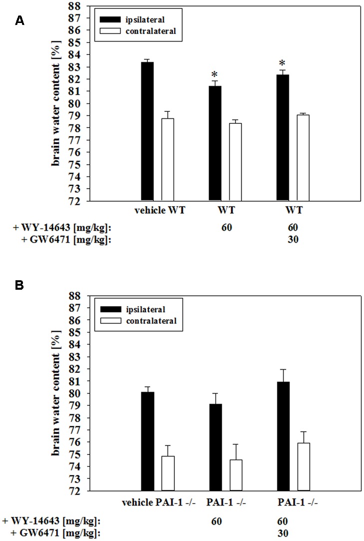
The blood-brain barrier (BBB) is damaged during ischemic insults such as traumatic brain injury or stroke. This contributes to vasogenic edema formation and deteriorate disease outcomes. Enormous efforts are pursued to understand underlying mechanisms of ischemic insults and develop novel therapeutic strategies. In the present study the effects of PPARα agonist WY-14643 were investigated to prevent BBB breakdown and reduce edema formation. WY-14643 inhibited barrier damage in a mouse BBB in vitro model of traumatic brain injury based on oxygen/glucose deprivation in a concentration dependent manner. This was linked to changes of the localization of tight junction proteins. Furthermore, WY-14643 altered phosphorylation of kinases ERK1/2, p38, and SAPK/JNK and was able to inhibit proteosomal activity. Moreover, addition of WY-14643 upregulated PAI-1 leading to decreased t-PA activity. Mouse in vivo experiments showed significantly decreased edema formation in a controlled cortical impact model of traumatic brain injury after WY-14643 application, which was not found in PAI-1 knockout mice. Generally, data suggested that WY-14643 induced cellular responses which were dependent as well as independent from PPARα mediated transcription. In conclusion, novel mechanisms of a PPARα agonist were elucidated to attenuate BBB breakdown during traumatic brain injury in vitro.
Keywords: PPARα; blood-brain barrier; ischemia; pirinixic acid; stroke; traumatic brain injury.
Figures
Similar articles
-
Induction of plasminogen activator inhibitor I by the PPARalpha ligand, Wy-14,643, is dependent on ERK1/2 signaling pathway.Thromb Haemost. 2003 Oct;90(4):611-9. doi: 10.1160/TH03-01-0059. Thromb Haemost. 2003. PMID: 14515181
-
bFGF Protects Against Blood-Brain Barrier Damage Through Junction Protein Regulation via PI3K-Akt-Rac1 Pathway Following Traumatic Brain Injury.Mol Neurobiol. 2016 Dec;53(10):7298-7311. doi: 10.1007/s12035-015-9583-6. Epub 2015 Dec 21. Mol Neurobiol. 2016. PMID: 26687235 Free PMC article.
-
Increased miR-21-3p in Injured Brain Microvascular Endothelial Cells after Traumatic Brain Injury Aggravates Blood-Brain Barrier Damage by Promoting Cellular Apoptosis and Inflammation through Targeting MAT2B.J Neurotrauma. 2019 Apr 15;36(8):1291-1305. doi: 10.1089/neu.2018.5728. Epub 2018 Dec 19. J Neurotrauma. 2019. PMID: 29695199
-
A Mouse Controlled Cortical Impact Model of Traumatic Brain Injury for Studying Blood-Brain Barrier Dysfunctions.Methods Mol Biol. 2018;1717:37-52. doi: 10.1007/978-1-4939-7526-6_4. Methods Mol Biol. 2018. PMID: 29468582 Review.
-
Angioneurins - Key regulators of blood-brain barrier integrity during hypoxic and ischemic brain injury.Prog Neurobiol. 2019 Jul;178:101611. doi: 10.1016/j.pneurobio.2019.03.004. Epub 2019 Apr 7. Prog Neurobiol. 2019. PMID: 30970273 Review.
Cited by
-
Hydroxyethylstarch revisited for acute brain injury treatment.Neural Regen Res. 2021 Jul;16(7):1372-1376. doi: 10.4103/1673-5374.300978. Neural Regen Res. 2021. PMID: 33318420 Free PMC article. Review.
-
Recombinant milk fat globule-EGF factor-8 reduces apoptosis via integrin β3/FAK/PI3K/AKT signaling pathway in rats after traumatic brain injury.Cell Death Dis. 2018 Aug 28;9(9):845. doi: 10.1038/s41419-018-0939-5. Cell Death Dis. 2018. PMID: 30154436 Free PMC article.
-
When Activator and Inhibitor of PPARα Do the Same: Consequence for Differentiation of Human Intestinal Cells.Biomedicines. 2021 Sep 17;9(9):1255. doi: 10.3390/biomedicines9091255. Biomedicines. 2021. PMID: 34572440 Free PMC article.
-
Fibrates Affect Levels of Phosphorylated p38 in Intestinal Cells in a Differentiation-Dependent Manner.Int J Mol Sci. 2023 Apr 22;24(9):7695. doi: 10.3390/ijms24097695. Int J Mol Sci. 2023. PMID: 37175404 Free PMC article.
-
The Role of Clusterin Transporter in the Pathogenesis of Alzheimer's Disease at the Blood-Brain Barrier Interface: A Systematic Review.Biomolecules. 2022 Oct 10;12(10):1452. doi: 10.3390/biom12101452. Biomolecules. 2022. PMID: 36291661 Free PMC article.
References
-
- Atlee J. (2007). Complications in Anesthesia. Philadelphia, PA: Elsevier Health Sciences.
LinkOut - more resources
Full Text Sources
Other Literature Sources
Molecular Biology Databases
Research Materials
Miscellaneous