. 2017 May 26:8:852.
doi: 10.3389/fpls.2017.00852. eCollection 2017.

Structural Characterization of Maize SIRK1 Kinase Domain Reveals an Unusual Architecture of the Activation Segment

Affiliations

Structural Characterization of Maize SIRK1 Kinase Domain Reveals an Unusual Architecture of the Activation Segment

Bruno Aquino et al. Front Plant Sci. .

Abstract

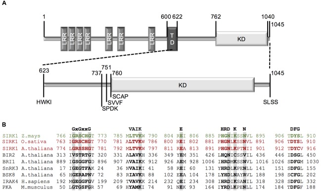
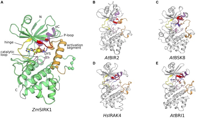
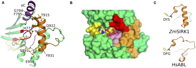
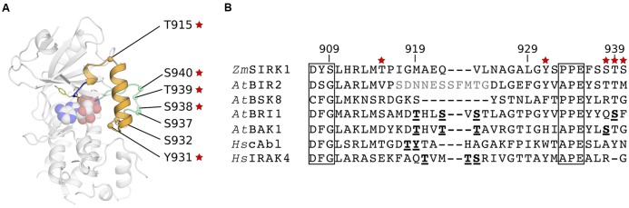
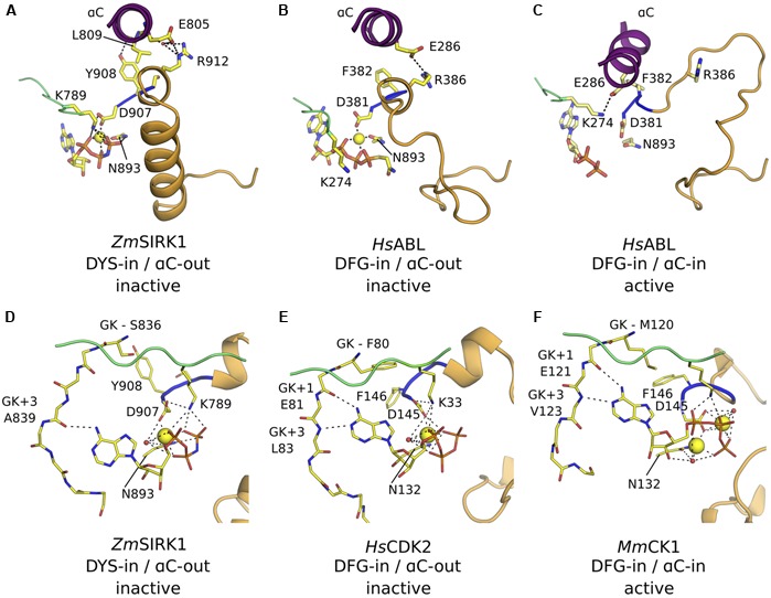
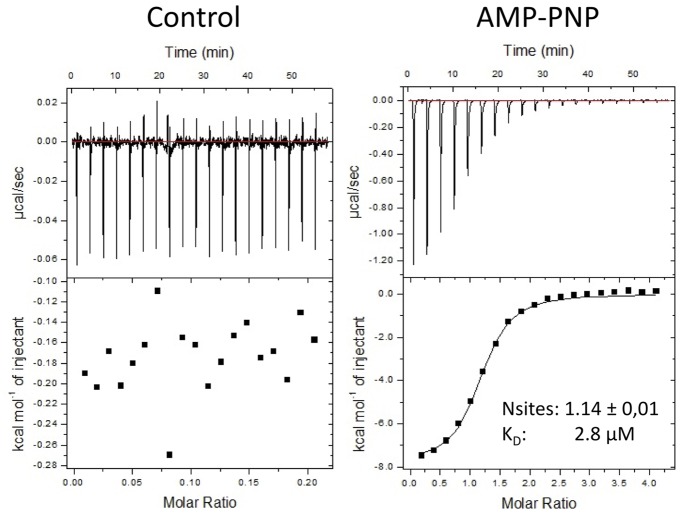
Kinases are primary regulators of plant metabolism and excellent targets for plant breeding. However, most kinases, including the abundant receptor-like kinases (RLK), have no assigned role. SIRK1 is a leucine-rich repeat receptor-like kinase (LRR-RLK), the largest family of RLK. In Arabidopsis thaliana, SIRK1 (AtSIRK1) is phosphorylated after sucrose is resupplied to sucrose-starved seedlings and it modulates the sugar response by phosphorylating several substrates. In maize, the ZmSIRK1 expression is altered in response to drought stress. In neither Arabidopsis nor in maize has the function of SIRK1 been completely elucidated. As a first step toward the biochemical characterization of ZmSIRK1, we obtained its recombinant kinase domain, demonstrated that it binds AMP-PNP, a non-hydrolysable ATP-analog, and solved the structure of ZmSIRK1- AMP-PNP co-crystal. The ZmSIRK1 crystal structure revealed a unique conformation for the activation segment. In an attempt to find inhibitors for ZmSIRK1, we screened a focused small molecule library and identified six compounds that stabilized ZmSIRK1 against thermal melt. ITC analysis confirmed that three of these compounds bound to ZmSIRK1 with low micromolar affinity. Solving the 3D structure of ZmSIRK1-AMP-PNP co-crystal provided information on the molecular mechanism of ZmSIRK1 activity. Furthermore, the identification of small molecules that bind this kinase can serve as initial backbone for development of new potent and selective ZmSIRK1 antagonists.

Keywords: SIRK1; ligand; maize; receptor-like kinase; structure.


Figures

FIGURE 1
FIGURE 1
Cloning strategy for ZmSIRK1. (A) Schematic representation of full-length ZmSIRK1. The extracellular domain (aa 1–600) contains seven leucine-rich repeats (LRR) and is separated from the intracellular domain by a transmembrane domain (TD) (aa 601–622). The intracellular domain (aa 623–1045) contains a kinase domain (KD) (aa 762–1040). Numbers represents the amino acid position and letters the amino acid sequence from the beginning of each construct used for recombinant protein expression. (B) Amino acid sequence alignment of conserved motifs within ZmSIRK1 kinase domains with other protein kinases. GxGxxG: glycine-rich loop; VAIK: motif harboring the catalytic lysine; E: conserved glutamate in helix αC; HRDxKxxN: catalytic loop containing the catalytic aspartate; DFG: DFG motif.
FIGURE 2
FIGURE 2
Co-crystal structure of ZmSIRK1 bound to AMP-PNP. (A) Cartoon representation of the ZmSIRK1-AMP-PNP structure. Protein regions were colored as follows: glycine-rich (P-)loop – red; hinge region – yellow; catalytic loop -pink; DYS (more commonly DFG) motif – blue; activation segment – orange; αC – purple. Other protein regions in green. AMP-PNP is shown in stick and Mg2+ ion as a yellow sphere. The Cαs from putative phosphorylation sites within and immediately C-terminal of ZmSIRK1 activation segment are shown as spheres. (B–E) Cartoon representation of ZmSIRK1 closest structural neighbors. (B) Crystal structure of AtBIR2 (PDB ID 4L68). (C) Crystal structure of AtBSK8 (PDB ID 4I92). (D) Crystal structure of HsIRAK4 (PDB ID 2NRY). (E) Crystal structure of AtBRI1 (PDB ID 5LPB). Protein regions colored as in (A). If present, ligands are represented as sticks and the Cαs from known phosphorylation sites as spheres.
FIGURE 3
FIGURE 3
ZmSIRK1 activation segment is highly structured. (A) Cartoon representation of ZmSIRK1 around the activation segment. Residues taking part in the stabilization of this segment are shown in stick. Possible hydrogen bonds are represented as dashed lines. (B) Surface representation of ZmSIRK1 nucleotide-binding site showing that the protein activation segment (orange) occludes the region normally available to interact with the target peptide. AMP-PNP is shown as spheres. (C) The 310 helix immediately C-terminal of ZmSIRK DYS motif (top) is also seen in inactive state mammalian kinase domains (bottom – human ABL; PDB ID 2G1T). The Cαs from putative phosphorylation sites in ZmSIRK1 and from known phosphorylation sites in HsABL are shown as spheres.
FIGURE 4
FIGURE 4
Conservation of possible phosphorylation sites within ZmSIRK1 activation segment. (A) Cartoon representation of ZmSIRK1 highlighting the position of possible phosphorylation sites within (orange) and C-terminal (green) of the protein activation segment. Red stars indicate sites with high (>80%) conservation within SIRK1 homologs. (B) Structure-based sequence alignment of the activation segment in ZmSIRK and its closest structural neighbors. Conserved kinase domain motifs (commonly DFG and APE) are boxed. Residues in bold and underlined are known phosphorylation sites. Residues in gray were not visible in the structure.
FIGURE 5
FIGURE 5
ZmSIRK1 adopts an inactive DFG-in/αC-out conformation. (A) Cartoon representation of ZmSIRK1 around αC, DYS motif and activation segment showing residues (stick) and interactions (dashed lines) that stabilize the inactive conformation in this protein. (B) Human proteins, such as HsABL (PDB ID 2G1T), can also adopt the DFG-in/αC-out conformation. (C) Phosphorylation of regulatory sites (not shown) within HsABL stabilizes the active (DFG-in/αC-in) conformation. (D) Details of ZmSIRK1 interaction with Mg2+ ion and AMP-PNP showing the unusual Mg2+ ion-coordination, ribose puckering and adenine ring interactions in ZmSIRK1-AMP-PNP co-structure. Possible hydrogen bonds are shown as dashes. (E) Inactive state human proteins, such as CDK2 (PDB ID 1HCK) can also display unusual Mg2+ ion coordination and sugar puckering. (F) Mg2+ ion coordination and nucleotide interaction for the prototypical active state kinase domain of Mus musculus (Ms)CK1 (PDB ID 1ATP). Residues in the hinge region are annotated relative to the gatekeeper residue (GK, GK+1, GK+3).
FIGURE 6
FIGURE 6
ZmSIRK1 binds to AMP-PNP in solution. Isothermal titration calorimetry (ITC) measurements for the interaction between AMP-PNP and ZmSIRK1. Protein (50 μM of ZmSIRK1737-1045) was titrated to AMP-PNP (1 mM) in ITC buffer (50 mM K-phosphate; 500 mM NaCl, 5% glycerol; 1 mM TCEP; 50 mM MgCl2). Y-axis in left (control) and right (AMP-PNP) panels are not in the same scale.

References

    1. Albert M., Jehle A. K., Mueller K., Eisele C., Lipschis M., Felix G. (2010). Arabidopsis thaliana pattern recognition receptors for bacterial elongation factor Tu and flagellin can be combined to form functional chimeric receptors. J. Biol. Chem. 285 19035–19042. 10.1074/jbc.M110.124800 - DOI - PMC - PubMed
    1. Apsel B., Blair J. A., Gonzalez B., Nazif T. M., Feldman M. E., Aizenstein B., et al. (2008). Targeted polypharmacology: discovery of dual inhibitors of tyrosine and phosphoinositide kinases. Nat. Chem. Biol. 4 691–699. 10.1038/nchembio.117 - DOI - PMC - PubMed
    1. Arrowsmith C. H., Audia J. E., Austin C., Baell J., Bennett J., Blagg J., et al. (2015). The promise and peril of chemical probes. Nat. Chem. Biol. 11 536–541. 10.1038/nchembio.1867 - DOI - PMC - PubMed
    1. Bain J., Plater L., Elliott M., Shpiro N., Hastie C. J., McLauchlan H., et al. (2007). The selectivity of protein kinase inhibitors: a further update. Biochem. J. 408 297–315. 10.1042/BJ20070797 - DOI - PMC - PubMed
    1. Blaum B. S., Mazzotta S., Noldeke E. R., Halter T., Madlung J., Kemmerling B., et al. (2014). Structure of the pseudokinase domain of BIR2, a regulator of BAK1-mediated immune signaling in Arabidopsis. J. Struct. Biol. 186 112–121. 10.1016/j.jsb.2014.02.005 - DOI - PubMed