

doi: 10.21769/BioProtoc.2294.
Isolation and Cultivation of Primary Brain Endothelial Cells from Adult Mice
Affiliations
- PMID: 28603749
- PMCID: PMC5464392
- DOI: 10.21769/BioProtoc.2294
Item in Clipboard
Isolation and Cultivation of Primary Brain Endothelial Cells from Adult Mice
Bio Protoc.
.
Abstract
Brain endothelial cells are the major building block of the blood-brain barrier. To study the role of brain endothelial cells in vitro, the isolation of primary cells is of critical value. Here, we describe a protocol in which vessel fragments are isolated from adult mice. After density centrifugation and mild digestion of the fragments, outgrowing endothelial cells are selected by puromycin treatment and grown to confluence within one week.
Keywords: Blood-brain barrier; CD31; Claudin-5; Occludin; Primary culture; Tight junctions; VE-cadherin; ZO-1.
Figures
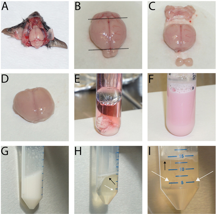
The images depict the first steps of the isolation procedure showing the brain in situ after removal of the skullcap (A), before (B. cut planes indicated by dashed line) and after removal of cerebellum, olfactory bulb (C) and the meninges (D). Then, collect the brains in a Dounce tissue grinder (E), homogenize them (F), and centrifuge the tissue homogenate. Next, resuspend the cells in the dextran solution and vortex extensively (G). Following the centrifugation, the resulting myelin layer is at the top while the vessel fragments collect around the edge of the tube bottom (H + I, black arrows: myelin layer, white arrows: pellet location). The size of the vessel fragment pellet depends on the number of brains used. In E-I, two brains were used for the preparation.
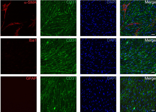
Cells were fixed with 4% paraformaldehyde 14 days after isolation and subsequently stained for CD31 (BD, 1:500) as an endothelial cell specific marker in combination with α-SMA (pericytes and smooth muscle cells, Acris, 1:200, upper row), Iba1 (microglia, Wako Pure Chemical Industries, 1:100, middle row) and GFAP (astrocytes, Millipore, 1:400, lower row). Scale bars represent 50 µm.
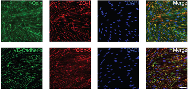
Cells were fixed 6-8 days after isolation with ice-cold methanol and subsequently stained for the tight junction proteins Ocln (Sigma-Aldrich, 1:500), ZO-1 (Thermo Fisher Scientific, 1:500), Cldn-5 (Thermo Fisher Scientific, 1:500) and the adherens junction protein VE-Cadherin (Santa Cruz Biotechnology, 1:500). Scale bars represent 50 µm.

Note the attached vessel fragment and its radially outgrowing endothelial cells after 2 days in culture. Scale bars represent 100 µm.
References
-
- Ridder D. A., Wenzel J., Müller K., Töllner K., Tong X. K., Assmann J. C., Stroobants S., Weber T., Niturad C., Fischer L., Lembrich B., Wolburg H., M. Grand'Maison, Papadopoulos P., Korpos E., Truchetet F., Rades D., Sorokin L. M., Schmidt-Supprian M., Bedell B. J., Pasparakis M., Balschun D., R. D’Hooge, Löscher W., Hamel E. and Schwaninger M.(2015). Brain endothelial TAK1 and NEMO safeguard the neurovascular unit. J Exp Med 212(10): 1529-1549. - PMC - PubMed
-
- Song L. and Pachter J. S.(2003). Culture of murine brain microvascular endothelial cells that maintain expression and cytoskeletal association of tight junction-associated proteins. In Vitro Cell Dev Biol Anim 39(7): 313-320. - PubMed
Grants and funding
LinkOut - more resources
Full Text Sources
Other Literature Sources
