. 2017 Jul 13;60(13):5826-5833.
doi: 10.1021/acs.jmedchem.7b00546. Epub 2017 Jun 21.

α-Conotoxin [S9A]TxID Potently Discriminates between α3β4 and α6/α3β4 Nicotinic Acetylcholine Receptors

Affiliations

α-Conotoxin [S9A]TxID Potently Discriminates between α3β4 and α6/α3β4 Nicotinic Acetylcholine Receptors

Yong Wu et al. J Med Chem. .

Abstract

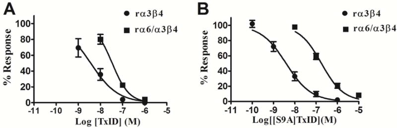
α3β4 nAChRs have been implicated in various pathophysiological conditions. However, the expression profile of α3β4 nAChRs and α6/α3β4 nAChRs overlap in a variety of tissues. To distinguish between these two subtypes, we redesigned peptide 1 (α-conotoxin TxID), which inhibits α3β4 and α6/α3β4 nAChR subtypes. We systematically mutated 1 to evaluate analogue selectivity for α3β4 vs α6/α3β4 nAChRs expressed in Xenopus laevis oocytes. One analogue, peptide 7 ([S9A]TxID), had 46-fold greater potency for α3β4 versus α6/α3β4 nAChRs. Peptide 7 had IC50s > 10 μM for other nAChR subtypes. Molecular dynamics simulations suggested that Ser-9 of TxID was involved in a weak hydrogen bond with β4 Lys-81 in the α6β4 binding site but not in the α3β4 binding site. When Ser-9 was substituted by an Ala, this hydrogen bond interaction was disrupted. These results provide further molecular insights into the selectivity of 7 and provide a guide for designing ligands that block α3β4 nAChRs.


Conflict of interest statement

Notes

The authors declare no competing financial interest.

Figures

Figure 1
Figure 1
The sequences of peptide 1 and its analogs. The disulfide connectivity of Cys (I–III, II–IV) is schematically illustrated with lines connecting the Cys residues. Substituted amino acids that were mutated to explore structure-activity relationships are marked in red. (The * indicates a C-terminal amide). Also shown for comparison is peptide 17 (α-conotoxin AuIB), for which we compare structure and activity data. aCompound number.
Figure 2
Figure 2
HPLC chromatograms and mass spectra of peptide 1 and 7. The peptides were analyzed on a reversed phase analytical Vydac C18 HPLC column using a linear gradient of 10% buffer B to 40% buffer B over 20 min, where buffer A = 0.65% trifluoroacetic acid (TFA), remainder H2O, B = 0.5% TFA, 90% acetonitrile, remainder H2O. Absorbance was monitored at 214 nm. (A) HPLC chromatogram of peptide 1; (B) electrospray ionization mass spectrometry (ESI-MS) data for peptide 1 with observed monoisotopic mass of 1488.6 Da; (C) HPLC chromatogram of peptide 7; (D) ESI-MS data for peptide 1 with an observed monoisotopic mass of 1472.6 Da.
Figure 3
Figure 3
Concentration-response analysis for inhibition of α3β4 and α6/α3β4 nAChRs by pepitde 1 and 7. (A) Pepitde 1 selectively blocks α3β4 nAChR vs α6/α3β4 nAChR by only 10-fold; (B) Peptide 7 selectively blocks α3β4 nAChR vs α6/α3β4 nAChR by 46-fold. The IC50 values are in Table 1. Error bars represent S.E.M. values from 4–5 oocytes for each experimental determination.
Figure 4
Figure 4
Block by peptide 1 and 7 of rat α3β4 and α6/α3β4 nAChRs. Xenopus laevis oocytes expressing the indicated nAChRs were voltage-clamped at −70 mV and subjected to a 1 s pulse of ACh every minute as described in the Experimental Section. A representative response in a single oocyte is shown. After control responses to ACh, the oocyte was exposed to 10 nM or 100 nM toxin for 5 min (arrow). The toxin was then washed out, and the response to ACh was again measured. “C” indicates control responses to ACh.
Figure 5
Figure 5
Comparison of NMR spectroscopy of the peptide 7 and wild-type peptide 1. (A) Secondary chemical shift analysis of peptide 7 isomer 1 (black) compared to that of isomer 1 of peptide 1 (grey). (B) Homology models of the two peptide 7 isomers (grey) compared to the NMR solution structures of the wild-type isomers (white). The amino acid sequence of peptide 7 is displayed, with the substituted residue for the mutant shown in brackets. The homology models were refined using extensive energy minimization in explicit water. The models suggest that the three-dimensional structure of the isomers is essentially not affected by the S9A substitution.
Figure 6
Figure 6
Comparison of molecular models of the binding mode of pepitde 1 at the α3β4 (A) and α6β4(B) interface, and distance between side chain hydroxyl of peptide 1 Ser-9 and side chain nitrogen of β4 Lys-81 during 50 ns molecular dynamics (MD) simulations. The molecular models shown are the final frames of the 50 ns MD simulations, and the α3 subunit is drawn in pink, the α6 is in blue and the β4 subunits are in green. A hydrogen bond between Ser-9 and Lys-81 is indicated with a dotted line. Positions of β4 subunits are numbered according to the full-length sequence of rat β4 subunit precursor sequence (UniProt identifier P12392).

Similar articles

Cited by

References

    1. Albuquerque EX, Pereira EF, Alkondon M, Rogers SW. Mammalian nicotinic acetylcholine receptors: from structure to function. Physiol Rev. 2009;89:73–120. - PMC - PubMed
    1. Gotti C, Clementi F. Neuronal nicotinic receptors: from structure to pathology. Prog Neurobiol. 2004;74:363–396. - PubMed
    1. Millar NS, Gotti C. Diversity of vertebrate nicotinic acetylcholine receptors. Neuropharmacology. 2009;56:237–246. - PubMed
    1. Hurst R, Rollema H, Bertrand D. Nicotinic acetylcholine receptors: from basic science to therapeutics. Pharmacol Ther. 2013;137:22–54. - PubMed
    1. Zoli M, Pistillo F, Gotti C. Diversity of native nicotinic receptor subtypes in mammalian brain. Neuropharmacology. 2015;96:302–311. - PubMed

Publication types

LinkOut - more resources