Review
. 2017 Jun 30;127(7):2492-2504.
doi: 10.1172/JCI90597. Epub 2017 Jun 12.

Antibody-mediated rejection across solid organ transplants: manifestations, mechanisms, and therapies

Review

Antibody-mediated rejection across solid organ transplants: manifestations, mechanisms, and therapies

Nicole M Valenzuela et al. J Clin Invest. .

Abstract

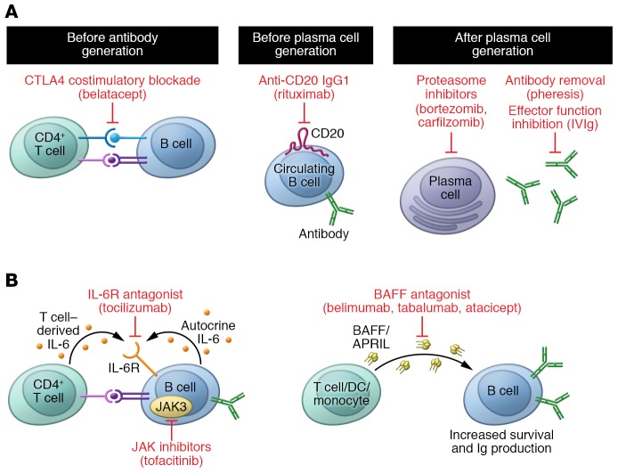
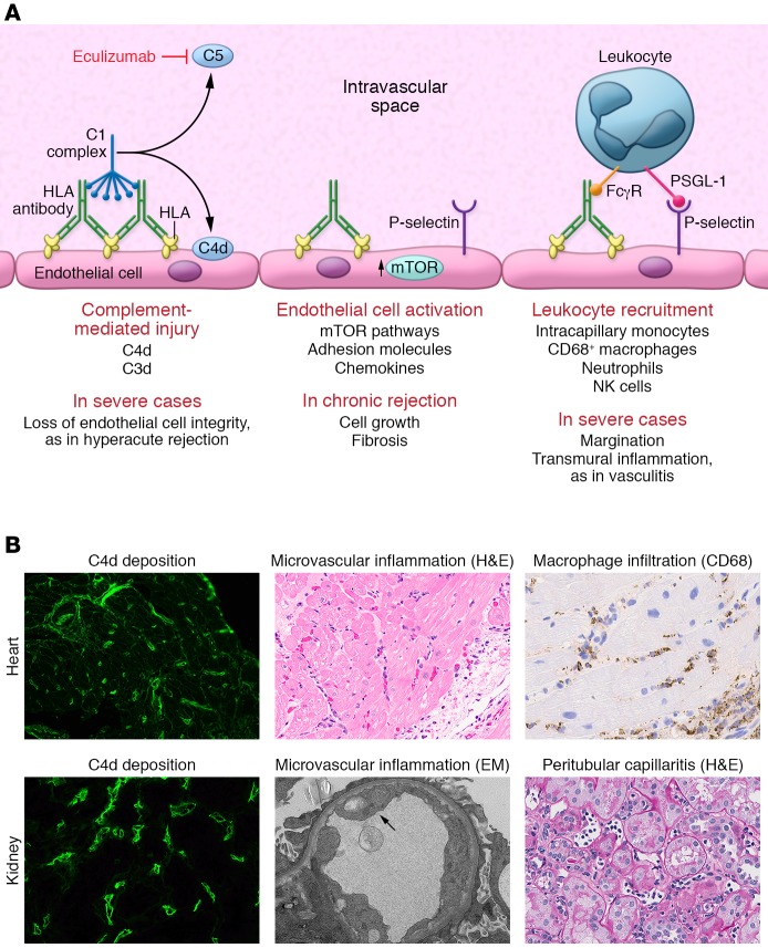
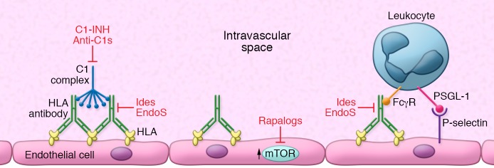
Solid organ transplantation is a curative therapy for hundreds of thousands of patients with end-stage organ failure. However, long-term outcomes have not improved, and nearly half of transplant recipients will lose their allografts by 10 years after transplant. One of the major challenges facing clinical transplantation is antibody-mediated rejection (AMR) caused by anti-donor HLA antibodies. AMR is highly associated with graft loss, but unfortunately there are few efficacious therapies to prevent and reverse AMR. This Review describes the clinical and histological manifestations of AMR, and discusses the immunopathological mechanisms contributing to antibody-mediated allograft injury as well as current and emerging therapies.


Conflict of interest statement

Conflict of interest: E.F. Reed received research grant funding from CSL Behring and Immucor Inc. E.F. Reed is an inventor on US Patent Publication No. 20170030928.

Figures

Figure 1
Figure 1. Current and emerging therapies to prevent HLA antibody production.
(A) Naive B cells rely on signals from CD4+ T cells for full activation, Ig class switching, and Ig production. Blockade of costimulation using the CTLA4 fusion protein belatacept inhibits this important signal and hampers B cell activation. Naive as well as antigen-experienced B cells may be depleted from the circulation with antibodies against CD20, such as rituximab. Once HLA antibody–producing B cells have differentiated into plasma cells, they are more difficult to target. Because these antibody “factories” are metabolically active and reliant on the function of the proteasome, inhibitors such as bortezomib and carfilzomib, which block the 20S proteasome, may trigger apoptosis of these cells. Bortezomib is a reversible inhibitor, while carfilzomib is irreversible. Antibodies may also be physically removed from the circulation with plasmapheresis, and their effector functions inhibited by intravenous immunoglobulin (IVIg). (B) Emerging therapies to reduce alloantibody production include antagonism of the IL-6 receptor (tocilizumab), BAFF (belimumab, tabalumab, atacicept), and inhibition of intracellular JAK (tofacitinib), which are needed for B cell activation and Ig production. BAFF/APRIL, B cell–activating factor/a proliferation-inducing ligand.
Figure 2
Figure 2. The known mechanisms of HLA antibody–mediated allograft injury and their therapeutic targets.
(A) HLA antibody binding to donor endothelial cells can trigger activation of the classical complement cascade. First, the complement C1 complex, which includes C1q, C1r, and C1s, binds to the IgG heavy chain Fc region. The C1 complex next sequentially activates serum complement proteins, catalyzing the generation of immunologically active split products. Activation of complement C5 protein occurs at the terminal stage of the signaling pathway, generating the highly potent anaphylatoxin C5a and initiating assembly of the membrane attack complex (MAC). In the process, the split product C4d becomes covalently bound to the endothelial cell surface and can be detected in biopsies of allografts undergoing rejection through immunofluorescent or immunohistochemical staining. Eculizumab is an mAb that prevents C5 cleavage. HLA binding also activates intracellular signaling within the donor endothelium, mainly via the mTOR signaling axis, and upregulation of the adhesion molecule P-selectin. P-selectin, in conjunction with the interaction between immune cell Fcγ receptors (FcγRs), enhances leukocyte-endothelial adhesion. PSGL-1, P-selectin glycoprotein 1. (B) Representative micrographs illustrating the three main histological features of AMR in heart (top panels) and kidney (bottom panels) allografts. Deposition of C4d within the microvasculature is visualized by immunofluorescence in green. H&E staining demonstrates increased capillary endothelial cell size and numerous leukocytes in the intravascular space. Immunohistochemical staining for CD68 highlights intracapillary macrophages (original magnification, ×400). EM, electron micrograph.
Figure 3
Figure 3. Emerging therapies to inhibit HLA antibody–induced allograft injury.
Emerging therapies for AMR include upstream complement inhibition with C1 esterase (C1-INH) or antibody against C1s. DSAs are capable of cross-linking HLA molecules on the donor endothelial and smooth muscle cells, resulting in activation of the mTOR signaling axis. Rapamycin (sirolimus) and other rapalogs (everolimus) inhibit mTOR and may dampen vascular cell growth and fibrosis, which contributes to chronic rejection. Finally, abolishing Fc-dependent antibody effector functions through cleavage of the Fc region with IdeS, or removal of the N-linked glycan with EndoS, is likely to hamper activation of the classical complement cascade as well as prevent interactions with FcγRs on monocytes, neutrophils, and NK cells.

Similar articles

Cited by

References

    1. Hart A, et al. OPTN/SRTR 2015 Annual Data Report: Kidney. Am J Transplant. 2017;17(suppl 1):21–116. - PMC - PubMed
    1. Patel R, Terasaki PI. Significance of the positive crossmatch test in kidney transplantation. N Engl J Med. 1969;280(14):735–739. doi: 10.1056/NEJM196904032801401. - DOI - PubMed
    1. Schinstock CA, et al. The value of protocol biopsies to identify patients with de novo donor-specific antibody at high risk for allograft loss. Am J Transplant. doi: 10.1111/ajt. [published online ahead of print December 15, 2016]. https://doi.org/10.1111/ajt.14161. - DOI - DOI - PMC - PubMed
    1. Everly MJ, et al. Incidence and impact of de novo donor-specific alloantibody in primary renal allografts. Transplantation. 2013;95(3):410–417. doi: 10.1097/TP.0b013e31827d62e3. - DOI - PubMed
    1. Viglietti D, et al. Value of donor-specific anti-HLA antibody monitoring and characterization for risk stratification of kidney allograft loss. J Am Soc Nephrol. 2017;28(2):702–715. doi: 10.1681/ASN.2016030368. - DOI - PMC - PubMed