. 2017 Aug;13(8):888-894.
doi: 10.1038/nchembio.2407. Epub 2017 Jun 12.

Oxidation of phosphorothioate DNA modifications leads to lethal genomic instability

Affiliations

Oxidation of phosphorothioate DNA modifications leads to lethal genomic instability

Stefanie Kellner et al. Nat Chem Biol. 2017 Aug.

Abstract

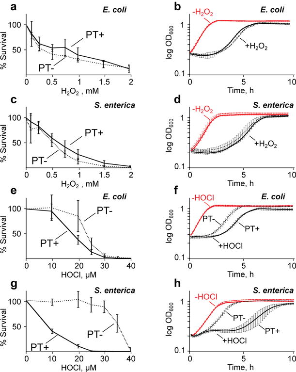
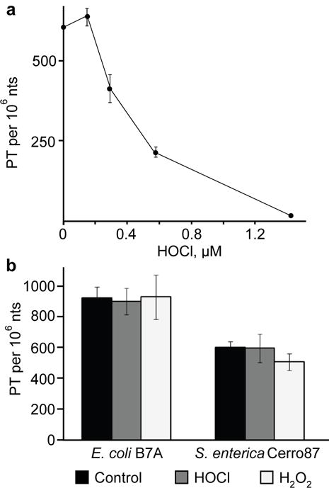
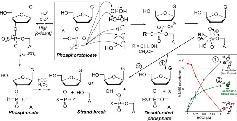
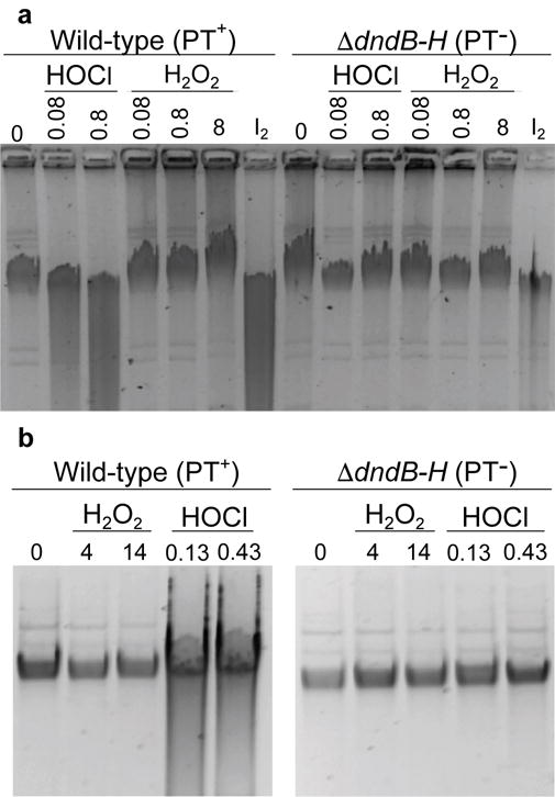
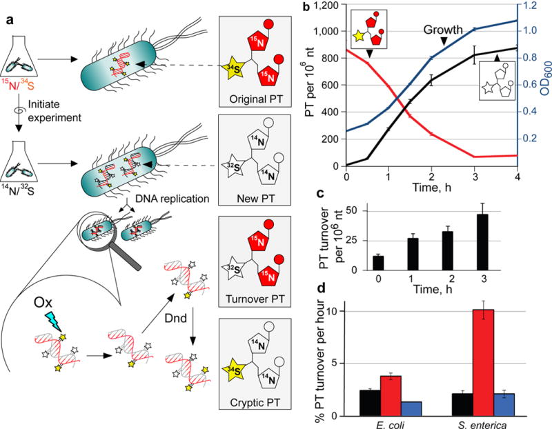
Genomic modification by sulfur in the form of phosphorothioate (PT) is widespread among prokaryotes, including human pathogens. Apart from its physiological functions, PT sulfur has redox and nucleophilic properties that suggest effects on bacterial fitness in stressful environments. Here we show that PTs are dynamic and labile DNA modifications that cause genomic instability during oxidative stress. In experiments involving isotopic labeling coupled with mass spectrometry, we observed sulfur replacement in PTs at a rate of ∼2% h-1 in unstressed Escherichia coli and Salmonella enterica. Whereas PT levels were unaffected by exposure to hydrogen peroxide (H2O2) or hypochlorous acid (HOCl), PT turnover increased to 3.8-10% h-1 after HOCl treatment and was unchanged by H2O2, consistent with the repair of HOCl-induced sulfur damage. PT-dependent sensitivity to HOCl extended to cytotoxicity and DNA strand breaks, which occurred at HOCl doses that were orders of magnitude lower than the corresponding doses of H2O2. The genotoxicity of HOCl in PT-containing bacteria suggests reduced fitness in competition with HOCl-producing organisms and during infections in humans.


Conflict of interest statement

COMPETING INTERESTS

The authors declare no competing financial interest.

Figures

Figure 1
Figure 1. The effect of PT modifications on survival and growth of bacteria following oxidant exposures
Wild-type (PT+, solid line) and ΔdndB–H (PT–, dashed line) strains of E. coli (A, B, E, F) and S. enterica (C, D, G, H) were exposed to H2O2 (A–D) or HOCl (E–H). Cytotoxicity assays (A, C, E, G) were performed with the indicated concentrations of H2O2 and HOCl. Growth curves (B, D, F, H) were then prepared using the LD80 doses of H2O2 (B, D) and HOCl (F, H). Red lines in the growth curves (B, D, F, H) indicate unexposed controls, with overlapping curves for wild-type and ΔdndB–H strains; black lines represent exposed bacteria, with data plotted in solid lines (PT+) distinguishing from dotted lines (PT–) only in panels F and H. Data represent mean ± SD for 3 biological replicates. Statistically significant differences among the data sets are discussed in the text.
Figure 2
Figure 2. PT reaction with HOCl leads to loss of PT and strand breaks in vitro and in vivo
(a) Quantification of PT in wild-type E. coli B7A DNA exposed to HOCl in vitro. Data represent mean ± SD for 3 biological replicates. (b) PT-containing dinucleotides were quantified by LC-MS/MS 3 hours (E. coli wild-type) and 5 hours (S. enterica wild-type) after exposure to their respective LD50 doses of either HOCl or H2O2 (as specified in Supplementary Table 3; data represent mean ± SD for 3 biological replicates).
Figure 3
Figure 3. Comprehensive model for PT oxidation and alkylation in DNA
Analysis of degradation products arising in reactions of HOCl and H2O2 with PT-containing dinucleotides (inset and Supplementary Fig. 4) revealed products consistent with the reaction model shown. The sulfur in PTs can undergo two types of reactions based on nucleophilic substitution (right side) and one-electron oxidation (left side). At low concentrations of HOCl or H2O2, sulfur reacts by nucleophilic substitution to generate sulfonyl chloride (Cl-S) or sulfenic acid (HO-S), which subsequently undergo hydrolysis to eliminate the modified sulfur (pathway 1; X = sulfur-containing product) or to form a DNA strand-break (pathway 2) possibly with a modified sulfur (X). At high concentrations (left side), both H2O2 and HOCl generate radicals (hydroxyl radical, hypochoryl radical) that perform a series of one-electron oxidations leading to phosphonate formation and subsequent DNA strand-breaks containing a phosphonate (X = H), phosphate (X = O), or other product. Inset: Concentration-dependent degradation of d(GPSA) by HOCl causes both desulfuration to a phosphate-linked dinucleotide and a strand-break with release of dG; data represent mean ± SD for 3 replicates.
Figure 4
Figure 4. PT reaction with HOCl leads to loss of PT and strand breaks in vitro and in vivo
(a) Isolated DNA from wild-type (PT+) and ΔdndB–H (PT–) E. coli were exposed to 0.08–0.8 mM HOCl or 0.08–8 mM H2O2; iodine (I2) exposure serves as a positive control for PT-dependent strand breaks. HOCl-induced strand-breaks are apparent as smearing in the lane for DNA containing PT (PT+), but not for DNA lacking PT (PT–); strand-breaks are not detectable for H2O2 exposure in any case. (b) WT and ΔdndB–H E. coli cells were exposed to 7.5- and 25-times the WT LD50 concentration of H2O2 or HOCl for 10 min (4 and 14 mM for H2O2; 0.13 and 0.43 for HOCl). Again, DNA isolated from HOCl-exposed WT bacteria, but not PT– bacteria, shows strand-breaks, while no strand breaks are apparent after H2O2 treatment in either strain. Gel images are cropped for clarity (see Supplementary Fig. 9 for unprocessed images). Data for LD50 and LD80 doses are presented in Supplementary Table 3.
Figure 5
Figure 5. Quantification of PT turnover by isotope-labeling and mass spectrometry
(a) Principle of PT turnover assay. Bacteria were grown in minimal medium containing [15N]/[34S]-labeled nutrients, with resulting bacterial DNA containing [15N]-labeled nucleobases with [34S]-labeled PTs (Original PT). To initiate an experiment, cells are transferred to minimal medium with [14N]/[32S]-labeled constituents and subjected to stresses. Newly synthesized PT-linked dinucleotides (New PT) are labeled with [14N] and [32S]. The [34S] in the original PTs was found to undergo replacement with [32S] (Turnover PT) in both untreated and oxidant-treated cells. We also observed low levels of PT dinucleotides labeled with [34S] and [14N] (Cryptic PT) resulting from reuse of [34S] from nutrient pools or other PT modification sites. In all cases, isolated DNA was hydrolyzed to canonical mononucleotides and PT-containing dinucleotides, and quantities of various isotope-labeled dinucleotides were determined by LC-MS/MS analysis. (b) Validation of PT turnover method. Cultures of wild-type E. coli B7A were grown with [15N]/[34S]-containing medium, and then cells were transferred to [14N]/[32S]-containing medium. At various times, genomic DNA was isolated and subjected to LC-MS/MS analysis of PT-containing dinucleotides. The plot shows cell growth (OD600; blue line), original PT (red line) and new PT (black line). (c) The level of [15N]/[33S]-labeled, PT-containing dinucleotides (“turnover” PT) were determined in wild-type, untreated E. coli B7A as a function of time. The level of this turnover PT as a function of total PT was used to calculate the %PT turnover. (d) Estimates of the rate of PT turnover per hour were calculated by performing linear regression analysis of plots of %PT turnover as a function of time in unstressed E. coli B7A and S. enterica Cerro 87 (black) and for bacteria exposed to HOCl (red) and H2O2 (blue), with regression analysis illustrated in Supplementary Figure 7 for wild-type, untreated Cerro 87. Data represent mean ± SD for 3 biological replicates and values for HOCl-treated samples are significantly different from untreated and H2)2-treated by Student’s t-test at P < 0.05.

References

    1. Bayliss CD. Determinants of phase variation rate and the fitness implications of differing rates for bacterial pathogens and commensals. FEMS Microbiol Rev. 2009;33:504–520. - PubMed
    1. Sanchez-Romero MA, Cota I, Casadesus J. DNA methylation in bacteria: from the methyl group to the methylome. Curr Opin Microbiol. 2015;25:9–16. - PubMed
    1. Tock MR, Dryden DT. The biology of restriction and anti-restriction. Curr Opin Microbiol. 2005;8:466–472. - PubMed
    1. Wilson GG, Murray NE. Restriction and modification systems. Ann Rev Genetics. 1991;25:585–627. - PubMed
    1. Thiaville JJ, et al. Novel genomic island modifies DNA with 7-deazaguanine derivatives. Proc Natl Acad Sci USA. 2016;113(11):E1452–E1459. - PMC - PubMed

ONLINE METHODS REFERENCES

    1. Perez-Cruz F, et al. Use of pyrogallol red and pyranine as probes to evaluate antioxidant capacities towards hypochlorite. Molecules. 2013;18:1638–1652. - PMC - PubMed
    1. Beers RF, Jr, Sizer IW. A spectrophotometric method for measuring the breakdown of hydrogen peroxide by catalase. J Biol Chem. 1952;195:133–140. - PubMed
    1. Held AM, Halko DJ, Hurst JK. Mechanisms of chlorine oxidation of hydrogen peroxide. J Am Chem Soc. 1978;100:5732–5740.
    1. Evans DF, Upton MW. Studies on singlet oxygen in aqueous solution. Part 1. Formation of singlet oxygen from hydrogen peroxide with two-electron oxidants. J Chem Soc Dalton Trans. 1985:1141–1145.
    1. Shams El Din AM, Mohammed RA. Kinetics of the reaction between hydrogen peroxide and hypochlorite. Desalination. 1998;115:145–153.

MeSH terms