. 2017 Jun 12;6(6):e348.
doi: 10.1038/oncsis.2017.45.

The deubiquitinating enzymes USP4 and USP17 target hyaluronan synthase 2 and differentially affect its function

Affiliations

The deubiquitinating enzymes USP4 and USP17 target hyaluronan synthase 2 and differentially affect its function

M Mehić et al. Oncogenesis. .

Abstract

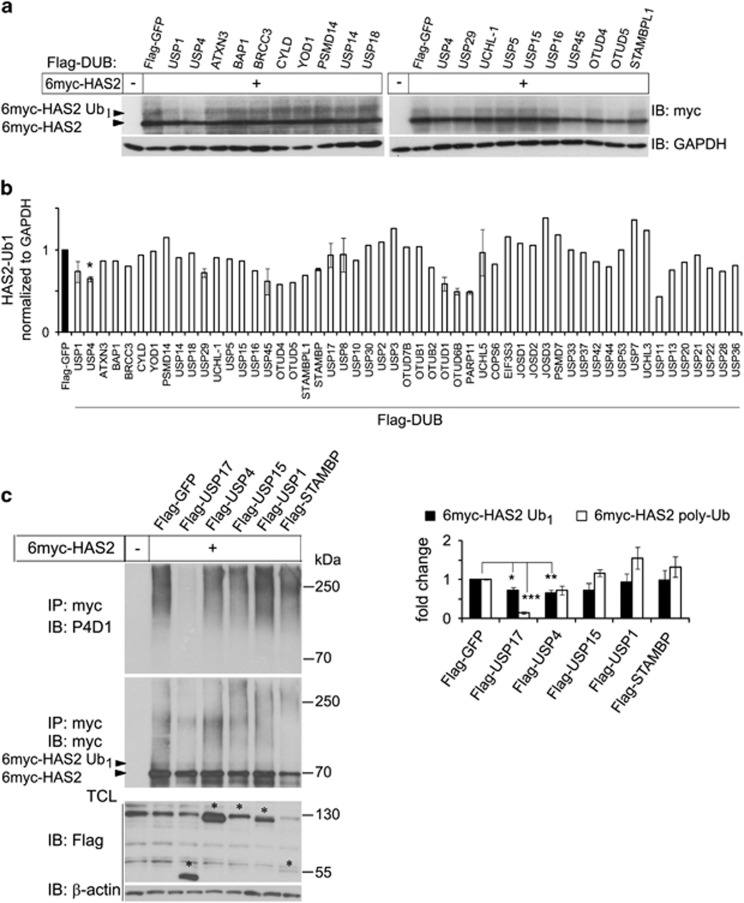
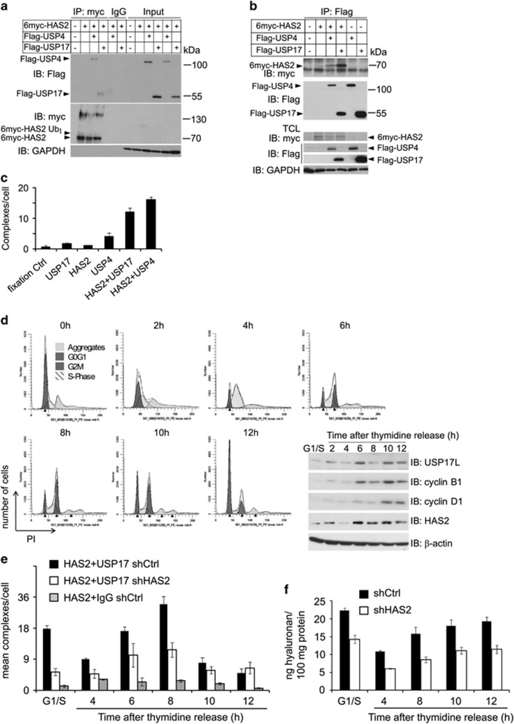
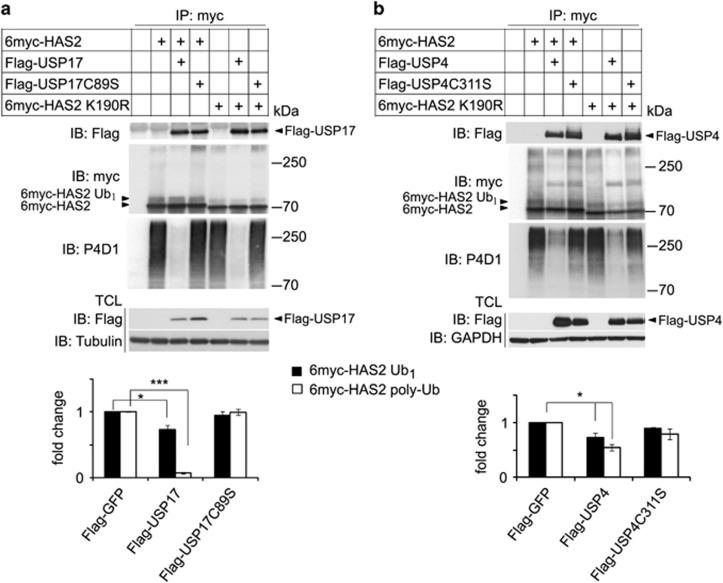
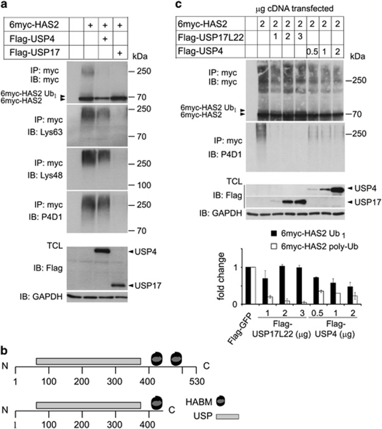
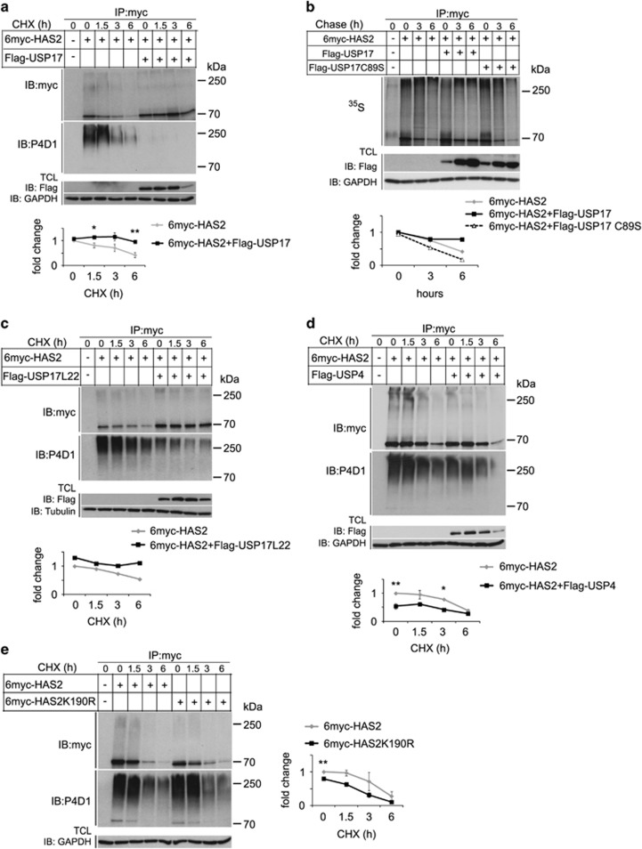
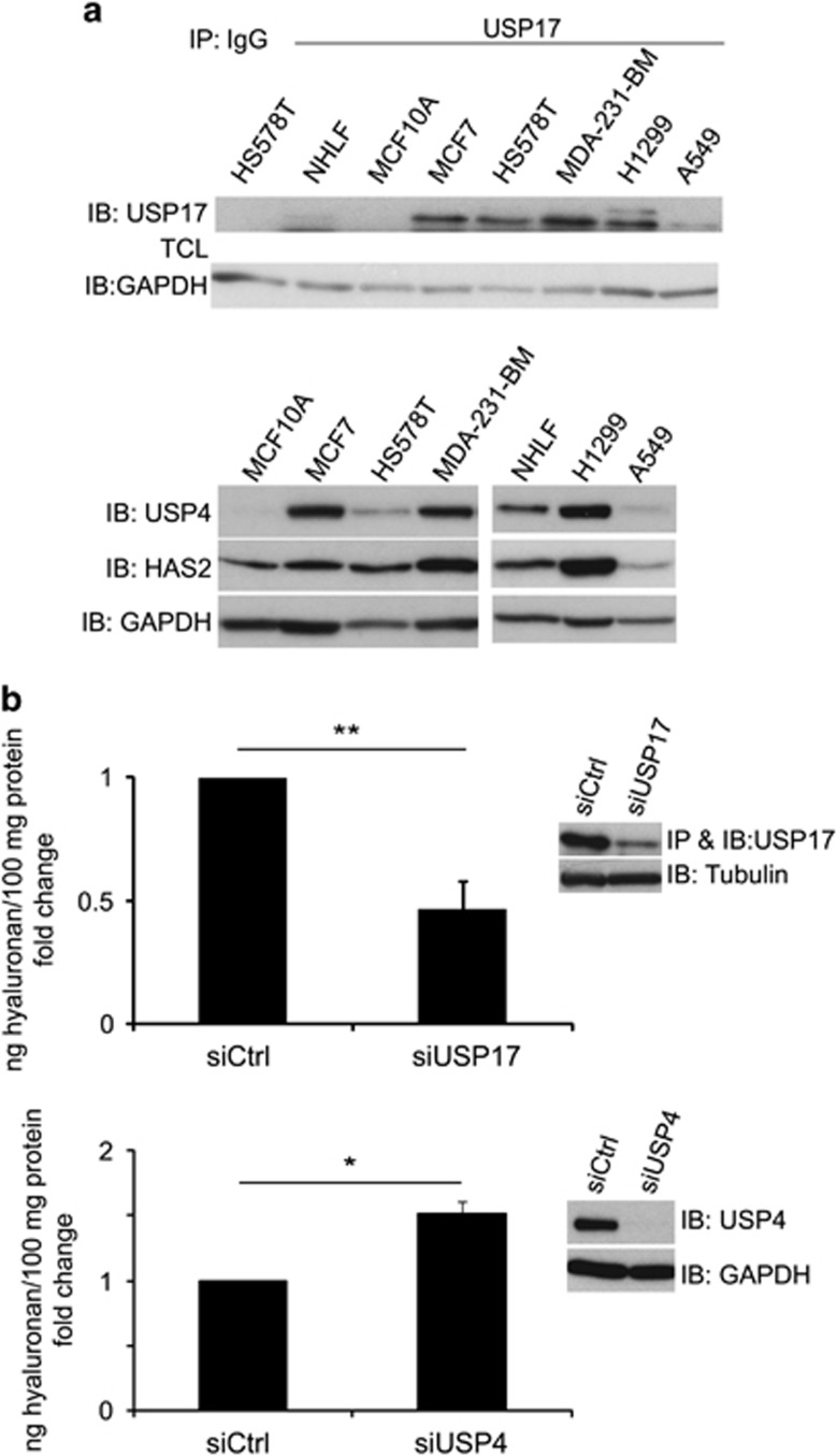
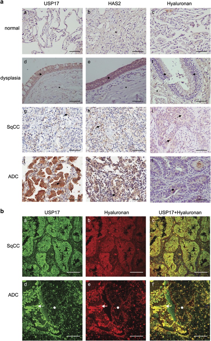
The levels of hyaluronan, a ubiquitous glycosaminoglycan prominent in the extracellular matrix, is balanced through the actions of hyaluronan-synthesizing enzymes (HAS1, 2 and 3) and degrading hyaluronidases (Hyal 1, 2, 3 and PH20). Hyaluronan accumulates in rapidly remodeling tissues, such as breast cancer, due to deregulated expression of the HAS2 gene and/or alterations of HAS2 activity. The activity of HAS2 is regulated by post-translational modifications, including ubiquitination. In order to identify deubiquitinating enzymes (DUBs) that are involved in de-ubiquitination of HAS2, a complementary (cDNA) library of 69 Flag-HA-tagged human DUBs cloned into retroviral vectors was screened in human embryonic kidney (HEK) 293T cells for their ability to de-ubiquitinate myc-tagged HAS2. Several DUBs were found to decrease the ubiquitination of 6myc-HAS2, among which, the most effective were USP17 and USP4. USP17 efficiently removed polyubiquitination, whereas USP4 preferentially removed monoubiquitination of 6myc-HAS2. Co-immunoprecipitation studies revealed interactions between HAS2 and USP17, as well as between HAS2 and USP4, in membrane preparations of HEK293T cells. USP17 significantly stabilized 6myc-HAS2 protein levels, whereas USP4 did not. The silencing of USP17 led to decreased hyaluronan production, whereas the suppression of USP4 increased hyaluronan synthesis. Importantly, high levels of USP17 and HAS2 were detected in a panel of cancer cell lines compared to normal cells, and immunohistochemical stainings revealed higher expression of USP17 and HAS2 in tissues of lung cancer patients compared to normal tissue. In conclusion, USP17 and USP4 differently affect HAS2 ubiquitination, and the stability and function of HAS2.


Conflict of interest statement

The authors declare no conflict of interest.

Figures

Figure 1
Figure 1
Identification of USP4 and USP17 as de-ubiquitinases of HAS2 by a DUB cDNA expression screen. (a) HEK293T cells (0.3 × 10⁶ cells per well in six-well plates) were co-transfected with 6myc-tagged HAS2 cDNA and individual Flag- and HA-tagged DUB cDNAs. 6myc-tagged empty vector and Flag-tagged vector encoding GFP were used as controls and to equalize the DNA load. Cell lysates were subjected to SDS–PAGE followed by immunoblotting with antibodies against myc as described in Materials and methods, to detect immunoreactive myc-tagged monoubiquitinated HAS2 (HAS2-Ub1; seen as a band of 5–10 kDa higher molecular mass than the 6myc-HAS2 band). (b) Quantification of HAS2-Ub1 of the immunoblots, using ImageJ. Asterisk indicates P<0.05 calculated with Student’s t-test (n=5), and error bars are the average of two experiments. (c) Re-screening of a subset of the DUBs described in a to determine the effects on polyubiquitination of HAS2; denaturated cell lysates were after dilution subjected to immunoprecipitation using a myc antibody followed by immunoblotting using the P4D1 antibody to detect polyubiquitinated HAS2. The right panel shows quantification of mono- as well as polyubiquitinated HAS2 after coexpression of different DUBs. Average±s.e.m. of three independent experiments is depicted. *P<0.05, **P<0.01 and ***P<0.0001, calculated with Student’s t-test.
Figure 2
Figure 2
HAS2 interacts with USP4 and USP17, and the interaction between USP17 and HAS2 is cell cycle-dependent. Aliquots of membrane fractions from HEK293T cells overexpressing Flag-tagged USP4 or USP17 with 6myc-tagged HAS2 were subjected to immunoprecipitation with a c-myc (a) or Flag (b) antibodies or IgG control, and proteins were separated by SDS–PAGE. Immunoblotting was performed with Flag-M2 or c-myc antibodies; whole-membrane lysates were run in parallel. (c) PLA was performed in MDA-MB-231-BM cells to detect the number of endogenous HAS2–USP17 and HAS2-USP4 complexes (presented by fluorescent dots) per cell. Number of dots detected when one of the primary antibodies were omitted represent background signals. (d) MDA-MB-231-BM cells were synchronized by double thymidine block, as described in Materials and methods; after release cells were analyzed for their cell cycle profile by FACS analysis, and the expression of HAS2, USP17, cyclin D1 and cyclin B1 were determined by immunoblotting. PLA (e) and hyaluronan (f) assays were performed at different time periods after release of MDA-MB-231-BM cells from the thymidine block. The number of fluorescent dots, representing complexes between HAS2 and USP17, and the levels of hyaluronan released, were quantified. (ad, f) Representative experiments out of two independently performed, whereas e is the mean of two experiments±variation.
Figure 3
Figure 3
Catalytically inactive USP17 and USP4 interact with, but do not de-ubiquitinate HAS2. HEK293T cells were co-transfected with 6myc-HAS2 or its K190R mutant, and wild-type USP17 or the catalytically inactive C89S mutant USP17 (a), as well as wild-type USP4 or the catalytically inactive C311S mutant Flag-USP4 (b). 6myc-tagged empty vector and Flag-tagged vector encoding GFP were used as control and to equalize the DNA load. HAS2 was immunoprecipitated, after denaturation, with a c-myc antibody, followed by SDS–PAGE and immunoblotting with the P4D1 antibody to detect mono- and polyubiquitination, and with the myc antibody to determine total HAS2 levels. Whole-cell lysates were probed with Flag-M2 antibody to verify DUB expression, and GAPDH or tubulin was used as loading controls. Monoubiquitinated (6myc-HAS2 Ub1) and polyubiquitinated HAS2 (6myc-HAS2 poly-Ub) were quantified with ImageJ and normalized to total HAS2. The average of three experiments is presented±s.e.m. *P<0.05 and ***P<0.001, calculated with Student’s t-test.
Figure 4
Figure 4
Lys48 and Lys63 polyubiquitin chains on HAS2 are efficiently removed by USP17, whereas USP4 also removes monoubiquitination. (a) HEK293T cells were co-transfected with 6myc-tagged HAS2 and Flag-tagged USP4 or USP17 cDNAs. 6myc-tagged empty vector and Flag-tagged vector encoding GFP were used as control and to equalize the DNA load. HAS2 was immunoprecipitated after denaturation and immunoblotting was performed with Lys63- or Lys48-specific polyubiquitin antibodies, as well as with P4D1 antibodies. Whole-cell lysates were probed with Flag-M2 antibody to verify DUB expression, and GAPDH was used as loading control. The data shown are a representative experiment out of three performed with similar results. (b) A schematic diagram of the USP17L22 and USP17 isoforms; the ubiquitin-specific protease domain (USP) and the two HABMs at positions 401–409 and 445–453, respectively, are depicted. (c) HEK293T cells were co-transfected with increasing amounts of Flag-USP17L22 (1–3 μg) or Flag-USP4 (0.5–2 μg), and 6myc-HAS2 (2 μg), and denaturated. Samples were subjected to immunoprecipitation with a myc antibody followed by immunoblotting with myc and P4D1 antibodies. A representative experiment out of two performed with similar results, and their quantification, is shown.
Figure 5
Figure 5
USP17, but not USP4, stabilizes 6myc-HAS2. HEK293T cells were transfected with 6myc-tagged HAS2 together with wild-type Flag-USP17 (a), wild-type or a catalytically deficient mutant of Flag-USP17 (b), Flag-USP17L22 (c), Flag-USP4 (d) or only HAS2 or the K190R mutant of HAS2 (e), and proteins were separated by SDS–PAGE. 6myc-tagged empty vector and Flag-tagged vector encoding GFP were used as control and to equalize the DNA load. Cells were left untreated or were treated with 20 μM cycloheximide (ae) for different time periods and the half-life of 6myc-HAS2 was quantified. (b) Pulse-chase analysis of 35S-labeled 6myc-HAS2 was performed as described in Materials and methods, and the half-life of radioactively labeled 6myc-HAS2 was quantified. Quantifications in b, c depict a representative experiment out of three performed with similar results. Quantifications in (a, d, e) represent average±s.e.m. of three independent experiments. *P<0.05 and **P<0.01, calculated with Student’s t-test.
Figure 6
Figure 6
The levels of USP17, USP4 and HAS2 are higher in malignant compared to normal cells, and the knockdown of USP17 or USP4 differentially affects hyaluronan production. (a) Normal human lung fibroblasts (NHLF), breast epithelial cells (MCF10A), as well as breast cancer (MCF7, HS578T, MDA-MB-231-BM) and lung cancer (H1299 and A549) cells were subjected to immunoblotting by specific antibodies against USP17, USP4 and HAS2; the immunodetection of USP17 was preceeded by immunoprecipitation. Data represent one out of three experiments with similar results. (b) USP17 or USP4 were silenced in MDA-MB-231-BM, and the secreted hyaluronan in the culture media was measured by a hyaluronan assay. The inserts depict immunoblots with anti-USP17 or anti-USP4 antibodies to confirm their knockdown efficiencies. The graphs represent the average of three experiments±s.e.m. *P<0.05 and **P<0.01, calculated with unpaired Student’s t-test.
Figure 7
Figure 7
Detection of USP17, HAS2 and hyaluronan in non-small cell lung cancer (NSCLC) tissue. (a) Immunohistochemical stainings of USP17, HAS2 and hyaluronan in normal lung tissue (a–c), lung tissue showing dysplasia (d–f), SqCC (g–i) and acinar ADC (j–l). Arrows indicate cellular expression in epithelial cells of pre-neoplastic and neoplastic tissue, and asterisks indicate expression in stromal cells. Scale bar: 200 μm. (b) Immunofluorescence staining of USP17 and hyaluronan using specific anti-USP17 antibodies and a biotinylated globular domain of aggrecan, respectively, in SqCC (a–c) and acinar ADC (d–f). Arrows indicate cellular staining foci cell–stroma interface, asterisks indicate stromal tissue signal. Scale bar: 100 μm.

References

    1. Watanabe K, Yamaguchi Y. Molecular identification of a putative human hyaluronan synthase. J Biol Chem 1996; 271: 22945–22948. - PubMed
    1. Shyjan AM, Heldin P, Butcher EC, Yoshino T, Briskin MJ. Functional cloning of the cDNA for a human hyaluronan synthase. J Biol Chem 1996; 271: 23395–23399. - PubMed
    1. Itano N, Kimata K. Molecular cloning of human hyaluronan synthase. Biochem Biophys Res Commun 1996; 222: 816–820. - PubMed
    1. Spicer AP, McDonald JA. Characterization and molecular evolution of a vertebrate hyaluronan synthase gene family. J Biol Chem 1998; 273: 1923–1932. - PubMed
    1. Spicer AP, Olson JS, McDonald JA. Molecular cloning and characterization of a cDNA encoding the third putative mammalian hyaluronan synthase. J Biol Chem 1997; 272: 8957–8961. - PubMed

LinkOut - more resources