. 2017 Jun 12;12(6):e0179184.
doi: 10.1371/journal.pone.0179184. eCollection 2017.

Prostaglandin E2 inhibits Tr1 cell differentiation through suppression of c-Maf

Affiliations

Prostaglandin E2 inhibits Tr1 cell differentiation through suppression of c-Maf

Kirsten Mary Hooper et al. PLoS One. .

Abstract

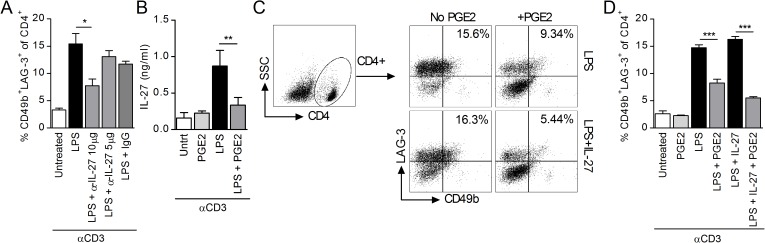
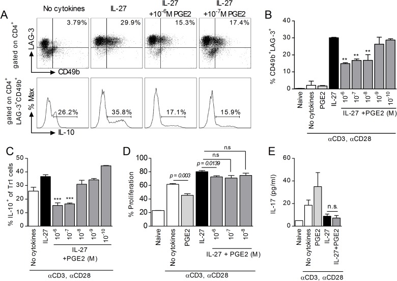
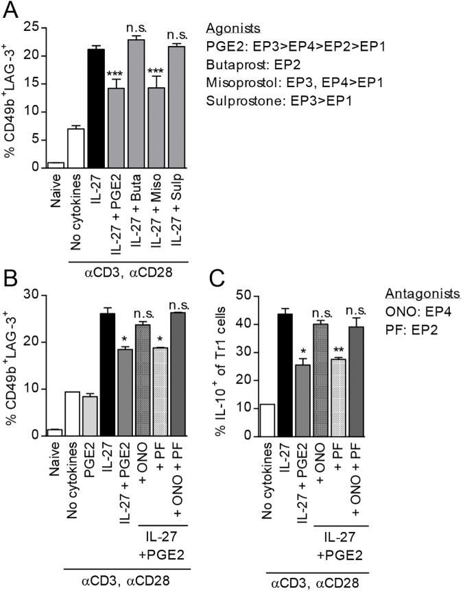
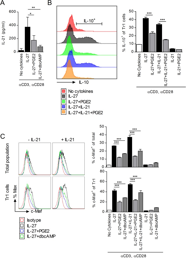
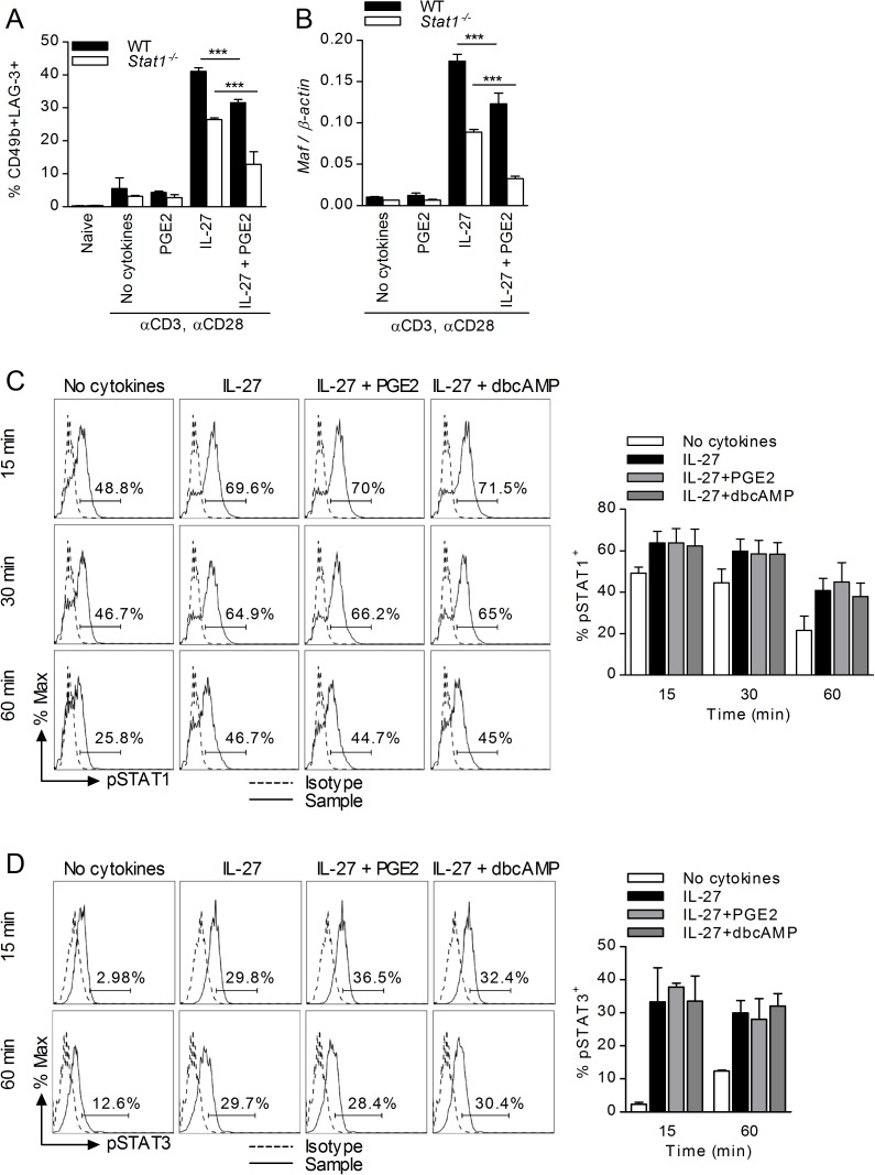
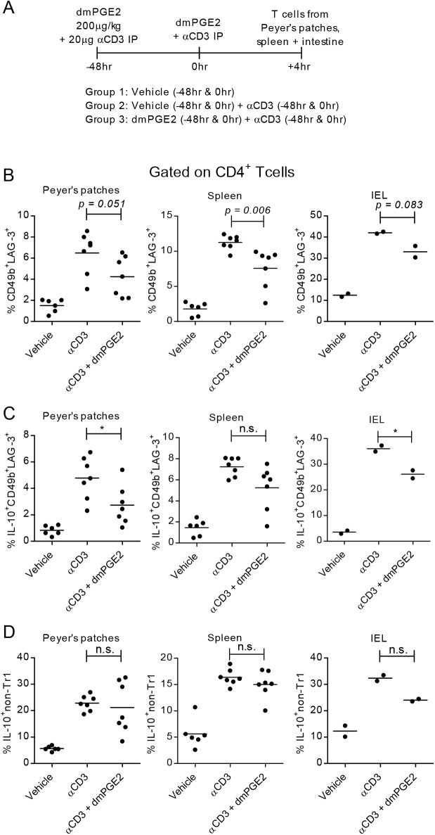
Prostaglandin E2 (PGE2), a major lipid mediator abundant at inflammatory sites, acts as a proinflammatory agent in models of inflammatory/autoimmune diseases by promoting CD4 Th1/Th17 differentiation. Regulatory T cells, including the IL-10 producing Tr1 cells counterbalance the proinflammatory activity of effector Th1/Th17 cells. Tr1 cell differentiation and function are induced by IL-27, and depend primarily on sustained expression of c-Maf in addition to AhR and Blimp-1. In agreement with the in vivo proinflammatory role of PGE2, here we report for the first time that PGE2 inhibits IL-27-induced differentiation and IL-10 production of murine CD4+CD49b+LAG-3+Foxp3- Tr1 cells. The inhibitory effect of PGE2 was mediated through EP4 receptors and induction of cAMP, leading to a significant reduction in c-Maf expression. Although PGE2 reduced IL-21 production in differentiating Tr1 cells, its inhibitory effect on Tr1 differentiation and c-Maf expression also occurred independent of IL-21 signaling. PGE2 did not affect STAT1/3 activation, AhR expression and only marginally reduced Egr-2/Blimp-1 expression. The effect of PGE2 on CD4+CD49b+LAG-3+ Tr1 differentiation was not associated with either induction of Foxp3 or IL-17 production, suggesting a lack of transdifferentiation into Foxp3+ Treg or effector Th17 cells. We recently reported that PGE2 inhibits the expression and production of IL-27 from activated conventional dendritic cells (cDC) in vivo and in vitro. The present study indicates that PGE2 also reduces murine Tr1 differentiation and function directly by acting on IL-27-differentiating Tr1 cells. Together, the ability of PGE2 to inhibit IL-27 production by cDC, and the direct inhibitory effect on Tr1 differentiation mediated through reduction in c-Maf expression, represent a new mechanistic perspective for the proinflammatory activity of PGE2.


Conflict of interest statement

Competing Interests: The authors have declared that no competing interests exist.

Figures

Fig 1
Fig 1. PGE2 inhibits Tr1 cell differentiation in splenocyte cultures.
(A) il10gfp splenocytes were stimulated with 3 μg/ml anti-CD3 and 1 μg LPS in the presence of neutralizing IL-27 antibodies (5 and 10 μg/ml) or IgG control (20 μg/ml). Cells were collected on day 3 and CD4+CD49b+LAG-3+ Tr1 cells were identified by FACS. Data are cumulative from two independent experiments. (B) Splenocytes were stimulated with anti-CD3 and LPS in the presence of PGE2 (10-6M). Supernatant was collected on day 3 and IL-27 levels analyzed by ELISA. (C-D) Splenocytes were stimulated with anti-CD3 and LPS in the presence or absence of IL-27 (50 ng/ml) and PGE2 (10-6M). Cells were collected on day 3 and CD4+CD49b+LAG-3+ Tr1 cells were identified by FACS. (C) Representative sample shows gating strategy to determine percentage of CD49b+LAG-3+ within CD4+ T cells and (D) graph presents representative data from three independent experiments. Each sample was tested in duplicate and results represent means ± SD. Significance was evaluated by one-way ANOVA; **P<0.01.
Fig 2
Fig 2. PGE2 inhibits IL-27 induced differentiation of Tr1 cells.
(A-C) Naïve CD4+CD62L+ cells from il10gfp mice were stimulated with plate-bound anti-CD3 (3 μg/ml) and soluble anti-CD28 (1 μg/ml) in the presence of 50 ng/ml IL-27 and various concentrations of PGE2 for three days. Cells were collected on day 3 and analyzed by FACS for Tr1 markers, and IL-10 within CD49b+LAG-3+ populations. Representative samples show CD49b+LAG-3+ populations (upper panel) and histograms of IL-10 (lower panel). Data are representative of four independent experiments. Each sample was tested in duplicate and results represent means ± SD. Significance was determined using one-way ANOVA and * represents the P value for a sample versus IL-27 control. **P<0.01, ***P<0.001. (D) For cell proliferation experiments, naïve CD4+CD62L+ cells were stained with CFSE per manufacturer’s instructions prior to stimulation with anti-CD3, anti-CD28, IL-27 and PGE2. Incorporation of CFSE was analyzed by FACS on day 3. Data are representative of two independent experiments. Each sample was tested in triplicate and results represent means ± SD. Significance was determined using unpaired t-test. (E) Naïve CD4+CD62L+ cells were stimulated in the presence of 50 ng/ml IL-27 and 10-6M PGE2. Supernatant was collected on day 3 and subjected to ELISA to determine IL-17 levels. Data are representative of three independent experiments. Each sample was tested in duplicate and results represent means ± SD. Significance was determined using one-way ANOVA.
Fig 3
Fig 3. PGE2 signals through EP4 to inhibit Tr1 cell differentiation and expression of IL-10.
(A) Naïve CD4+CD62L+ cells were stimulated in the presence of IL-27 and 10-6M PGE2 or 10-5M of EP receptor agonists: Butaprost (EP2), Misoprostol (EP3, EP4>EP1) or Sulprostone (EP3>EP1). Cells were collected on day 3 and analyzed by FACS for Tr1 cell markers. (B,C) Naïve CD4+CD62L+ cells from il10gfp mice were pretreated for 30 minutes with 10-6M EP receptor antagonists, ONO-AE3-208 (ONO; EP4) or PF-04418948 (PF; EP2) prior to treatment with 50 ng/ml IL-27 and 10-7M PGE2. Cells were collected on day 3 and analyzed by FACS for Tr1 markers and IL-10 expression. Data are representative of three independent experiments. Each sample was tested in duplicate and results represent means ± SD. Significance was evaluated by one-way ANOVA and * symbolizes significance of experimental condition versus IL-27 control; *P<0.05, **P<0.01, ***P<0.001.
Fig 4
Fig 4. PGE2 signals through cAMP, but not EPAC or PKA, to inhibit IL-27 mediated Tr1 differentiation.
(A) il10gfp naïve CD4+CD62L+ cells were pretreated with PI3K inhibitor LY294002 (LY) for 30 min prior to stimulation in the presence of IL-27 and PGE2. (B) Naïve CD4+CD62L+ cells were stimulated as described in the presence of dbcAMP. (C) Cells were stimulated in the presence of a cell-permeable EPAC activator 8-pCPT-2′-O-Me-cAMP (8-CPT). Cells were collected on day 3 and analyzed by FACS for Tr1 markers. (D, E) Naïve CD4+CD62L+ cells were pretreated with PKA inhibitors PKI 6–22 and PKI 5–24 (2x10⁻⁵M) for 30 minutes prior to stimulation with IL-27 and PGE2 and analyzed on day 3 for Tr1 markers (D) and IL-10 within Tr1 cell populations (E). Data are representative of three independent experiments. Each sample was tested in duplicate and results represent means ± SD. Significance was evaluated by one-way ANOVA and * symbolizes significance of experimental condition versus IL-27 control (B); *P<0.05, **P<0.01, ***P<0.001.
Fig 5
Fig 5. PGE2 inhibits c-Maf in CD4 T cells differentiated in the presence of IL-27.
(A) Naïve CD4+CD62L+ cells were stimulated in the presence of IL-27 and PGE2. RNA was collected at 24, 48 and 72 hr and analyzed by RT-PCR for Maf and Ahr expression. (B) Naïve CD4+CD62L+ cells were stimulated in the presence of IL-27 and PGE2 or dbcAMP. Cells were collected on day 3 and analyzed by RT-PCR for Maf expression. (C) Splenocytes from il10gfp mice were stimulated with 3 μg/ml anti-CD3 and 1 μg LPS in the presence of IL-27 and PGE2. RNA was collected on day 3 and Maf expression was analyzed by RT-PCR. (D) Naïve CD4+CD62L+ cells were stimulated in the presence of IL-27 and PGE2 or dbcAMP. Cells were collected on day 3 and analyzed by FACS for intracellular c-Maf within the whole cell population (upper panel) and within CD49b+LAG-3+ populations (lower panel). Graphical representation of data is on the right. (E) Naïve CD4+CD62L+ cells were stimulated as in D. Cells were collected on day 3 and analyzed by FACS for intracellular Egr-2 within the whole cell population (upper panel) and within CD49b+LAG-3+ populations (lower panel). (F) Naïve CD4+CD62L+ cells were stimulated as in D. Cells were collected on day 3 and analyzed by FACS for intracellular Blimp-1 within the whole cell population and within CD49b+LAG-3+ populations. Data are representative of two to three independent experiments. Each sample was tested in duplicate and results represent means ± SD. Significance was evaluated by one-way ANOVA and * symbolizes significance of experimental condition versus IL-27 control (B, D, E); *P<0.05, **P<0.01, ***P<0.001.
Fig 6
Fig 6. Inhibition of c-Maf by PGE2 is independent of IL-21.
(A) Naïve CD4+CD62L+ cells were stimulated in the presence of IL-27 and PGE2 or dbcAMP. Supernatant was collected on day 3 and analyzed by ELISA for IL-21. Data are cumulative from two independent experiments. (B) Naïve CD4+CD62L+ cells from il10gfp mice were stimulated in the presence of IL-27, 100 ng/ml IL-21 and PGE2. Cells were collected on day 3 and analyzed by FACS to determine IL-10 production within CD49b+LAG-3+ CD4 T cells. Data are representative of two independent experiments. (C) Naïve CD4+CD62L+ cells from C57BL/6 mice were stimulated as above. Cells were collected on day 3 and analyzed by FACS to determine presence of intracellular c-Maf within CD4 T cells (top) and CD49b+LAG-3+ CD4 T cells (bottom). Histograms show representative samples, while graphs present data from one of three independent experiments. Each sample was tested in duplicate and results represent means ± SD. Significance was determine by one-way ANOVA; **P<0.01, ***P<0.001.
Fig 7
Fig 7. Inhibition of Tr1 differentiation by PGE2 is independent of STAT1/3.
Naïve CD4+CD62L+ cells from wildtype 129S6 (WT) and Stat1-/- mice were stimulated in the presence of IL-27 and PGE2. (A) Samples were collected on day 3 and cells were analyzed for Tr1 markers. (B) Cells were subjected to RNA extraction and subsequent analysis by RT-PCR for Maf expression. (C) Naïve CD4+CD62L+ cells from C57BL/6 mice were stimulated in the presence of IL-27, PGE2 and 10-4M dbcAMP. Cells were collected 15, 30 60 minutes later and analyzed by FACS for intracellular phospho-STAT1 (Tyr701). (D) Naïve CD4+CD62L+ cells from C57BL/6 mice were stimulated as described in (C) for 15 and 60 minutes. Cells were analyzed by FACS for intracellular phospho-STAT3 (Tyr705). Data are cumulative from three independent experiments. Each sample was tested in duplicate and results represent means ± SD. Significance was determined using one-way ANOVA; ***P<0.001.
Fig 8
Fig 8. PGE2 inhibits Tr1 differentiation in vivo.
(A) Schematic of timeline and experimental groups. Il10gfp mice were injected i.p. with vehicle, 20 μg/ml anti-CD3 or anti-CD3 and 4μg dmPGE2 at 0hr and 48hr. Peyer’s patches, spleen, and intraepithelial lymphocyte populations were collected. (B) CD4+ populations were analyzed by FACS for Tr1 cell populations (CD49b+LAG-3+). (C-D) CD4+ populations were analyzed by FACS for IL-10 expression in both Tr1 populations and non-Tr1 populations (including CD49b+LAG-3-, CD49b-LAG-3+ and CD49b-LAG-3- populations). Data are cumulative of two independent experiments. Significance was evaluated by unpaired t-test.

Similar articles

Cited by

References

    1. Sakaguchi S, Sakaguchi N, Asano M, Itoh M, Toda M. Immunologic self-tolerance maintained by activated T cells expressing IL-2 receptor alpha-chains (CD25). Breakdown of a single mechanism of self-tolerance causes various autoimmune diseases. J Immunol. 1995;155(3):1151–64. - PubMed
    1. Brunkow ME, Jeffery EW, Hjerrild KA, Paeper B, Clark LB, Yasayko SA, et al. Disruption of a new forkhead/winged-helix protein, scurfin, results in the fatal lymphoproliferative disorder of the scurfy mouse. Nat Genet. 2001;27(1):68–73. doi: 10.1038/83784 - DOI - PubMed
    1. Groux H, O'Garra A, Bigler M, Rouleau M, Antonenko s, de Vries JE, et al. A CD4+ T-cell subset inhibits antigen-specific T-cell responses and prevents colitis. Nature. 1997;389(6652):737–42. doi: 10.1038/39614 - DOI - PubMed
    1. Gagliani N, Magnani CF, Huber S, Gianolini ME, Pala M, Licona-Limon P, et al. Coexpression of CD49b and LAG-3 identifies human and mouse T regulatory type 1 cells. Nat Med. 2013;19(6):739–46. doi: 10.1038/nm.3179 - DOI - PubMed
    1. Gagliani N, Jofra T, Stabilini A, Valle A, Atkinson M, Roncarolo MG, et al. Antigen-specific dependence of Tr1-cell therapy in preclinical models of islet transplant. Diabetes. 2010;59(2):433–9. doi: 10.2337/db09-1168 - DOI - PMC - PubMed

MeSH terms